THE DIABETIC FOOT
•Ali akbar Beigi M.D
General and Vascular
Surgeon Taleghani
Hospital Shahid Beheshti
U.M.S
3.
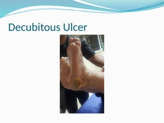
Diabetic Foot
Definition:
Le pieddiabétique regroupe toute infection, ulcération ou destruction des
tissus profonds du pied associées à une neuropathie et/ou une artériopathie
périphérique des membres inférieurs chez le diabétique [1]. C'est une
complication fréquente et grave du diabète avec un taux d´amputation de
membres inférieurs très élevé et des conséquences souvent dramatiques
sur le plan socio-économique et psychologique [2]. En Afrique, les lésions du
pied chez le diabétique sont malheureusement très courantes. Elles sont à
l'origine de 15% à 25% des hospitalisations chez les diabétiques [2,3].
Souvent, la pauvreté, le manque d´hygiène et la marche à pieds nus
interagissent pour aggraver l´impact des lésions du pied causées par le
diabète
1. International Working Group on the Diabetic Foot
4.
• Le pieddiabétique est une complication
fréquente et grave du diabète;
• Le pied diabétique est la principale cause
d'amputation non traumatique chez le
diabétique.
5.
Diabetic Foot
Definition:
Infection, ulcerationor destruction of deep
tissues associated with neurological
abnormalities & various degrees of peripheral
vascular diseases in the lower limb
(based on WHO definition)
6.
Epidemiology
15% is theprevalence of foot ulcer in diabetics
Patients intheir lifetime
40% - 60% of all non traumatic amputation in
lower limb
85% of diabetic related foot amputation are
preceded by foot ulcer
4 out of 5 ulcer in diabetics are precipitated by
trauma
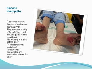
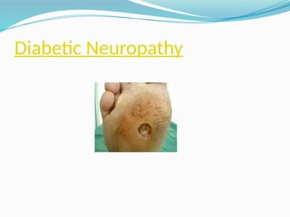
Diabetic
Neuropathy
History & careful
footexamination are
mandatory to
diagnose neuropathy
Up to 50%of type2
diabetic patient have
significant
neuropathy & at risk
of foot ulcer
Sensorimotor &
peripheral
sympathatic
neuropathy are
major risk factors for
ulcer
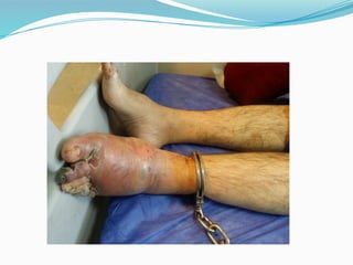
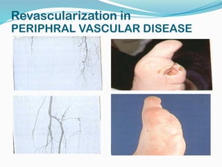
Periphral vascular disease&diabetic
PVD
PVD is the most important factors related to
outcome of diabetic foot ulcer
PVD is diagnosed by simple clinical examination
Symptoms of ischemia may be masked by
neuropathy
Microangiopathy shouldn't be accepted as primary
cause of ulcer
non invasive vascular test determines probability of
healing Ankle Brachial index&Trans cutaneous
oxigenometry
Outcome of revascularization is similar to that in
non-diabetic
21.
Examination
Neurological examination
◦Vibration perception – tuning
fork at 128 Hz
◦ Light pressure - Simmes –
Weinstein 10 gram
monofilament
◦ Light touch
◦ Two point discrimination
◦ Pain
◦ Temperature perception
◦ Deep tendon reflexes
◦ Clonus
◦ Babinski test
◦ Romberg test
Vascular Examination
◦ Palpation of pulses
◦ Skin/limb colour
changes
◦ Presence of edema
◦ Temperature gradient
◦ Skin changes
Abnormal wrinkling
Absence of hair
Onychodystrophy
◦ Venous filling time
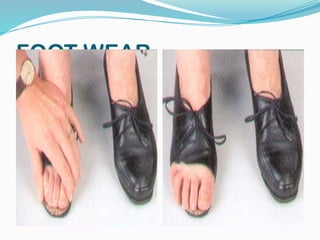
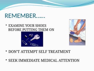
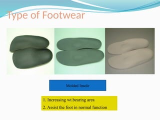
Preventive care
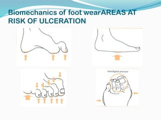
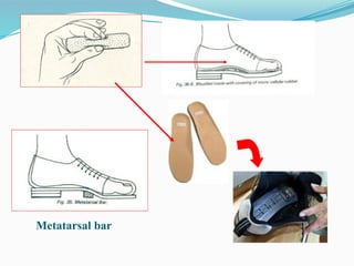
Biomechanics offoot wear
Biomechanical abnormalities are
consequence of neuropathy, they lead to
abnormal foot pressure
Foot deformity & neuropathy increase the
risk of ulcer
Pressure relief is essential for ulcer healing
and/or prevention
Frequent inspection of shoes & insoles is
mandatory
Appropriate foot wear significantly reduce
ulcer recurrence
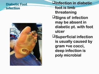
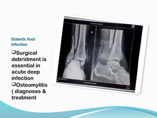
Infection in diabetic
footis limb
threatening
Signs of infection
may be absent in
diabetic pt. with foot
ulcer
Superficial infection
is usually caused by
gram +ve cocci,
deep infection is
poly microbial
Diabetic Foot
Infection
27.
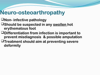
Neuro-osteoarthropathy
Non- infective pathology
Shouldbe suspected in any swollen hot
erythematous foot
Differentiation from infection is important to
prevent misdiagnosis & possible amputation
Treatment should aim at preventing severe
deformity
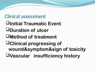
Clinical assessment
Initial TraumaticEvent
Duration of ulcer
Method of treatment
Clinical progressing of
wound&symptom&sign of toxicity
Vascular insufficiency history
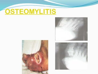
32.
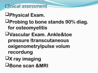
Clinical assessment
Physical Exam.
Probingto bone stands 90% diag.
for osteomyelitis
Vascular Exam. Ankle&toe
pressure ltranscutaneous
oxigenometrylpulse volum
recordung
X ray imaging
Bone scan &MRI
33.
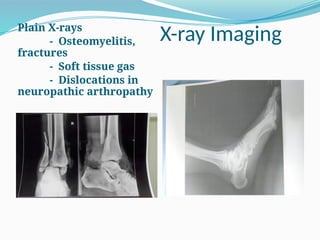
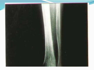
X-ray Imaging
Plain X-rays
-Osteomyelitis,
fractures
- Soft tissue gas
- Dislocations in
neuropathic arthropathy
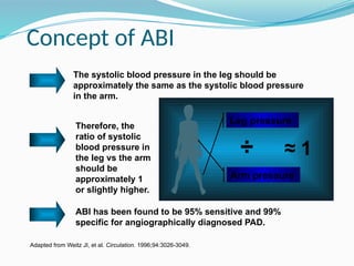
Concept of ABI
ABIhas been found to be 95% sensitive and 99%
specific for angiographically diagnosed PAD.
The systolic blood pressure in the leg should be
approximately the same as the systolic blood pressure
in the arm.
Therefore, the
ratio of systolic
blood pressure in
the leg vs the arm
should be
approximately 1
or slightly higher.
Adapted from Weitz JI, et al. Circulation. 1996;94:3026-3049.
Arm pressure
Leg pressure
÷ ≈ 1
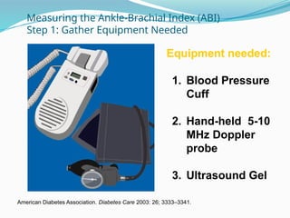
37.
Equipment needed:
1. BloodPressure
Cuff
2. Hand-held 5-10
MHz Doppler
probe
3. Ultrasound Gel
American Diabetes Association. Diabetes Care 2003: 26; 3333–3341.
Measuring the Ankle-Brachial Index (ABI)
Step 1: Gather Equipment Needed
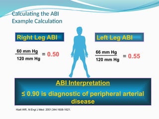
Calculating the ABI
ExampleCalculation
66 mm Hg
120 mm Hg
Hiatt WR. N Engl J Med. 2001;344:1608-1621.
= 0.50 = 0.55
Right Leg ABI Left Leg ABI
60 mm Hg
120 mm Hg
Right Leg ABI
ABI Interpretation
≤ 0.90 is diagnostic of peripheral arterial
disease
40.
An Auto ABIDevice
Systolic Pressures and ABI PVR Waveforms
41.
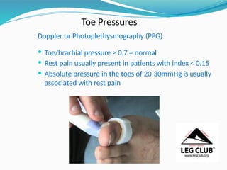
Toe Pressures
Doppler orPhotoplethysmography (PPG)
Toe/brachial pressure > 0.7 = normal
Rest pain usually present in patients with index < 0.15
Absolute pressure in the toes of 20-30mmHg is usually
associated with rest pain
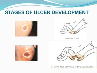
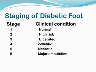
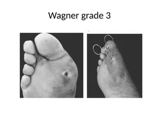
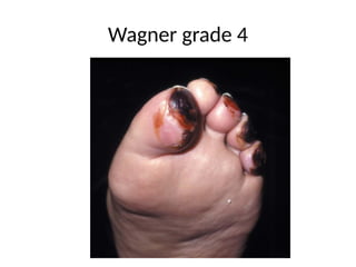
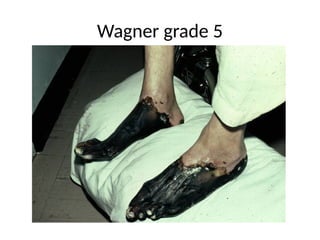
Staging of DiabeticFoot
Stage Clinical condition
1 Normal
2 High risk
3 Ulcerated
4 cellulitic
5 Necrotic
6 Major amputation
44.
Diabetic Foot UlcerTreatment
Modalities
Microbiological control
Wound control
Vascular control
Mechanical control
Metabolic control
Educational control
45.
Diabetic Foot UlcerTreatment
Multidisciplenary approach
Staging dictate the treatment
option
Continuity of care & life long
observation
46.
Diabetic Foot UlcerTreatment
Rigid shoeing System for repeated local
trauma ulcer l total contact Cast 90% healing
in non vascular ulcer
Off loading of ulcer/Half shoes/felted foam
plantar dressing
Non weight bearing ambulation/bed rest
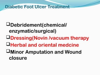
Diabetic Foot UlcerTreatment
Debridement(chemical/
enzymatic/surgical)
Dressing(Novin /vacuum therapy
Herbal and oriental medcine
Minor Amputation and Wound
closure
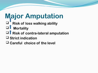
50.
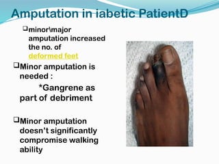
Amputation in iabeticPatientD
minormajor
amputation increased
the no. of
deformed feet
Minor amputation is
needed :
*Gangrene as
part of debriment
Minor amputation
doesn’t significantly
compromise walking
ability
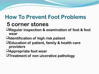
How To PreventFoot Problems
5 corner stones
Regular inspection & examination of foot & foot
wear
Identification of high risk patient
Education of patient, family & health care
providers
Appropriate foot wear
Treatment of non ulcerative pathology
Diabetic foot
Introduction
Diabetesmellitus is a group of metabolic diseases
characterized by hyperglycemia resulting from
defects in insulin secretion, insulin action, or both.
The chronic hyperglycemia of diabetes is associated
with long-term damage, dysfunction, and failure of
various organs, especially the eyes, kidneys, nerves,
heart, and blood vessels.
Diabetic foot is defined as any foot pathology that
results directly from diabetes or its long term
complications
Two types of diabetes: type I and type II diabetes
71.
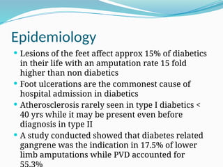
Epidemiology
Lesions ofthe feet affect approx 15% of diabetics
in their life with an amputation rate 15 fold
higher than non diabetics
Foot ulcerations are the commonest cause of
hospital admission in diabetics
Atherosclerosis rarely seen in type I diabetics <
40 yrs while it may be present even before
diagnosis in type II
A study conducted showed that diabetes related
gangrene was the indication in 17.5% of lower
limb amputations while PVD accounted for
72.
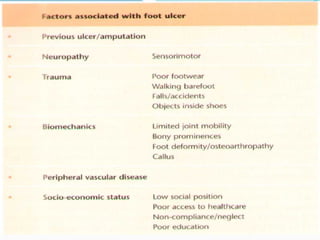
Epidemiology – riskfactors
Male sex
DM > 10 years duration
Peripheral neuropathy
Abnormal foot structure
Peripheral arterial disease
Smoking
H/O previous ulceration / amputation
Poor glycemic control (HbA1c > 7%)
73.
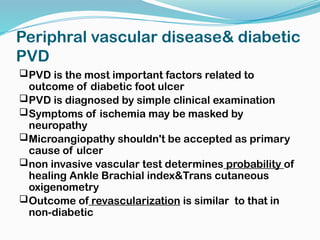
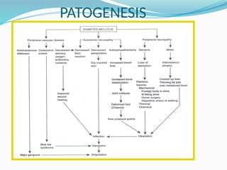
Pathophysiology
Factors leadingto development of diabetic foot:
Diabetic macroangiopathy – peripheral arterial
occlusive disease
Diabetic microangiopathy – thickening of
basement membranes
Diabetic polyneuropathy
Diabetic osteoathropathy – abnormal foot
biomechanics
Reduced resistance to infection
Delayed wound healing
Reduced rate of collateral vessel formation
74.
Diabetic angiopathy
Diabeticmacroangiopathy is histologically
similar to non diabetic atherosclerosis but
distributed in the distal segments of the lower
extremities (calf and foot arteries)
Arterial calcification readily detectable on plain
x ray with constriction noted on angiography.
This compromises oxygen supply to the
periphery
Gas exchange is compromised by marked
thickening of the capillary basement membrane
– a feature of diabetic microangiopathy
75.
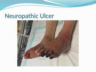
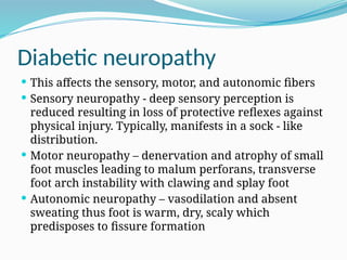
Diabetic neuropathy
Thisaffects the sensory, motor, and autonomic fibers
Sensory neuropathy - deep sensory perception is
reduced resulting in loss of protective reflexes against
physical injury. Typically, manifests in a sock - like
distribution.
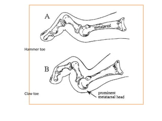
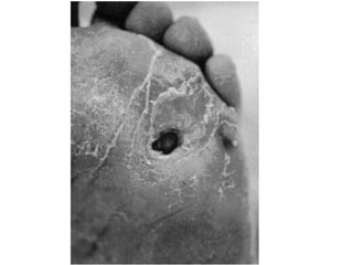
Motor neuropathy – denervation and atrophy of small
foot muscles leading to malum perforans, transverse
foot arch instability with clawing and splay foot
Autonomic neuropathy – vasodilation and absent
sweating thus foot is warm, dry, scaly which
predisposes to fissure formation
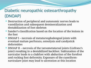
Diabetic neuropathic osteoarthropathy
(DNOAP)
Destruction of peripheral and autonomic nerves leads to
vasodilation and subsequent demineralization and
destabilization of foot skeleton
Sander’s classification based on the location of the lesions in
the foot
DNOAP I – necrosis of metatarsophalengeal joints with
eventual malum perforans, osteolysis and candystick
deformities
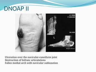
DNOAP II – necrosis of the tarsometatarsal joints (Lisfranc’s
joint) resulting in a destabilized backfoot. Subluxation of the
navicular leads to a clubfoot with abduction of the forefoot
and rocking foot deformity. Exposure of the cuneiform-
naviculare joint may lead to ulceration at this location
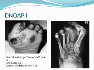
82.
DNOAP I
Central malumperforans – MT I and
IV
Osteolysis MT II
Candystick deformity MT III
83.
DNOAP II
Ulceration overthe navicular-cuneiform joint
Destruction of lisfranc articulations
Fallen medial arch with navicular subluxation
84.
DNOAP
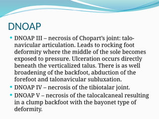
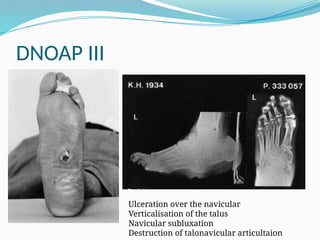
DNOAP III– necrosis of Chopart’s joint: talo-
navicular articulation. Leads to rocking foot
deformity where the middle of the sole becomes
exposed to pressure. Ulceration occurs directly
beneath the verticalized talus. There is as well
broadening of the backfoot, abduction of the
forefoot and talonavicular subluxation.
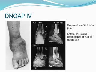
DNOAP IV – necrosis of the tibiotalar joint.
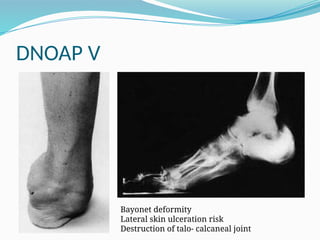
DNOAP V – necrosis of the talocalcaneal resulting
in a clump backfoot with the bayonet type of
deformity.
85.
DNOAP III
Ulceration overthe navicular
Verticalisation of the talus
Navicular subluxation
Destruction of talonavicular articultaion
Increased infection rate
Skin fissurations predisposes to penetration of
infectious microbes
Polymorphonuclear granulocyte chemotaxis and
phagocytosis is impaired
Polyneuropathy predisposes to deep seated
infections due to impaired pain sensation
Both anaerobe and aerobe infections are
implicated in diabetic foot infections
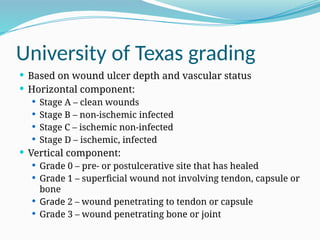
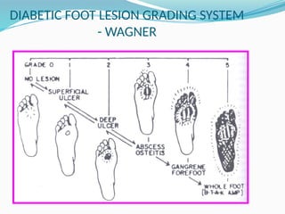
University of Texasgrading
Based on wound ulcer depth and vascular status
Horizontal component:
Stage A – clean wounds
Stage B – non-ischemic infected
Stage C – ischemic non-infected
Stage D – ischemic, infected
Vertical component:
Grade 0 – pre- or postulcerative site that has healed
Grade 1 – superficial wound not involving tendon, capsule or
bone
Grade 2 – wound penetrating to tendon or capsule
Grade 3 – wound penetrating bone or joint
Diabetic foot infection
Divided into uncomplicated non limb
threatening infection - superficial cellulitis of
limited extension that can be treated on an
outpatient basis
Complicated limb threatening infections are
more extended and penetrate to deeper tissues,
such as tendons, joint capsules, bone or
articulations. They require inpatient treatment
with surgical debridement and intravenous
antibiotics
Osteomyelitis has therapeutic implications such
as prolonged antibiotic courses and need for
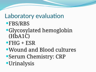
101.
Diabetic foot infection
Superficial swabs overestimate the number of
likely microorganisms therefore a deep tissue
specimen is preferred as it is more
representative
Aerobic gram +ve cocci most common infecting
organisms: S. aureus and β-hemolytic streptococci
(especially group B)
Chronic wounds have more complex flora:
enterococci, enterobactereciae, obligate
anaerobes, P. aeuroginosa and other non-
fermentative gram negative rods
102.
Management
Preventative footcare
Diabetic foot ulcer (DFU) care
Ischemia management
Neuropathy management
Surgery
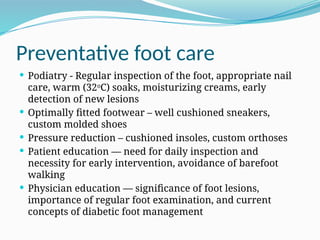
103.
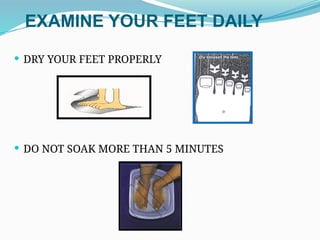
Preventative foot care
Podiatry - Regular inspection of the foot, appropriate nail
care, warm (32o
C) soaks, moisturizing creams, early
detection of new lesions
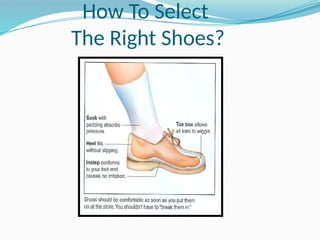
Optimally fitted footwear – well cushioned sneakers,
custom molded shoes
Pressure reduction – cushioned insoles, custom orthoses
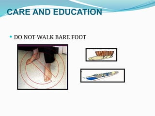
Patient education — need for daily inspection and
necessity for early intervention, avoidance of barefoot
walking
Physician education — significance of foot lesions,
importance of regular foot examination, and current
concepts of diabetic foot management
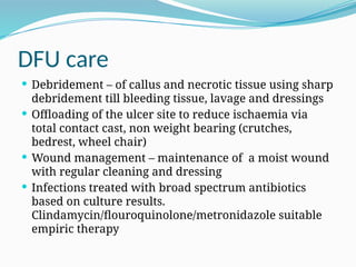
104.
DFU care
Debridement– of callus and necrotic tissue using sharp
debridement till bleeding tissue, lavage and dressings
Offloading of the ulcer site to reduce ischaemia via
total contact cast, non weight bearing (crutches,
bedrest, wheel chair)
Wound management – maintenance of a moist wound
with regular cleaning and dressing
Infections treated with broad spectrum antibiotics
based on culture results.
Clindamycin/flouroquinolone/metronidazole suitable
empiric therapy
105.
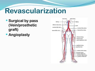
Ischemia/neuropathy
Angiography evaluatesfor chance of catheter
intervention or vascular surgery
Vascular bypass surgery successful if occlusion is
supramalleolar but less so in inframalleolar
PAOD
Aspirin is useful for primary and secondary
prevention
Neuropathy treated pharmacologically with
agents such as carbamazepine, gabapentin and
pregabalin and prevention of minor trauma that
will go undetected due to insensate foot
106.
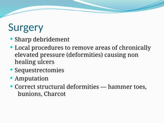
Surgery
Sharp debridement
Local procedures to remove areas of chronically
elevated pressure (deformities) causing non
healing ulcers
Sequestrectomies
Amputation
Correct structural deformities — hammer toes,
bunions, Charcot
107.
Indications for amputation
Uncontrollable infection or sepsis
Inability to obtain a plantar grade, dry foot that
can tolerate weight bearing
Non ambulatory patient
108.
Other peripheral vasculardiseases
Peripheral arterial occlusive disease (PAOD)
Post thrombotic syndrome
Chronic venous insufficiency
109.
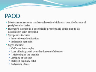
PAOD
Most commoncause is atheroclerosis which narrows the lumen of
peripheral arteries
Buerger’s disease is a potentially preventable cause due to its
association with smoking
Symptoms include:
Intermittent claudication
Ischaemic rest pain
Signs include:
Calf muscles atrophy
Loss of hair growth over the dorsum of the toes
Thickening of the toenails
Atrophy of the skin
Delayed capillary refill
Ischaemic ulcers
110.
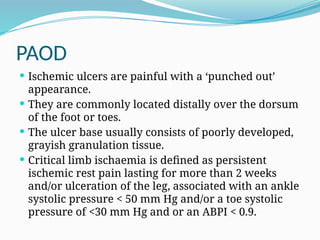
PAOD
Ischemic ulcersare painful with a ‘punched out’
appearance.
They are commonly located distally over the dorsum
of the foot or toes.
The ulcer base usually consists of poorly developed,
grayish granulation tissue.
Critical limb ischaemia is defined as persistent
ischemic rest pain lasting for more than 2 weeks
and/or ulceration of the leg, associated with an ankle
systolic pressure < 50 mm Hg and/or a toe systolic
pressure of <30 mm Hg and or an ABPI < 0.9.
111.
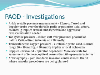
PAOD - Investigations
Ankle systolic pressure measurement – 12cm cuff used and
doppler probe over the dorsalis pedis or posterior tibial artery.
<50mmHg implies critical limb ischemia and aggressive
revascularisation needed
Toe systolic pressure – 25mm cuff over proximal phalanx of
hallux. Critical limb ischemia at < 30mmHg
Transcutaneous oxygen pressure – electronic probe used. Normal
range 30 – 50 mmHg. < 30 mmHg implies critical ischaemia
Doppler ultrasound – operator dependent. More accurate for
assessment of femoropopliteal vessels than tibioperoneal arteries
Arteriography – gold standard, invasive, contrast used. Useful
where vascular procedures are being planned
112.
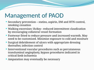
Management of PAOD
Secondary prevention – statins, aspirin, DM and HTN control,
smoking cessation
Walking excersises 1h/day– reduced intermittent claudication
by encouraging collateral vessel formation
Footwear fitted to reduce pressure and increased warmth. May
need to be customised. Minimise exposure to cold and mositure
Surgical debridement of ulcers with appropriate dressing
thereafter, infection control
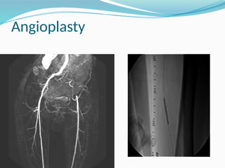
Interventional vascular procedures such as percutaneous
transluminal angioplasty, bypass procedures indicated in
critical limb ischaemia
Amputation may eventually be necessary
113.
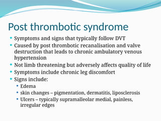
Post thrombotic syndrome
Symptoms and signs that typically follow DVT
Caused by post thrombotic recanalisation and valve
destruction that leads to chronic ambulatory venous
hypertension
Not limb threatening but adversely affects quality of life
Symptoms include chronic leg discomfort
Signs include:
Edema
skin changes – pigmentation, dermatitis, liposclerosis
Ulcers – typically supramalleolar medial, painless,
irregular edges
114.
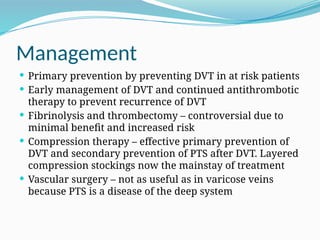
Management
Primary preventionby preventing DVT in at risk patients
Early management of DVT and continued antithrombotic
therapy to prevent recurrence of DVT
Fibrinolysis and thrombectomy – controversial due to
minimal benefit and increased risk
Compression therapy – effective primary prevention of
DVT and secondary prevention of PTS after DVT. Layered
compression stockings now the mainstay of treatment
Vascular surgery – not as useful as in varicose veins
because PTS is a disease of the deep system
115.
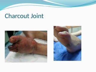
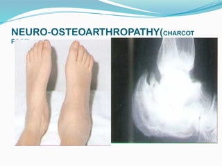
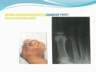
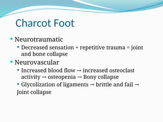
Charcot Foot
Neurotraumatic
Decreased sensation + repetitive trauma = joint
and bone collapse
Neurovascular
Increased blood flow increased osteoclast
→
activity osteopenia Bony collapse
→ →
Glycolization of ligaments brittle and fail
→ →
Joint collapse
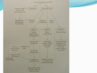
116.
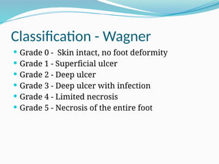
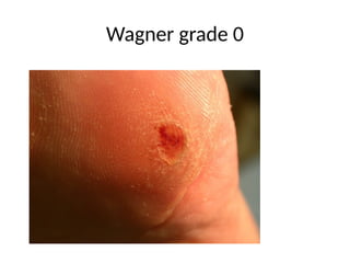
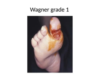
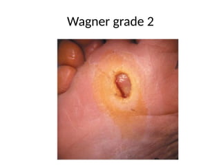
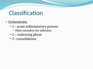
Classification
Eichenholtz
1– acute inflammatory process
Often mistaken for infection
2 – coalescing phase
3 - consolidation
117.
Classification
Location
Forefoot,midfoot (most common) , hindfoot
Atrophic or hypertrophic
Radiographic finding
Little treatment implication
Indications for Amputation
Uncontrollable infection or sepsis
Inability to obtain a plantar grade, dry foot that
can tolerate weight bearing
Non-ambulatory patient
Decision not always straightforward
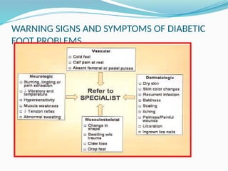
141.
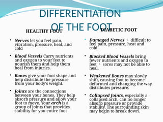
DIFFERENTIATION
OF THE FOOT
HEALTHYFOOT
• Nerves let you feel pain,
vibration, pressure, heat, and
cold
• Blood Vessels Carry nutrients
and oxygen to your feet to
nourish them and help them
heal from injuries.
• Bones give your foot shape and
help distribute the pressure
from your body's weight.
• Joints are the connections
between your bones. They help
absorb pressure and allow your
foot to move. Your arch is a
group of joints that provides
stability for you entire foot
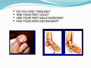
DIABETIC FOOT
• Damaged Nerves difficult to
feel pain, pressure, heat and
cold.
• Blocked Blood Vessels bring
fewer nutrients and oxygen to
feet sores may not be able to
heal.
• Weakened Bones may slowly
shift, causing foot to become
deformed and changing the way
distributes pressure.
• Collapsed Joints, especially a
collapsed arch, can no longer
absorb pressure or provide
stability. The surrounding skin
may begin to break down.
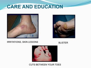
142.
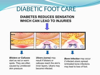
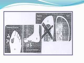
DIABETIC FOOT CARE
DIABETESREDUCES SENSATION
WHICH CAN LEAD TO INJURIES
Blisters or Calluses
start as red or warm
spots. They are often
caused by unrelieved
skin pressure
Ulcers (sores) may
result if blisters or
calluses reach the skin's
inner layers. Ulcers may
become infected.
Bone Infection may occur
if infected ulcers spread.
Untreated bone infections
may lead to loss of foot.
143.
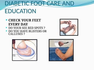
DIABETIC FOOT CAREAND
EDUCATION
CHECK YOUR FEET
EVERY DAY
DO YOUR SEE RED SPOTS ?
DO YOU HAVE BLISTERS OR
CALLUSES ?
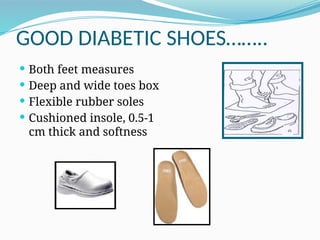
GOOD DIABETIC SHOES……..
Both feet measures
Deep and wide toes box
Flexible rubber soles
Cushioned insole, 0.5-1
cm thick and softness
153.
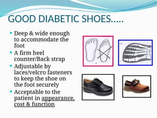
GOOD DIABETIC SHOES…..
Deep & wide enough
to accommodate the
foot
A firm heel
counter/Back strap
Adjustable by
laces/velcro fasteners
to keep the shoe on
the foot securely
Acceptable to the
patient in appearance,
cost & function
Conclusion
Multi-disciplinary approachneeded
Going to be an increasing problem
High morbidity and cost
Solution is probably in prevention
Most feet can be spared…at least for a while
#36 The ankle-brachial index is an easy-to-use tool that can performed in an any clinician’s office with a stethoscope and a hand-held Doppler device. The concept of the ABI is as follows:
The systolic blood pressure in the leg should be approximately the same as the systolic blood pressure in the arm.
Therefore, the ratio of systolic blood pressure in the leg vs the arm should be approximately 1.
ABI has been found to be 95% sensitive and 99% specific for angiographically
diagnosed PAD.
Weitz JI, et al. Diagnosis and treatment of chronic arterial insufficiency of the lower extremities: a critical review. Circulation. 1996;94:3026-3049.
#37 Step 1: You will need the following equipment to measure the ankle-brachial index (ABI): a blood pressure cuff, and a hand-held 5-10 MHz Doppler probe.
#39 Here we see the calculation worked through again with data.
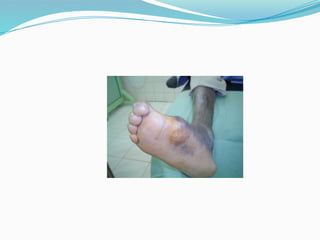
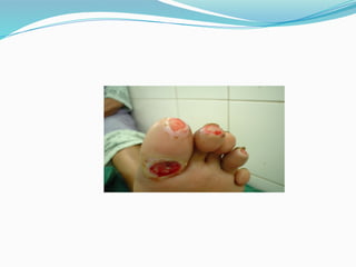
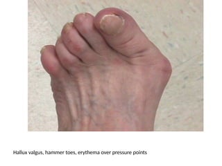
#77 A marked Hallux valgus deformity and early hammer-toe deformities from diabetic motor neuropathy.
Note the areas of persistent erythema over pressure points on the first MTP joint and on the dorsum of the proximal phalanges.
This patient requires a modification of shoe gear to relieve pressure and prevent callus and ulcer formation.
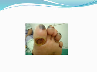
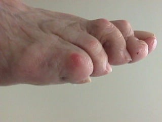
#78 Severe hammer and claw-toe deformities.
There are areas of persistent erythema on the dorsum of the fourth and fifth toes.
The consequences of the ill-fitting shoe gear have now progressed to marked callus formation at the peak of the hammer toe deformities on the dorsum of the second and third toes.
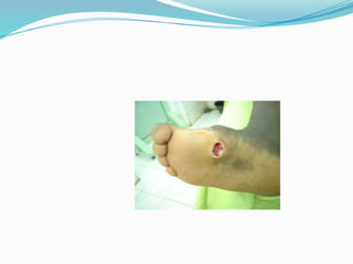
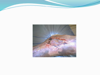
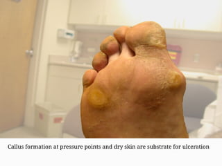
#79 Diabetic motor neuropathy has resulted in hammer and claw-toe deformities and very prominent metatarsal heads on the plantar surface of the foot.
Excessive pressure on the metatarsal heads and inadequate shoe gear have resulted in marked callus build-up that is further accelerated by the dry skin.
The patient is at high risk for ulceration at these sites.
Ted: Can we remove background and put neutral color or blue background instead so foot shows up better?
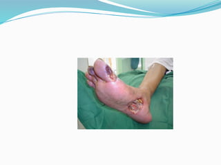
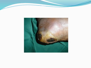
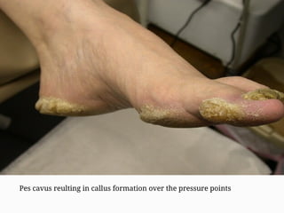
#80 This patient has a pes cavus or high plantar arch deformity that has resulted in pressure points and callus formation over the heels, metatarsal heads, and along the medial aspect of the great toe.
Extensive callus increases the subcutaneous pressure immediately beneath the callus and can result in a subcutaneous hemorrhage, the so-called “pre-ulcer.”
Note the extensive nail pathology.
Ted: Can you provide a color background?


![Diabetic Foot
Definition:
Le pied diabétique regroupe toute infection, ulcération ou destruction des
tissus profonds du pied associées à une neuropathie et/ou une artériopathie
périphérique des membres inférieurs chez le diabétique [1]. C'est une
complication fréquente et grave du diabète avec un taux d´amputation de
membres inférieurs très élevé et des conséquences souvent dramatiques
sur le plan socio-économique et psychologique [2]. En Afrique, les lésions du
pied chez le diabétique sont malheureusement très courantes. Elles sont à
l'origine de 15% à 25% des hospitalisations chez les diabétiques [2,3].
Souvent, la pauvreté, le manque d´hygiène et la marche à pieds nus
interagissent pour aggraver l´impact des lésions du pied causées par le
diabète
1. International Working Group on the Diabetic Foot](https://image.slidesharecdn.com/diabeticfoot-251118012302-112e23c3/85/Diabetic-Foot-introduction-description-3-320.jpg)