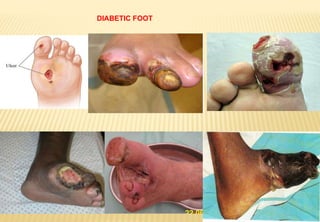
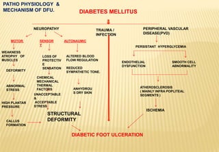
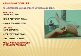
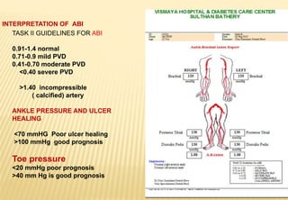
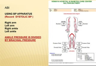
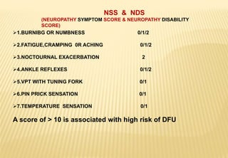
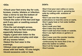
2. Diabetic foot complication is a challenge to the health
care professionals.
This particular complication accounts for more
hospital admissions than any other complication of
Diabetes with variable morbidity and mortality.
The risk of lower extremity amputation is 10 times
higher in patients with diabetes than those without
Diabetes.
But…. fortunately …early detection and timely
intervention promises almost 85 % reduction in the
incident of lower limb amputations.
5. RISK FACTORS FOR
DIABETIC FOOT LESIONS.
POOR GLYCEMIC CONTROL
PREVIOUS AMPUTATION.
PAST HISTORY OF DFU
DPN, PVD.
FOOT DEFORMITY.
VISUAL IMPAIRMENT
MALE SEX—SMOKING ,
ALCOHOL.
DIABETIC KIDNEY DISEASE
ESPECIALLY ON DIALYSIS.
PRECIPITATING FACTORS
TRAUMA FROM INAPPROPRIATE SHOES.
BARE-FOOT WALKING
IMPROPER CUTTING OF NAILS.
(INGROWING TOE NAILS ,PARONYCHIA)
INTERTRIGO.
HOME SURGERY OF CORNS AND CALLUS.
6. Epidemiology
GLOBALLY > 7 % OF THE POPULATION IS DIABETIC
15% OF DIABETICS WILL DEVELOP A FOOT ULCER
FOOT ULCERATIONS IS THE MOST COMMON CAUSE OF
CHRONIC HOSPITALIZED CARE AND EXPENSIVE TO TREAT.
50% NON -TRAUMATIC AMPUTATIONS ARE DUE TO DIABETIC
FOOT COMPLICATION
AFTER AMPUTATION 30% LOSE OTHER LIMB IN 3 YRS & 2/3 DIE IN 5
YRS
EARLY DETECTION AND TIMELY INTERVENTION COULD REDUCE
LIMB THREATENING AND LIFE THREATENING COMPLICATIONS.
8. DIABETES MELLITUS
NEUROPATHY TRAUMA /
INFECTION
PERIPHERAL VASCULAR
DISEASE(PVD)
DIABETIC FOOT ULCERATION
MOTOR SENSOR
Y
AUTONAUMIC
WEAKNESS
ATROPHY OF
MUSCLES
DEFORMITY
ABNORMAL
STRESS
HIGH PLANTAR
PRESSURE
CALLUS
FORMATION
LOSS OF
PROTECTIV
E
SENSATION
ANHYDROU
S DRY SKIN
ALTERED BLOOD
FLOW REGULATION
REDUCED
SYMPATHETIC TONE.
PERSISTANT HYPERGLYCEMIA
ENDOTHELIAL
DYSFUNCTION
SMOOTH CELL
ABNORMALITY
ATHEROSCLEROSIS
( MAINLY INFRA POPLITEAL
SEGMENTS )
PATHO PHYSIOLOGY &
MECHANISM OF DFU.
STRUCTURAL
DEFORMITY
UNACCEPTABLE
&
ACCEPTABLE
STRESS
ISCHEMIA
CHEMICAL
MECHANICAL
THERMAL
FACTORS
9. CLASSIFICATION
The examination of an Ulcer must provide a detailed
account of the ulcer characteristics .
-site
-Size
-Depth
-appearance
It helps for an appropriate management plan.
1.Wagners diabetic ulcer classification
2.University of Texas diabetic wound classification
system
10. Wagner’s Diabetic Ulcer Classification
(Based on wound depth and extent of tissue necrosis)
grade 0 – Intact skin (impending ulcer)
grade 1 – superficial
grade 2 – deep to tendon bone or ligament
grade 3 - osteomyelitis
grade 4 – gangrene of toes or forefoot
grade 5 – gangrene of entire foot
This classification has not taken into consideration the
presence of Infection or Ischemia.
11. STAGE GRADE
0 i ii iii
A
( No infection or
ischemia )
Post ulcerative
lesion completely
epithelialized
Superficial wound
not involving deeper
structures
Wound penetrating
to tendon or capsule
Wound penetrating
to bone or joint
B Infection Infection Infection Infection
C Ischemia Ischemia Ischemia Ischemia
D Ischemia and
infection
Ischemia and
infection
Ischemia and
infection
Ischemia and
infection
UNIVERSITY OF TEXAS DIABETIC WOUND CLASSIFICATION SYSTEM
The two critical factors- Infection and Ischemia have
been addressed here.
12. EVALUATION OF DIABETIC FOOT
HISTORY
Tingling , numbness, burning in feet
Fatigue, cramping and aching
Nocturnal exacerbation,
Previous foot ulceration/ amputation
Claudication pain in legs
Difficulty in walking
Associated vision loss /chronic kidney disease
Use of tobacco
13. GLYCEMIC EVALUATION
Epidemiological studies revealed correlation between
hyperglycemia, DPN and DFU
There is Linear association between HBA1c values and
amputation
57% reduction in DPN in patients treated intensively.
DCCT reveals that 1% mean reduction in HBA1c produces
26% reduction in micro vascular complications.
There is also a decline in percentage of amputations by 36 % in
the intensively treated group
14. INSPECTION
Look for
nail dystrophies ,
erythema,
prominent veins,
callus,
foot ulcer,
inter digital
mycosis,
swelling of foot,
fissures ,
dermopathy,
dryness of skin etc.
Look for skin color ,
turgor,
shiny thin papery onion skin,
bluish discoloration ,
impending gangrene etc.
Foot deformities like claw
toes,
hammer toes , bunions,
gross deformities of mid foot
( Charcot foot )
15. Lower 1/3 Tibia bisects vertical
Knee joint Sub talar joint axis and supporting surface are in
parallel
Both lateral and medial malleoli are in the same line.
Calcaneus bisects vertical
NORMAL FOOT
16. NORMAL BIO –MECHANICS OF
FOOT.
Heel strike is central
Pronation is controlled by small muscles.
First MTP contact time is optimum.
Weight shifts from 1st MTP to other MTPs
Supination of fore-foot completes stance
phase.
21. CHARCOT FOOT
Neuro –arthropathy
Insensitive foot –repetitive stress leading
to microfaracture and bone destruction
and remodelling of foot into ‘bag of bones’
Mistaken often for osteomyelitis /cellulitis.
-X- ray shows
‘pencil pointing’ and ‘sucked candy
‘appearance of metatarsal heads and
shafts.
ROCKER
BOTTOM
FOOT
22. PALPATION
Temperature difference in feet is evaluated.
- Cold feet in AN, Ischemia
Peripheral pulsation (dorsalis pedis, posterior tibial)
Bony prominences on the plantar aspect
Limited joint mobility of 1st MTP.
LJM leads to hallux rigidus.
This can be diagnosed by
asking the patients to
Dorsi flex hallux
while the foot is weight bearing.
24. 10 GRAMS SEMMES-WEINSTEIN MONOFILAMENT
It is a Nylon monofilament of 5.07 size, equalent
to 10 grams of linear force.
It tests sense of touch( large nerve fibre function )
Site of testing : plantar and dorsum
Before testing demonstrate the sensation on the
hand or palm.
Place the monofilament perpendicular to the
test site.
Bow into C shape for one second
Test six sites of the foot . It predicts 95 %
ulcer formers
25. Biothesiometer
Used to assess the VPT.
When applied to the foot
it delivers a vibratory
stimulus which increases
as the voltage is increased.
VPT beyond 25 is 7 times
prone to DFU
Beyond 42 is 23 times prone
to DFU.
1. Distal part of great
toe
2. First metatarsal
head
3. Third metatarsal
head
4 .Fifth metatarsal
head
5. Instep
6. Heel
26. VASCULAR ASSESSMENT
i.PALPATION OF PERIPHERAL
VASCULAR PULSE
- Dorsalis pedis artery
- Posteriar tibial
ii. ABI (anke brachial index )
-using normal BP apparatus
-Using DOPPLER probes
iii. ANGIOGRAPHY
MR angiography
CT angiography.
27. ABI-- USING DOPPLER
BP IS MEASURED USING DOPPLER ULTRASONIC PROBE .
RIGHT SIDE
-RIGHT BRACHIAL
-RIGHT POSTERIAR TIBIAL
- RIGHT DORSALIS PEDIS
LEFT SIDE
-LEFT BRACHIAL
-LEFT POSTERIAR TIBIAL
-LEFT DORSALIS PEDIS
ANKLE PRESSURE IS DIVIDED
BY BRACHIAL PRESSURE
28. TASK II GUIDELINES FOR ABI
0.91-1.4 normal
0.71-0.9 mild PVD
0.41-0.70 moderate PVD
<0.40 severe PVD
>1.40 incompressible
( calcified) artery
ANKLE PRESSURE AND ULCER
HEALING
<70 mmHG Poor ulcer healing
>100 mmHg good prognosis
Toe pressure
<20 mmHg poor prognosis
>40 mm Hg is good prognosis
INTERPRETATION OF ABI
29. ABI
USING BP APPARATUS
(Record SYSTOLIC BP )
Right arm
Left arm
Right ankle
Left ankle
ANKLE PRESSURE IS DIVIDED
BY BRACHIAL PRESSURE
30. 1.BURNIBG OR NUMBNESS 0/1/2
2.FATIGUE,CRAMPING 0R ACHING 0/1/2
3.NOCTOURNAL EXACERBATION 2
4.ANKLE REFLEXES 0/1/2
5.VPT WITH TUNING FORK 0/1
6.PIN PRICK SENSATION 0/1
7.TEMPERATURE SENSATION 0/1
A score of > 10 is associated with high risk of DFU
NSS & NDS
(NEUROPATHY SYMPTOM SCORE & NEUROPATHY DISABILITY
SCORE)
31. ASSESSMENT OF FOOT AT RISK
GAIT OBSERVED WHILE THE
PATIENT
ENTERING INTO THE CHAMPER
SUBTALAR POINT POSITION AT
REST
1ST MTP JOINT MOBILITY
HEEL STRIKE
CALLUS AT PLANTAR AREA
PALPATE MTP HEADS
32. FOOT WEAR OF THE PATIENT
Insist on removal of the foot wear
Inspect the foot wear-–in-shoe foot prints-
- as they can often be the cause of foot
ulcer.
Abnormal foot biomechanics can cause
uneven pattern of wear and tear and make
the shoes troublesome for use.
This makes the need for regular review of
foot wear
Look for shoes that are worn out, too
small, too narrow, high heels with too low
toe box
Insist on avoiding Hawaii slippers
33. GOOD FOOT WEAR SHOULD HELP
Prevent ulceration
Protection of wound
Easy to apply
Frequent wound inspection
should be possible
No interference with mobility
Cosmetically acceptable
Safe (no complications )
34. PLANTAR PRESSURE
ASSESSMENT
HARRIS MATE ( FOOT IMPRINTER )
INEXPENSIVE
DETECTS THE PRESSURE PONTS IN THE FOOT.
HELPS TO MAKE A PROPER FOOT WEAR.
We can assess
Static pressure Imprint
Dynamic pressure Imprint
The pressure gradient is
expressed in intensity gradient
35. PODIA SCAN (software with
scanner)
USING THE HARRIS
IMPRINTER PAPER WE
RECORD THE IMPRESSION
SCAN IT AND TAKE THE
COLOR PRINT -OUT
THE PRESSURE
GRADIENT IS EXPRESSED
IN THE COLOR
DIFFERENCE.
36. Off loading --Foot wear.
The podiatrist’s most important
treatment arsenal is off-loading.
The insensitive feet are prone to
be affected by unfelt and
unperceived peak plantar
pressures which is responsible
for precipitating and
perpetuating the plantar ulcers.
37. Patient education
Foot care tips
Exam of foot
Dos and don’ts
Foot wear at home
and office
Exercise tips
1.Walking
2.Jogging
3.Running
4.Static cycling
5.Swimmig
Low risk feet
Very important
Allowed
Allowed
Allowed
Allowed
Allowed
High risk feet
Very important
Avoid brisk walking
Avoid
Avoid
Recommended
Recommended
39. Skin temperature monitoring
The elevation in skin temperature of the
pre-ulcerative inflammatory site as compared to
the contra lateral limb is predictive of an area at
risk
-liquid crystal thermography
-infra red thermometry
-cutaneous thermal discrimination thresh hold
-electrical contact thermometry
Currently being used for evaluation.
40. Surgical intervention
Aimed at eliminating deformities responsible for
increased plantar pressure
-short Achilles tendon could be corrected
by lengthening the tendon
-removal of osseous prominences
-reconstruction of deformed foot/ankle
-vascular surgery to improve the blood flow.
Evidence based studies are still lacking to support
these prophylactic measures.
42. DOs
Check your feet every day for cuts,
bruises, cracks, blisters or infection.
Use a mirror to see the bottom of
your feet if u cant lift them up.
Check the color of the feet and legs
and if any pain or swelling consult
the doctor.
Trim the nail straight across.
Wash and dry the feet everyday
especially between toes.
Apply a good skin lotion to the
heels and soles everyday and wipe
off any excess lotion.
Select the foot wear in the
evenings.
Always wear good supportive
shoes with low heels. <5 cms height.
Avoid excessive cold or heat.
DONTs
Don’t Cut your own callus or corns.
Don’t treat your own in -growing toe
nails by cutting with scissors or
blades.
Don’t use over the counter
medications to treat the corns or warts.
Don’t apply heat or hot water bottles
on the feet.
Don’t soak your feet.
Don’t take very hot bath.
Don’t walk bare-foot inside or outside.
Don’t use tight shoes or garters or
elastics.
Don’t wear over the counter insoles.
Don’t wear the new shoes
continuously for more than 2 hrs to
allow the leather to soften.
Don’t sit for longer periods .
Don’t smoke.
43. RISK CATEGORIZATION —PERIODICITY OF CHECK
UP.
CATOGORY RISK PROFILE CHECK UP FREQUENCY
0 NO SENSORY NEUROPATHY ONCE A YEAR
1 SENSORY NEUROPATHY EVERY SIX MONTHS
2 SENSORY NEUROPATHY
PVD
FOOT DEFORMITIES
EVERY THREE MONTHS
3 PREVIOUS ULCER /AMPUTATION ONCE IN EVERY 1-3 MONTHS
44. e
Glycemic
control
Evaluation and
management of
PVD
Evaluation and
management
of DPN
Self
examination
of foot daily.
Annual foot
examination
Screening and
management of
elevated plantar
pressure
Education
intervention
Skin
temperature
monitoring
Surgical
intervention
-
deformity
correction
Prevention
of DFU
COMPREHENSIV
E
FOOT CARE