
Recommended
More Related Content
What's hot
What's hot (20)
Similar to Diabetic foot Wilson N. M.D. MBBS
Similar to Diabetic foot Wilson N. M.D. MBBS (20)
Recently uploaded
Recently uploaded (20)
Diabetic foot Wilson N. M.D. MBBS
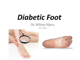
- 1. Diabetic Foot Dr. Wilson Njeru M.D. MBBS
- 2. Flow of our presentation • Introduction • Definition • Classification & Staging • Pathogenesis • Clinical features • Management • Prevention guidelines • A quick recap • Conclusion • Literature
- 3. Introduction • From foot deformities, to ulceration, infection, necrosis, gangrene & eventually amputation, “diabetic foot” is by far the most COSTLY complication of DM (according to WHO).
- 4. Introduction • Diabetes is the most common cause of non- traumatic amputations of lower limbs. • It accounts for >70% of lower limb amputations. • This leads to long periods of hospitalization, rehabilitation, not to mention disability & its impending socio-economical & psychological impacts. • I can not emphasize enough on this morbidity!
- 6. A quick review on all complications of DM; Complications of DM Acute complications Chronic complications “emergencies” Diabetic - -DKA *MACROangiopathies (CAD, CVD, PAD) -NKHHS *Microangiopathies (Retinopathy, Nephropathy) -Lactic Acidosis *Neuropathy (Central & Peripheral) -Hypoglycemia *Diabetic foot {mainly caused by peripheral polyneuropathy or angiopathy}
- 7. Definition • Diabetic foot (DF) is a pathological condition of the foot in diabetic patients, xrised by skin & soft tissue lesions, damage to joints & bones, presenting as formation of ulcers, joint deformities, infection, necrotic and gangrenous processes. • It is one of the several chronic complications of DM, but it stems from the others, as we’ve already seen in the previous slide.
- 10. Classification & Staging • A standard classification of DF is useful in: Assessing the etiology Designing appropriate Rx Assessing prognosis Monitoring the course of disease/progress
- 11. Classification on basis of etiology: 1. Neuropathic foot 2. Ischemic foot 3. Neuro-ischemic foot (mixed)
- 12. Classification on basis of etiology: Neuropathic foot o Due to peripheral PolyNeuropathy i.e. “somatic + autonomic”, both sensory & motor neuropathies. • W or w/o CHARCOT joint disease (i.e. neuro-OSTEOARTHROPATHY) Ischemic foot oMainly macroangiopathy i.e. PAD of lower limbs • W or w/o infection Neuro-ischemic foot (mixed)
- 14. Staging according to clinical condition (ME Edmond et AV Foster staging): Stage Clinical condition of FOOT I. Normal foot II. HIGH risk foot III. Ulcerated foot IV. Cellulitic foot V. Necrotic foot VI. Major Amputation
- 16. Wagner’s classification of diabetic foot ulcer (2001) The most widely used classification Grades diabetic foot based on severity of ulcer penetration
- 17. Wagner’s classification of diabetic foot ulcer (2001) Grade Physical Findings Description 0. INTACT skin No ulcer (but HIGH risk foot i.e. deformities, callus, or insensitivity) 1. Superficial ULCER Only skin involvement 2. Deep ULCER Involving tendons or ligaments 3. OsteoMyelitis Deeper ULCER with bone involvement 4. Partial GANGRENE Of toes or forefoot 5. Total GANGRENE Of entire foot
- 19. Pathogenesis
- 20. Pathogenesis • Development of DF is multi-factorial & complex. • First & foremost it involves longstanding high levels of HYPERglycemia. This in turn contributes to the following disorders, through various mechanisms: 1. Neuropathy 2. Angiopathy 3. Immune dysfunction
- 21. Neuropathy a) Somatic Sensory neuropathy Loss of pain sensation Unnoticed trauma (mechanical, thermal, chemical) Unchecked worsening of the lesion Formation of Callus Tissue damage & necrosis beneath the callus Dev of cavity filled with serous fluid Cavity erupts to the surface Resulting in ULCERation
- 23. Neuropathy b) Somatic Motor neuropathy Weakness & decreased contraction of foot muscles Atrophy /wasting of these muscles Foot deformity Abnormal gait Easy ULCERation
- 24. Foot deformities that predispose to ULCERATION include: » Clawed toes » Hammer toes » Pes cavus » Pes planus » Charcot joint » Talipes equinus (ankle joint rigidity) » Hallux varus/valgus/rigidus » Bunions (bony bump of hallux joint) » Nail deformities » Deformities from previous trauma/surgery
- 32. Neuropathy c) Autonomic neuropathy Decreased sweat secretion Dry & brittle skin Easily cracks & fissures Infection occurs ULCERation
- 34. In general, Factors contributing to foot ulceration include
- 35. MACROangiopathy Atherosclerosis of large arteries (i.e. PAD) Increased peripheral resistance Microangiopathy Thickening of basement membranes of capillaries Decreased capillary permeability Angiopathy Decreased perfusion of foot structures Decreased supply of immune components & Antibiotics Poor wound healing GANGRENE
- 39. Immune dysfunction • Impaired defenses against infections: Decreased Leukocyte migration Decreased Phagocytosis Decreased Intracellular killing Decreased Chemotaxis
- 46. Ddx in clinical forms of Diabetic Foot Xristic Neuropathic Ischemic Skin Temp Warm Cold Color Not altered Pink Cyanotic Pulse BOUNDING pulse DIMINISHED Pulseless Sensation Painless Painful Skin Dry, thickened, fissures Atrophic, thin, Callus + at pressure points Usually Absent Ulceration On pressure points On toe tips, heels, foot margins Complication Charcot foot (osteoarthropathy) Gangrene
- 48. Management
- 49. Management includes 5 aspects: 1. Metabolic control -effective blood sugar control, neuropathy Rx 2. Mechanical control -reduce risk of trauma, treat deformities 3. Vascular management 4. Infection Prophx and Rx 5. Patient EDUCATION
- 77. A Quick Recap
- 78. “Diabetic foot” is a pathological condition of the foot in people with longstanding diabetes. It is by far the most COSTLY complication of diabetes (according to WHO). It has 3 main etiological factors, and many predisposing risk factors. Pathogenesis is multi-factorial. Management is case dependant.
- 79. Conclusions
- 80. • Close coordination between a physician, nurse, surgeon, orthopedic, physiotherapist, psychotherapist & the patient is vital in the care of a diabetic foot. • Most foot problems can be prevented. • Each & every patient should be educated & issued with a pamphlet containing straightforward safety & footcare instructions!
- 81. Literature • WHO guidelines on diabetes (WDF – world diabetes foundation) • National clinical guidelines for management of DM (MoH, GOK) • DM textbook for overseas medical students (Nizhny Novgorod State Medical University, 2015 edition) • Diabetic foot by Dr. Hardik Pawar, Orthopedic specialist • Many images thanks to GOOGLE.