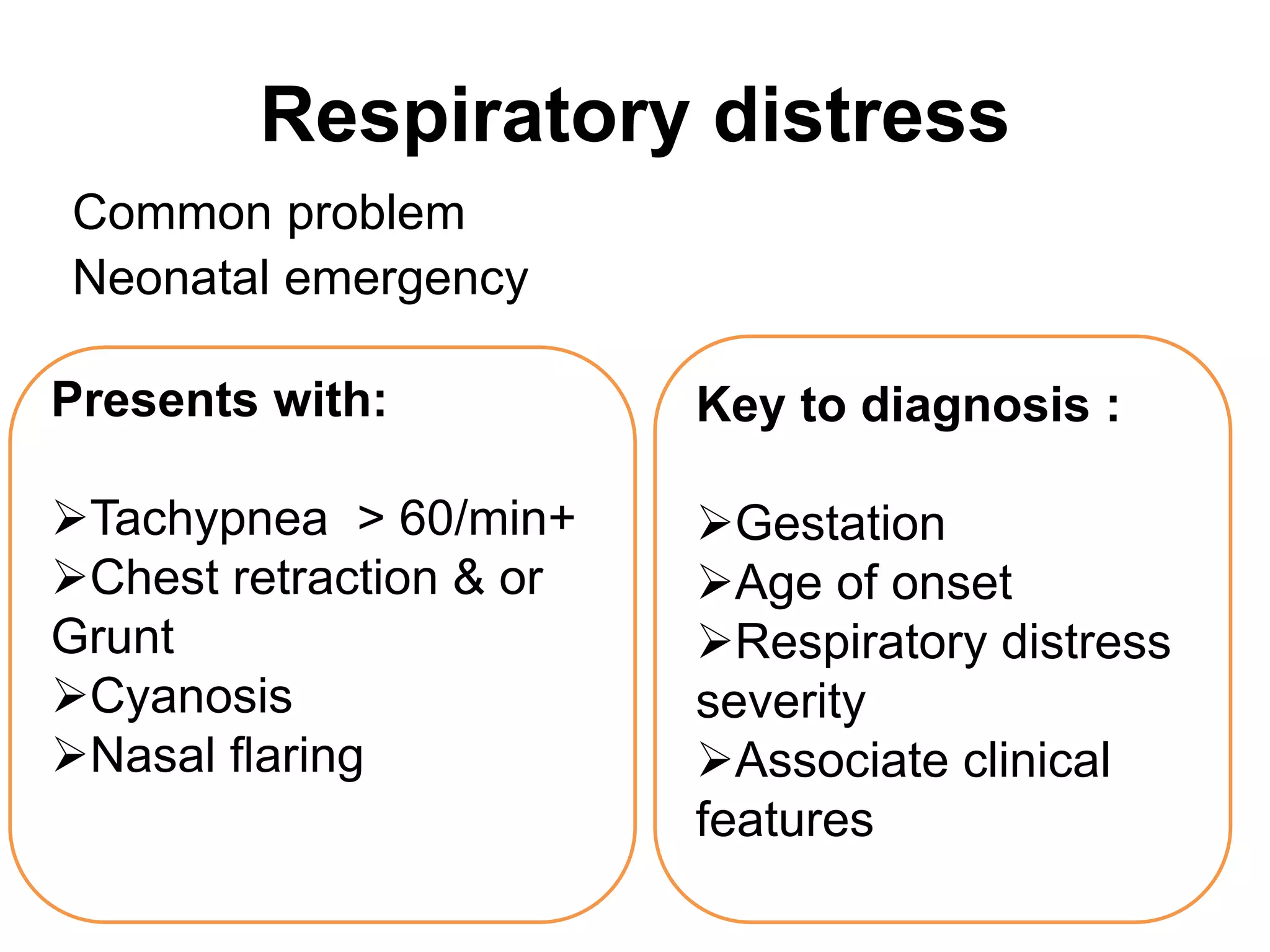
Respiratory Distress in a
Neonate
Presented by :
Tek Bdr. Khadka
MBBS CMC 2nd Batch
Respiratory distress
Common problem
Neonatal emergency
Presents with:
Tachypnea > 60/min+
Chest retraction & or
Grunt
Cyanosis
Nasal flaring
Key to diagnosis :
Gestation
Age of onset
Respiratory distress
severity
Associate clinical
features
Etiology of respiratory distress
A. Respiratory
causes:
 RDS
 MAS
 Pneumonia
 TTN
 PPH
 Pneumothorax
 TOF
 Diaphragmatic
hernia
 Lobar emphysema
B. Non- pulmonary causes:
Cardiac: CCF, congenital
heart disease
Metabolic: Metabolic
acidosis, hypoglycemia,
hypothermia
CNS: Asphyxia, meningitis,
cerebral edema, hemorrhage
Chest wall: Thoracic
dystrophy
 Also known as Hyaline membrane disease
 Preterm birth is the most common etiological factor
 Incidence inversely related to gestational age and
birth weight.
Incidence
 Occurs in 60-80% of infants <28 wk of gestational age
 15-30% of those between 32-36 wks
 Rarely >37 wk
 Incidence highest in preterm male or white infants
Respiratory Distress Syndrome
Risk of RDS increases
Maternal diabetes,
Multiple births,
Cesarean delivery,
Precipitous delivery,
Asphyxia,
Cold stress,
Maternal history of
previously affected infants.
Risk of RDS reduced in
pregnancies with
Chronic or PIH
Prolong rupture of
membrane
Antenatal corticosteroid
prophylaxis
Prenatal diagnosis to identify risk, prevention of disease,
antenatal administration of glucocorticoids, improvement in
prenatal and neonatal care, advances in respiratory
support and surfactant replacement therapy
reduce mortality of RDS.
Etiology and Pathophysiology
 Surfactant deficiency (decreased production and
secretion) - primary cause of RDS
• Surfactant is a complex lipoprotein,Contains :
70-80% phospholipids,
8-10% protein, and
10% neutral lipids, primarily cholesterol.
• Major constituents of surfactant are:
Dipalmitoyl phosphatidylcholine (DPPC) or lecithin, is
functionally the principle phospholipid,
Phosphatidylglycerol,
Apoproteins (surfactant proteins SP-A, SP-B, SP-C, and
SP-D), and
Cholesterol
Bar chart demonstrates the composition of lung surfactant.
About 1% of the 10% protein component comprises
surfactant apoproteins; the remaining proteins are derived
from alveolar exudate.
Synthesis:
Synthesized in the Golgi apparatus of the
endoplasmic reticulum
Packaged in multilamellar vesicles in the
cytoplasm of the type II alveolar cell and
Secreted by a process of exocytosis.
• Surfactant present in high concentrations in fetal
lung homogenates by 20 wks of gestation, but
does not reach the surface of the lungs until later.
• Increasing amounts of phospholipids are
synthesized & stored with advancing gestational
age.
• Synthesis of surfactant depends on normal pH,
temperature, and perfusion
• Asphyxia, hypoxemia, and pulmonary ischemia,
particularly in association with hypovolemia,
hypotension, and cold stress, may suppress
surfactant synthesis.
• Surface-active agents in alveoli:
reduce surface tension and
maintain alveolar stability by preventing the
collapse of small air spaces at end-
expiration.
• Due to immaturity, the amounts produced or
released may be insufficient to meet postnatal
demands.
• Alveolar atelectasis,hyaline membrane
formation,and interstitial edema make the lungs
less compliant in RDS,so greater pressure is
required to expand the alveoli and small
airways.
Hypoxemia & acidosis
Pulmonary vasoconstriction
Rt to Lt shunt across foramen ovale
Respiratory failure
Identification and Prevention
• Perinatal risk factors
• Prenatal prediction
• Postnatal diagnosis
1.Avoidance of unnecessary or poorly timed cesarean
section.
2.Appropriate management of high-risk pregnancy and
labor
 Assessment of lung maturity by testing amniotic fluid
obtained by amniocentesis
– Lecithin/Sphingomyelin(L/S) - risk low if L/S > 2
except in Diabetes mother, IUGR, pre eclampsia.
– Foam stability index
– Presence of phosphatidylglycerol : appears late in
maturation process in lung
– Lamellar body count :LB are phospholipids
produced by type II alveolar cells
3.Antenatal and intrapartum fetal monitoring may decrease
the risk of fetal asphyxia
4. Administration of antenatal corticosteroids to
women between 24 and 34 wk of gestation
significantly reduces the incidence and mortality of
RDS as well as overall neonatal mortality.
• Antenatal steroids reduce
(1) Need for and duration of ventilatory support and
admission to a neonatal intensive care unit (NICU)
(2) Incidence of severe IVH, necrotizing enterocolitis,
early-onset sepsis, and developmental delay.
Corticosteroid administration is recommended for all
women in preterm labor (24-34 wk of gestation) who
are likely to deliver a fetus within 1 wk.
• Betamethasone(12 mg im 24 hourly interval-2 doses)
• Dexamethasone(6 mg im 12 hourly interval- 4 doses)
used antenatally. C/I chorioamniotis
• Dexamethasone - lower incidence of IVH but higher
periventricular white matter injury than
betamethasone.
5. Administration of a 1st dose of surfactant into the
trachea of symptomatic premature infants immediately
after birth (prophylactic) or during the 1st few hours of
life (early rescue) reduces air leak and mortality from
RDS but does not alter the incidence of BPD.
Chest radiographs in a premature infant with respiratory
distress syndrome before and after surfactant treatment.
Left Initial radiograph shows poor lung expansion, air
bronchogram, and reticular granular appearance. Right
Repeat chest radiograph obtained when the neonate is
aged 3 hours and after surfactant therapy demonstrates
marked improvement.
Clinical presentation
• Signs of RDS usually appear within minutes of birth.
• Rapid, shallow respirations to 60 breaths/min or
greater within hours.
• Initial severe respiratory distress (especially with a
birth weight < 1,000 g) – requiring resuscitation
• Characterized by
Tachypnea,
Prominent (often audible) grunting,
Intercostal and subcostal retractions,
Nasal flaring, and
Cyanosis.
• Breath sounds - normal or diminished with a
harsh tubular quality, fine rales.
• Apnea and irregular respirations
• Edema, ileus, and oliguria.
• Respiratory failure may occur in infants with
rapid progression of the disease.
• Natural course of untreated RDS characterized
by progressive worsening of cyanosis and
dyspnea.
• Worsening characterized By - fall in blood
pressure; cyanosis and pallor increase, and
grunting decreases or disappears.
• Improvement seen by spontaneous diuresis and
improved blood gas values at lower inspired
oxygen levels and/or lower ventilator support.
• Death due to severe impairment of gas
exchange, alveolar air leaks (interstitial
emphysema, pneumothorax), pulmonary
hemorrhage, or IVH.
Diagnosis
Based on:
Clinical course,
Chest radiographic findings, and
Blood gas and acid-base values
• Laboratory findings are characterized by
hypoxemia
hypercapnia, and
variable metabolic acidosis.
Characteristic but
not pathognomonic
appearance:
 A fine reticular
granularity of the
parenchyma
 Air bronchograms,
 Low lung volume
 Ground glass
opacity
 White out lungs.
Initial radiographic
appearance
occasionally normal,
with the typical
pattern developing at
6-12 hr.
Differential diagnosis
• Early-onset sepsis
• Pneumonia manifested at birth
• Cyanotic heart disease (total anomalous pulmonary
venous return
• Persistent pulmonary hypertension
• Aspiration (meconium, amniotic fluid) syndromes
• Spontaneous pneumothorax
• Pleural effusions
• Congenital anomalies such as cystic adenomatoid
malformation, pulmonary lymphangiectasia,
diaphragmatic hernia, and lobar emphysema
• Transient tachypnea
Management
Goal of treatment:
 Minimize abnormal
physiologic variations
and iatrogenic
problems.
RDS – managed in NICU
 Oxygen
 Continuous Positive
Airway Pressure(CPAP)
 Surfactant replacement
 Mechanical ventilation
 Supportive therapy
• Inadequate pulmonary exchange of oxygen and
carbon dioxide; metabolic acidosis and
circulatory insufficiency - treated
• Early supportive care of premature infants,
especially treatment of acidosis, hypoxia,
hypotension, and hypothermia, may decrease
the severity of RDS.
• Careful and frequent monitoring of heart and
respiratory rates, oxygen saturation, Pao2,
Paco2, pH, serum bicarbonate, electrolytes,
glucose, and hematocrit, blood pressure, and
temperature.
Oxygen
• Warm humidified oxygen provided to keep
arterial oxygen pressure between 40-
70 mm Hg (85-95% saturation) in order to
maintain normal tissue oxygenation while
minimizing the risk of oxygen toxicity.
• If SPO2 cannot be kept > 85% at inspired oxygen
concentrations of 40-70% or greater, applying
CPAP at a pressure of 5-10 cm H2O via nasal
prongs indicated
• CPAP prevents collapse of surfactant-deficient
alveoli and improves ventilation-perfusion
matching.
Continuous Positive Airway Pressure(CPAP)
• Early use at-risk VLBW infants beginning as early as in
the delivery room reduces ventilatory needs.
• Another approach - InSurE approach and begin CPAP
• Infant with RDS undergoing CPAP cannot keep oxygen
saturation > 85% while breathing 40-70% oxygen,
assisted ventilation and surfactant are indicated.
• Methods of administering : begin CPAP with nasal
prong with continuous flow ventilator 5-7cm of H2O
using high flow enough to avoid rebreathing(5-
10L/min)& pressure increment of 1-2 cm of H20 to
maxm 8cm of H2O.
• Orogastric tube is used to decompress swallowed air
Mechanical ventilation
• Infants with respiratory failure or persistent apnea
require assisted mechanical ventilation.
• Goal of mechanical ventilation: improve oxygenation
and elimination of carbon dioxide without causing
pulmonary injury or oxygen toxicity.
1. Intermittent positive pressure ventilation
delivered by time-cycled, pressure-limited,
continuous flow ventilators - method of
conventional ventilation for newborns.
Synchronized intermittent mechanical ventilation
(SIMV), which synchronizes with the infant's own
respiratory effort, is preferred.
2. High-frequency ventilation (HFV) achieves
desired alveolar ventilation by using smaller tidal
volumes and higher rates (300-1,200 breaths/min
or 5-20 Hz. useful to minimize lung injury in very
small and/or sick infants who require high peak
inspiratory pressures and oxygen concentration to
maintain adequate gas exchange.
• Adjustment of MV by seeing ABG, maintain
PaCO2 in lung at 45-55mm of HG, acidosis
exacerbate RDS so relative hypercapnia to
minimize lung injury and metabolic acidosis.
• Observe : Colour, chest motion and respiratory effort
and listen breath sound and observe changes in O2
saturation.
Raising PaCO2 indicate onset of complication
including atelectasis, air leak or symptomatic PDA
PaO2 usually rises in response to increase in FiO2 or
mean airway pressure
• Weaning : As the infant shows signs of improvement,
weaning from the ventilator should be attempted.
• Care of infants receiving ventilator therapy :
check FiO2 and ventilator setting,
O2 saturation,
ABG 4-6hr during active illness and 30 min following
adjustment of setting.
Airway secretions require periodic suctioning.
Complications of MV
• Air leak
• Infection
• Intracranial hemorrhage
• PDA
Long term ->
Broncho-pulmonary dysplasia,
Neurodevelopmental impairement and
Retinopathy of prematurity
Surfactant replacement
• Surfactant deficiency - the primary pathophysiology
of RDS.
• Surfactant replacement therapy results improved
alveolar-arterial oxygen gradients, reduced ventilatory
support, increased pulmonary compliance, and
improved chest radiograph appearance .
• Treatment initiated as soon as possible in the hours
after birth.
Route: Intratracheal
Technique: InSurE
• Repeated dosing is given via the endotracheal tube
every 6-12 hr for a total of 2 to 4 doses, depending on
the preparation.
• Surfactant preparations:
Synthetic surfactants and
Natural surfactants derived from animal sources.
• Exosurf - a synthetic surfactant.
• Natural surfactants include Survanta (bovine) -
4ml/kg, Infasurf (calf) -3ml/kg,and Curosurf (porcine)
-2.5ml/kg.
• Prophylactic(all neonates <28 wks) and rescue(when
RDS actually develops) administrations of synthetic
and natural surfactants.
• Prophylactic administration of both types of
surfactants decreases the risk for pneumothorax and
pulmonary interstitial emphysema.
Supportive therapy
1.Temp: Incubator or radiant warmer
• Avoid hypothermia and minimize oxygen consumption
– infant placed in incubator or radiant warmer, and
core temperature maintained between 36.5 and 37⁰C
• Incubator use - preferable in VLBW infants.
2. Fluids and nutrition:
• Calories and fluids should initially provided
intravenously.
• For the 1st 24 hr, 10% glucose and water should be
infused through a peripheral vein at a rate of 65-
75 mL/kg/24 hr.
• Electrolytes added on day 2 in the most mature
infants and on days 3 to 7 in the more immature ones.
Fluid volume increased gradually over the 1st
week. Excessive fluids (> 140 mL/kg/day) contribute
to the development of patent ductus arteriosus (PDA).
3.Diuresis and improvement in pul compliance
occurs around 2nd-4th day but sooner in surfactant
treatment.
• If diuresis and improvement in lung compliance
doesn’t occur by 1-2wk this indicates onset of
bronchopulmonary dysplasia.
4. Dopamine 5µg/kg/min to maintain BP, CO and
tissue perfusion and avoid acidosis.
5. Possible infection:with broad spectrum antibiotics
Complications of RDS
• Septicemia
• Bronchopulmonary dysplasia (BPD)
• Patent ductus arteriosus (PDA)
• Complications of tracheal intubation
• Pulmonary hemorrhage
• Necrotizing enterocolitis (NEC)
• Intraventricular hemorrhage (IVH)
• Death
Prognosis
• Antenatal steroids, postnatal surfactant use,
and improved modes of ventilation have
resulted in low mortality from RDS (≈10%).
• References
Manual of Neonatal Care 7th ed. - Cloherty
Nelson Textbook of Pediatrics 20th Edition
Ghai Essential pediatrics 8th Edition
http://emedicine.medscape.com/article/97603
4-overview
THANK YOU

Respiratory Distress(RDS)

  • 1.
    Respiratory Distress ina Neonate Presented by : Tek Bdr. Khadka MBBS CMC 2nd Batch
  • 2.
    Respiratory distress Common problem Neonatalemergency Presents with: Tachypnea > 60/min+ Chest retraction & or Grunt Cyanosis Nasal flaring Key to diagnosis : Gestation Age of onset Respiratory distress severity Associate clinical features
  • 3.
    Etiology of respiratorydistress A. Respiratory causes:  RDS  MAS  Pneumonia  TTN  PPH  Pneumothorax  TOF  Diaphragmatic hernia  Lobar emphysema B. Non- pulmonary causes: Cardiac: CCF, congenital heart disease Metabolic: Metabolic acidosis, hypoglycemia, hypothermia CNS: Asphyxia, meningitis, cerebral edema, hemorrhage Chest wall: Thoracic dystrophy
  • 4.
     Also knownas Hyaline membrane disease  Preterm birth is the most common etiological factor  Incidence inversely related to gestational age and birth weight. Incidence  Occurs in 60-80% of infants <28 wk of gestational age  15-30% of those between 32-36 wks  Rarely >37 wk  Incidence highest in preterm male or white infants Respiratory Distress Syndrome
  • 5.
    Risk of RDSincreases Maternal diabetes, Multiple births, Cesarean delivery, Precipitous delivery, Asphyxia, Cold stress, Maternal history of previously affected infants. Risk of RDS reduced in pregnancies with Chronic or PIH Prolong rupture of membrane Antenatal corticosteroid prophylaxis Prenatal diagnosis to identify risk, prevention of disease, antenatal administration of glucocorticoids, improvement in prenatal and neonatal care, advances in respiratory support and surfactant replacement therapy reduce mortality of RDS.
  • 6.
    Etiology and Pathophysiology Surfactant deficiency (decreased production and secretion) - primary cause of RDS • Surfactant is a complex lipoprotein,Contains : 70-80% phospholipids, 8-10% protein, and 10% neutral lipids, primarily cholesterol. • Major constituents of surfactant are: Dipalmitoyl phosphatidylcholine (DPPC) or lecithin, is functionally the principle phospholipid, Phosphatidylglycerol, Apoproteins (surfactant proteins SP-A, SP-B, SP-C, and SP-D), and Cholesterol
  • 7.
    Bar chart demonstratesthe composition of lung surfactant. About 1% of the 10% protein component comprises surfactant apoproteins; the remaining proteins are derived from alveolar exudate.
  • 8.
    Synthesis: Synthesized in theGolgi apparatus of the endoplasmic reticulum Packaged in multilamellar vesicles in the cytoplasm of the type II alveolar cell and Secreted by a process of exocytosis.
  • 10.
    • Surfactant presentin high concentrations in fetal lung homogenates by 20 wks of gestation, but does not reach the surface of the lungs until later. • Increasing amounts of phospholipids are synthesized & stored with advancing gestational age. • Synthesis of surfactant depends on normal pH, temperature, and perfusion • Asphyxia, hypoxemia, and pulmonary ischemia, particularly in association with hypovolemia, hypotension, and cold stress, may suppress surfactant synthesis.
  • 11.
    • Surface-active agentsin alveoli: reduce surface tension and maintain alveolar stability by preventing the collapse of small air spaces at end- expiration. • Due to immaturity, the amounts produced or released may be insufficient to meet postnatal demands. • Alveolar atelectasis,hyaline membrane formation,and interstitial edema make the lungs less compliant in RDS,so greater pressure is required to expand the alveoli and small airways.
  • 12.
    Hypoxemia & acidosis Pulmonaryvasoconstriction Rt to Lt shunt across foramen ovale Respiratory failure
  • 13.
    Identification and Prevention •Perinatal risk factors • Prenatal prediction • Postnatal diagnosis
  • 14.
    1.Avoidance of unnecessaryor poorly timed cesarean section. 2.Appropriate management of high-risk pregnancy and labor  Assessment of lung maturity by testing amniotic fluid obtained by amniocentesis – Lecithin/Sphingomyelin(L/S) - risk low if L/S > 2 except in Diabetes mother, IUGR, pre eclampsia. – Foam stability index – Presence of phosphatidylglycerol : appears late in maturation process in lung – Lamellar body count :LB are phospholipids produced by type II alveolar cells 3.Antenatal and intrapartum fetal monitoring may decrease the risk of fetal asphyxia
  • 15.
    4. Administration ofantenatal corticosteroids to women between 24 and 34 wk of gestation significantly reduces the incidence and mortality of RDS as well as overall neonatal mortality. • Antenatal steroids reduce (1) Need for and duration of ventilatory support and admission to a neonatal intensive care unit (NICU) (2) Incidence of severe IVH, necrotizing enterocolitis, early-onset sepsis, and developmental delay. Corticosteroid administration is recommended for all women in preterm labor (24-34 wk of gestation) who are likely to deliver a fetus within 1 wk.
  • 16.
    • Betamethasone(12 mgim 24 hourly interval-2 doses) • Dexamethasone(6 mg im 12 hourly interval- 4 doses) used antenatally. C/I chorioamniotis • Dexamethasone - lower incidence of IVH but higher periventricular white matter injury than betamethasone. 5. Administration of a 1st dose of surfactant into the trachea of symptomatic premature infants immediately after birth (prophylactic) or during the 1st few hours of life (early rescue) reduces air leak and mortality from RDS but does not alter the incidence of BPD.
  • 17.
    Chest radiographs ina premature infant with respiratory distress syndrome before and after surfactant treatment. Left Initial radiograph shows poor lung expansion, air bronchogram, and reticular granular appearance. Right Repeat chest radiograph obtained when the neonate is aged 3 hours and after surfactant therapy demonstrates marked improvement.
  • 18.
    Clinical presentation • Signsof RDS usually appear within minutes of birth. • Rapid, shallow respirations to 60 breaths/min or greater within hours. • Initial severe respiratory distress (especially with a birth weight < 1,000 g) – requiring resuscitation • Characterized by Tachypnea, Prominent (often audible) grunting, Intercostal and subcostal retractions, Nasal flaring, and Cyanosis.
  • 19.
    • Breath sounds- normal or diminished with a harsh tubular quality, fine rales. • Apnea and irregular respirations • Edema, ileus, and oliguria. • Respiratory failure may occur in infants with rapid progression of the disease. • Natural course of untreated RDS characterized by progressive worsening of cyanosis and dyspnea.
  • 20.
    • Worsening characterizedBy - fall in blood pressure; cyanosis and pallor increase, and grunting decreases or disappears. • Improvement seen by spontaneous diuresis and improved blood gas values at lower inspired oxygen levels and/or lower ventilator support. • Death due to severe impairment of gas exchange, alveolar air leaks (interstitial emphysema, pneumothorax), pulmonary hemorrhage, or IVH.
  • 21.
    Diagnosis Based on: Clinical course, Chestradiographic findings, and Blood gas and acid-base values • Laboratory findings are characterized by hypoxemia hypercapnia, and variable metabolic acidosis.
  • 22.
    Characteristic but not pathognomonic appearance: A fine reticular granularity of the parenchyma  Air bronchograms,  Low lung volume  Ground glass opacity  White out lungs. Initial radiographic appearance occasionally normal, with the typical pattern developing at 6-12 hr.
  • 23.
    Differential diagnosis • Early-onsetsepsis • Pneumonia manifested at birth • Cyanotic heart disease (total anomalous pulmonary venous return • Persistent pulmonary hypertension • Aspiration (meconium, amniotic fluid) syndromes • Spontaneous pneumothorax • Pleural effusions • Congenital anomalies such as cystic adenomatoid malformation, pulmonary lymphangiectasia, diaphragmatic hernia, and lobar emphysema • Transient tachypnea
  • 24.
    Management Goal of treatment: Minimize abnormal physiologic variations and iatrogenic problems. RDS – managed in NICU  Oxygen  Continuous Positive Airway Pressure(CPAP)  Surfactant replacement  Mechanical ventilation  Supportive therapy
  • 25.
    • Inadequate pulmonaryexchange of oxygen and carbon dioxide; metabolic acidosis and circulatory insufficiency - treated • Early supportive care of premature infants, especially treatment of acidosis, hypoxia, hypotension, and hypothermia, may decrease the severity of RDS. • Careful and frequent monitoring of heart and respiratory rates, oxygen saturation, Pao2, Paco2, pH, serum bicarbonate, electrolytes, glucose, and hematocrit, blood pressure, and temperature.
  • 26.
    Oxygen • Warm humidifiedoxygen provided to keep arterial oxygen pressure between 40- 70 mm Hg (85-95% saturation) in order to maintain normal tissue oxygenation while minimizing the risk of oxygen toxicity. • If SPO2 cannot be kept > 85% at inspired oxygen concentrations of 40-70% or greater, applying CPAP at a pressure of 5-10 cm H2O via nasal prongs indicated • CPAP prevents collapse of surfactant-deficient alveoli and improves ventilation-perfusion matching.
  • 27.
    Continuous Positive AirwayPressure(CPAP) • Early use at-risk VLBW infants beginning as early as in the delivery room reduces ventilatory needs. • Another approach - InSurE approach and begin CPAP • Infant with RDS undergoing CPAP cannot keep oxygen saturation > 85% while breathing 40-70% oxygen, assisted ventilation and surfactant are indicated. • Methods of administering : begin CPAP with nasal prong with continuous flow ventilator 5-7cm of H2O using high flow enough to avoid rebreathing(5- 10L/min)& pressure increment of 1-2 cm of H20 to maxm 8cm of H2O. • Orogastric tube is used to decompress swallowed air
  • 28.
    Mechanical ventilation • Infantswith respiratory failure or persistent apnea require assisted mechanical ventilation. • Goal of mechanical ventilation: improve oxygenation and elimination of carbon dioxide without causing pulmonary injury or oxygen toxicity. 1. Intermittent positive pressure ventilation delivered by time-cycled, pressure-limited, continuous flow ventilators - method of conventional ventilation for newborns. Synchronized intermittent mechanical ventilation (SIMV), which synchronizes with the infant's own respiratory effort, is preferred.
  • 29.
    2. High-frequency ventilation(HFV) achieves desired alveolar ventilation by using smaller tidal volumes and higher rates (300-1,200 breaths/min or 5-20 Hz. useful to minimize lung injury in very small and/or sick infants who require high peak inspiratory pressures and oxygen concentration to maintain adequate gas exchange. • Adjustment of MV by seeing ABG, maintain PaCO2 in lung at 45-55mm of HG, acidosis exacerbate RDS so relative hypercapnia to minimize lung injury and metabolic acidosis.
  • 30.
    • Observe :Colour, chest motion and respiratory effort and listen breath sound and observe changes in O2 saturation. Raising PaCO2 indicate onset of complication including atelectasis, air leak or symptomatic PDA PaO2 usually rises in response to increase in FiO2 or mean airway pressure • Weaning : As the infant shows signs of improvement, weaning from the ventilator should be attempted. • Care of infants receiving ventilator therapy : check FiO2 and ventilator setting, O2 saturation, ABG 4-6hr during active illness and 30 min following adjustment of setting. Airway secretions require periodic suctioning.
  • 31.
    Complications of MV •Air leak • Infection • Intracranial hemorrhage • PDA Long term -> Broncho-pulmonary dysplasia, Neurodevelopmental impairement and Retinopathy of prematurity
  • 32.
    Surfactant replacement • Surfactantdeficiency - the primary pathophysiology of RDS. • Surfactant replacement therapy results improved alveolar-arterial oxygen gradients, reduced ventilatory support, increased pulmonary compliance, and improved chest radiograph appearance . • Treatment initiated as soon as possible in the hours after birth. Route: Intratracheal Technique: InSurE • Repeated dosing is given via the endotracheal tube every 6-12 hr for a total of 2 to 4 doses, depending on the preparation.
  • 33.
    • Surfactant preparations: Syntheticsurfactants and Natural surfactants derived from animal sources. • Exosurf - a synthetic surfactant. • Natural surfactants include Survanta (bovine) - 4ml/kg, Infasurf (calf) -3ml/kg,and Curosurf (porcine) -2.5ml/kg. • Prophylactic(all neonates <28 wks) and rescue(when RDS actually develops) administrations of synthetic and natural surfactants. • Prophylactic administration of both types of surfactants decreases the risk for pneumothorax and pulmonary interstitial emphysema.
  • 35.
    Supportive therapy 1.Temp: Incubatoror radiant warmer • Avoid hypothermia and minimize oxygen consumption – infant placed in incubator or radiant warmer, and core temperature maintained between 36.5 and 37⁰C • Incubator use - preferable in VLBW infants. 2. Fluids and nutrition: • Calories and fluids should initially provided intravenously. • For the 1st 24 hr, 10% glucose and water should be infused through a peripheral vein at a rate of 65- 75 mL/kg/24 hr. • Electrolytes added on day 2 in the most mature infants and on days 3 to 7 in the more immature ones.
  • 36.
    Fluid volume increasedgradually over the 1st week. Excessive fluids (> 140 mL/kg/day) contribute to the development of patent ductus arteriosus (PDA). 3.Diuresis and improvement in pul compliance occurs around 2nd-4th day but sooner in surfactant treatment. • If diuresis and improvement in lung compliance doesn’t occur by 1-2wk this indicates onset of bronchopulmonary dysplasia. 4. Dopamine 5µg/kg/min to maintain BP, CO and tissue perfusion and avoid acidosis. 5. Possible infection:with broad spectrum antibiotics
  • 37.
    Complications of RDS •Septicemia • Bronchopulmonary dysplasia (BPD) • Patent ductus arteriosus (PDA) • Complications of tracheal intubation • Pulmonary hemorrhage • Necrotizing enterocolitis (NEC) • Intraventricular hemorrhage (IVH) • Death
  • 38.
    Prognosis • Antenatal steroids,postnatal surfactant use, and improved modes of ventilation have resulted in low mortality from RDS (≈10%).
  • 39.
    • References Manual ofNeonatal Care 7th ed. - Cloherty Nelson Textbook of Pediatrics 20th Edition Ghai Essential pediatrics 8th Edition http://emedicine.medscape.com/article/97603 4-overview
  • 40.