Downloaded 85 times
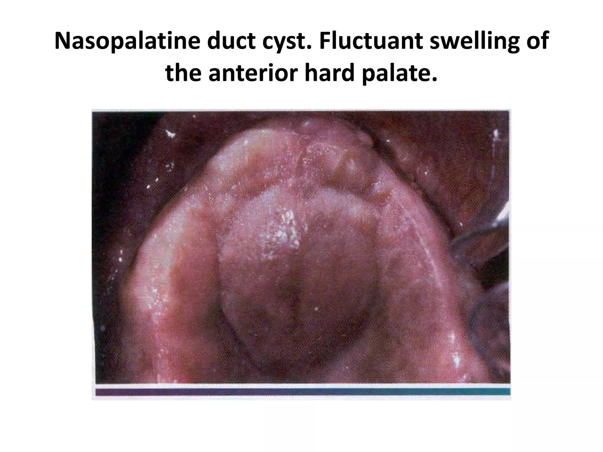
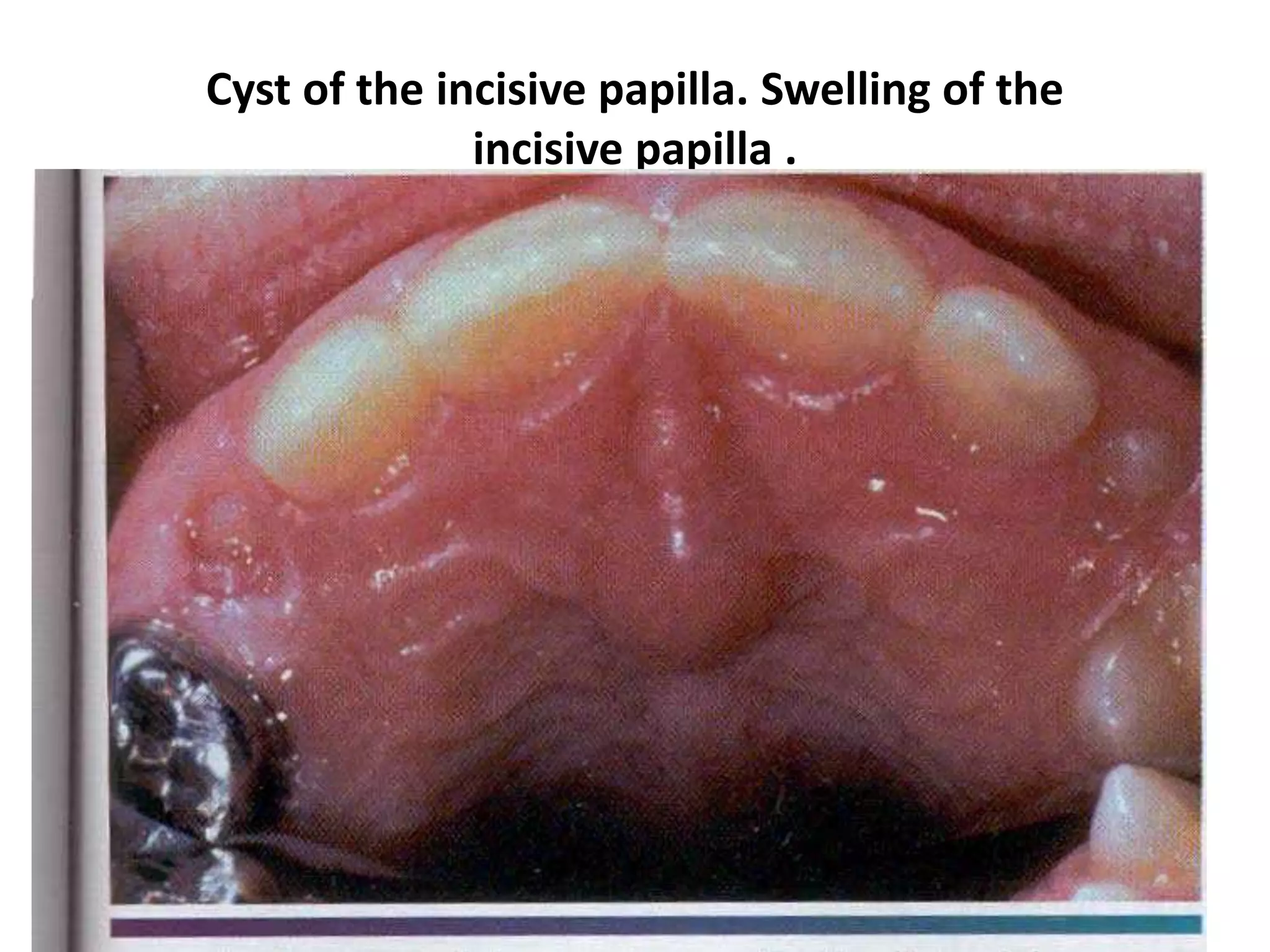
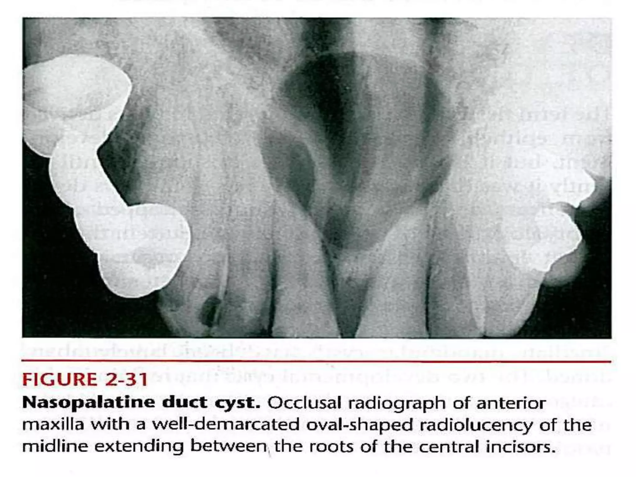
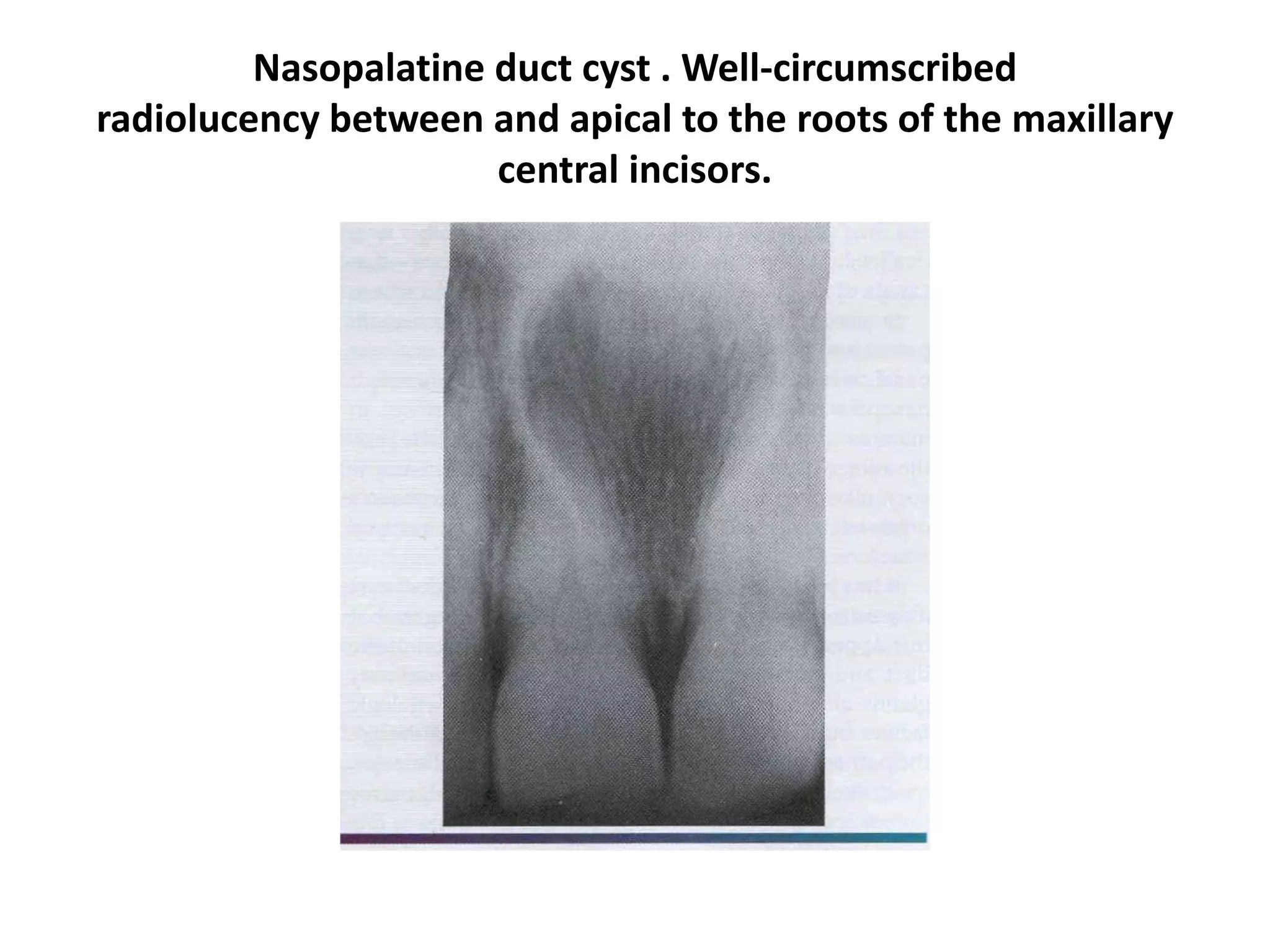

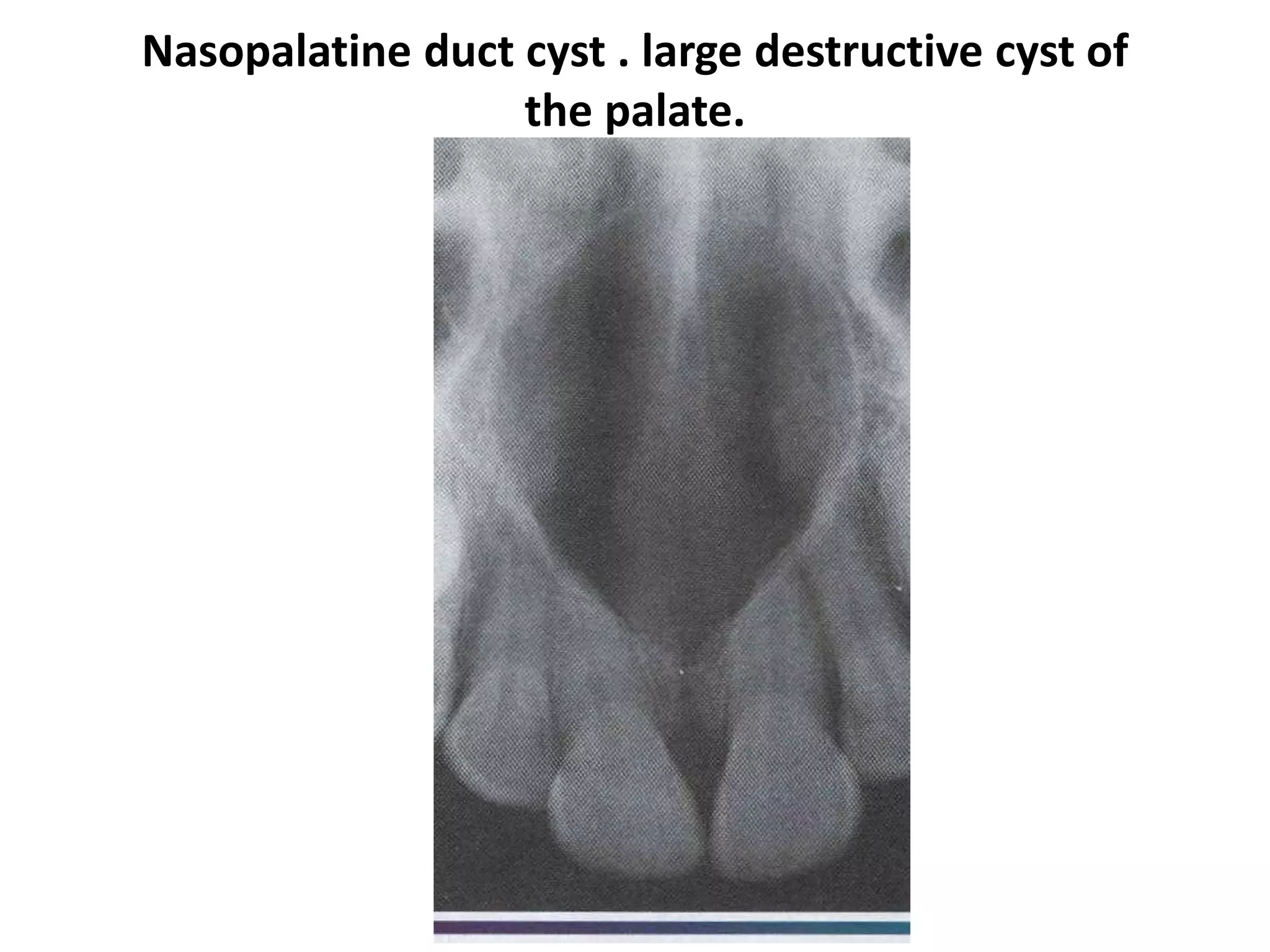
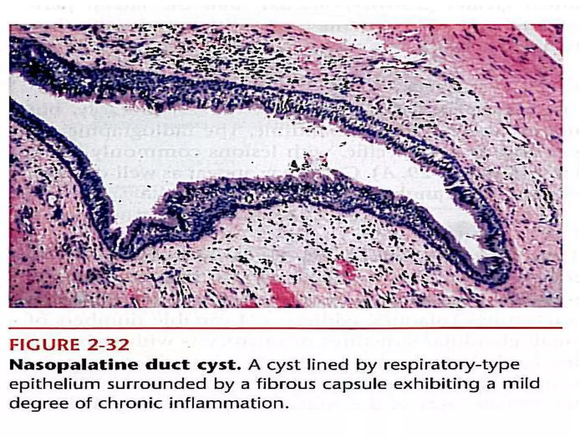
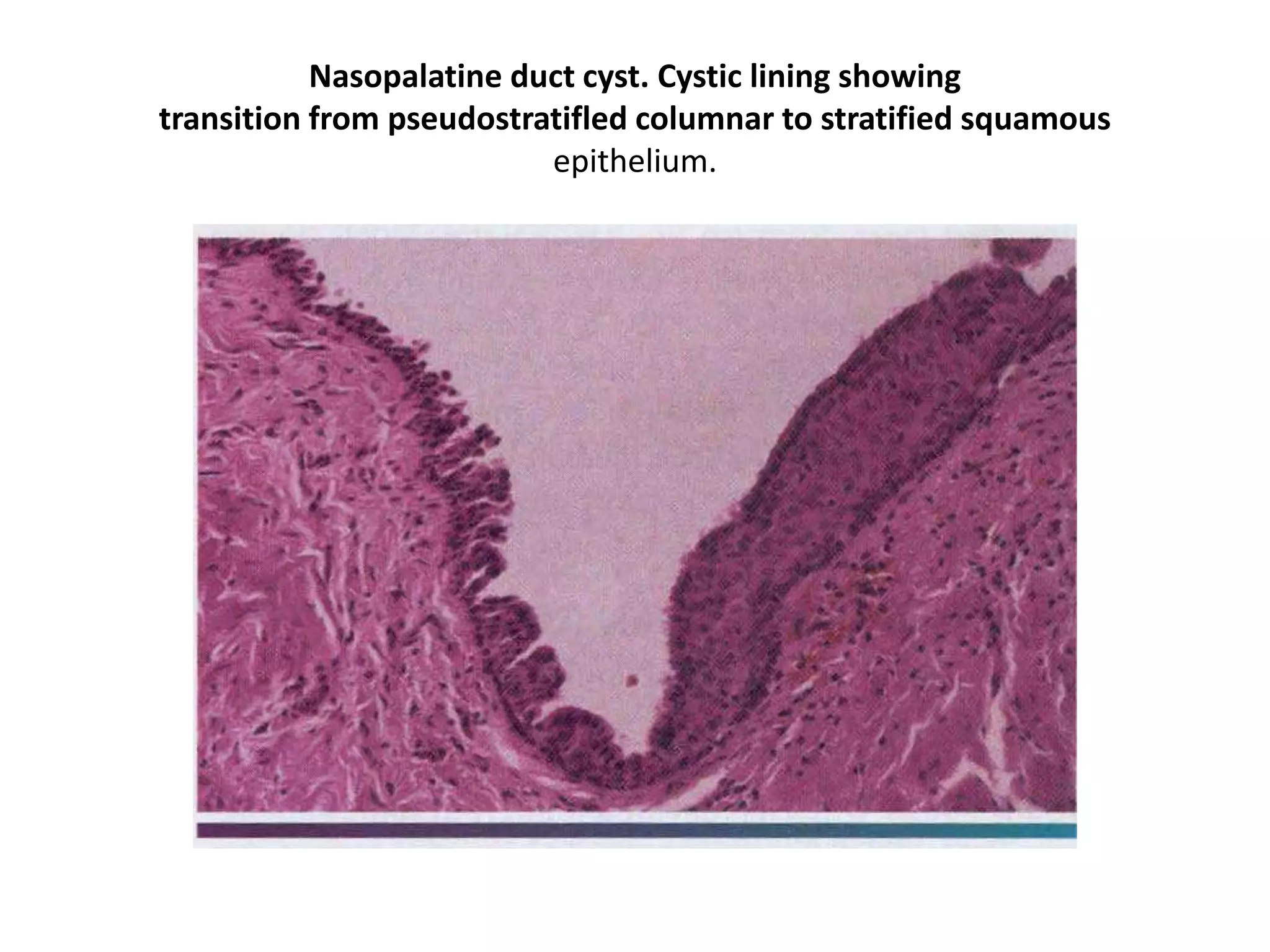
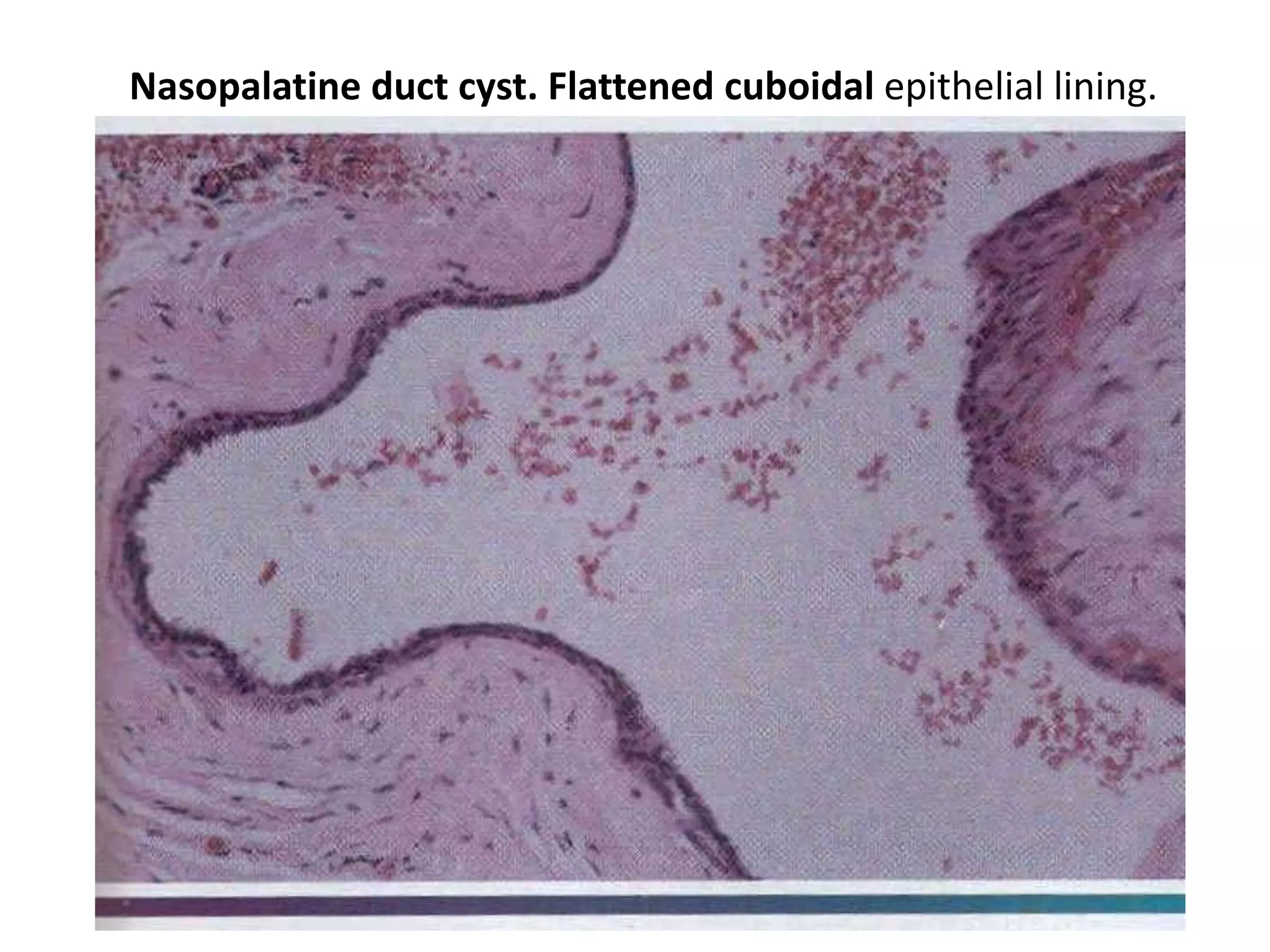
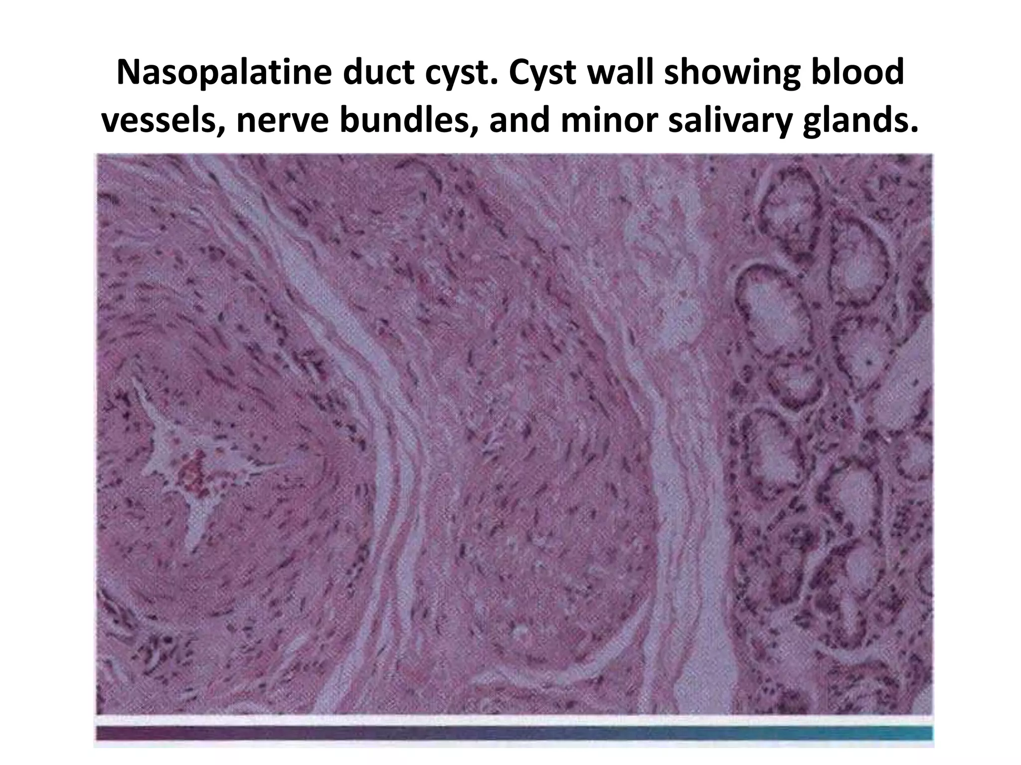
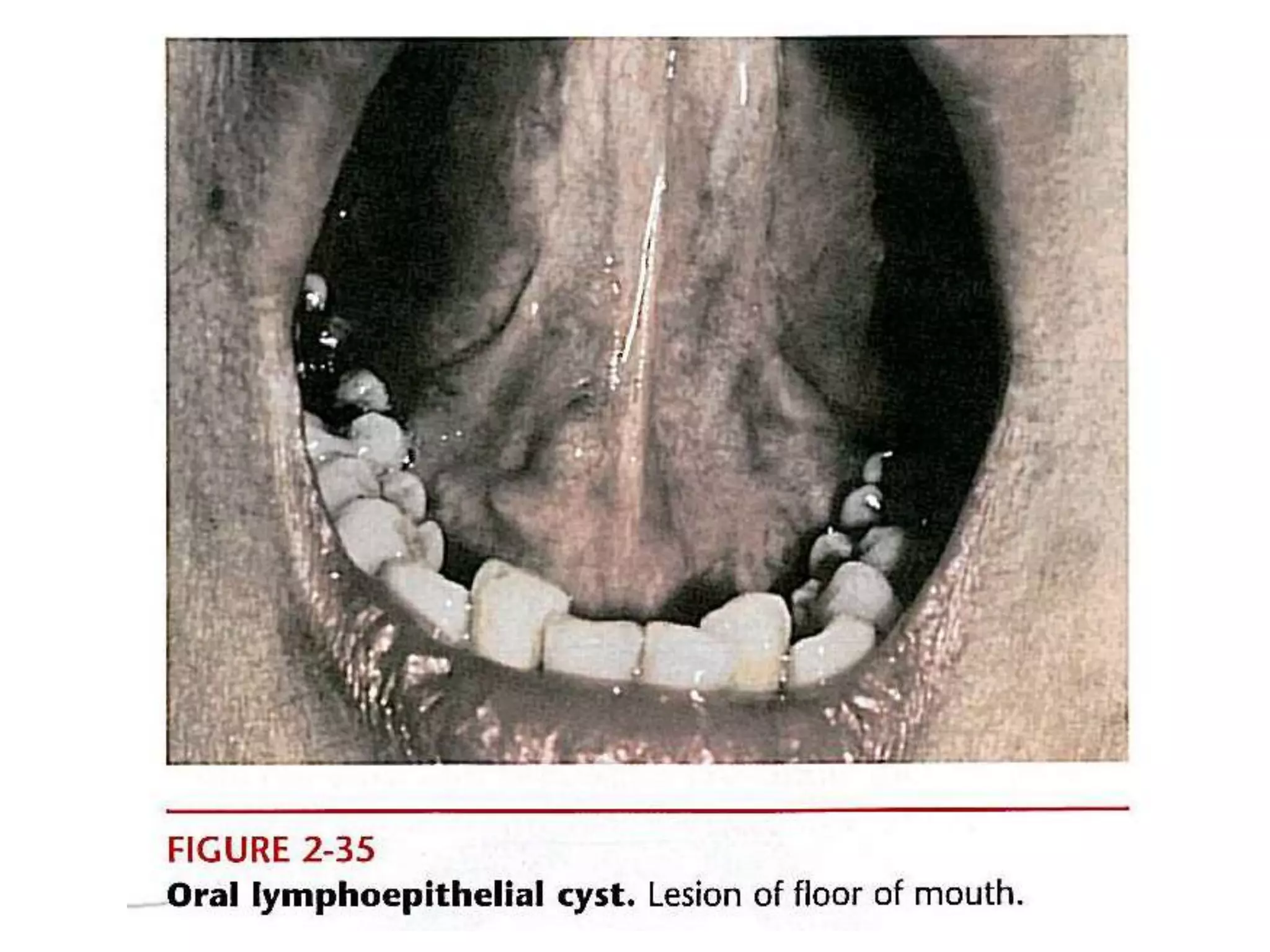
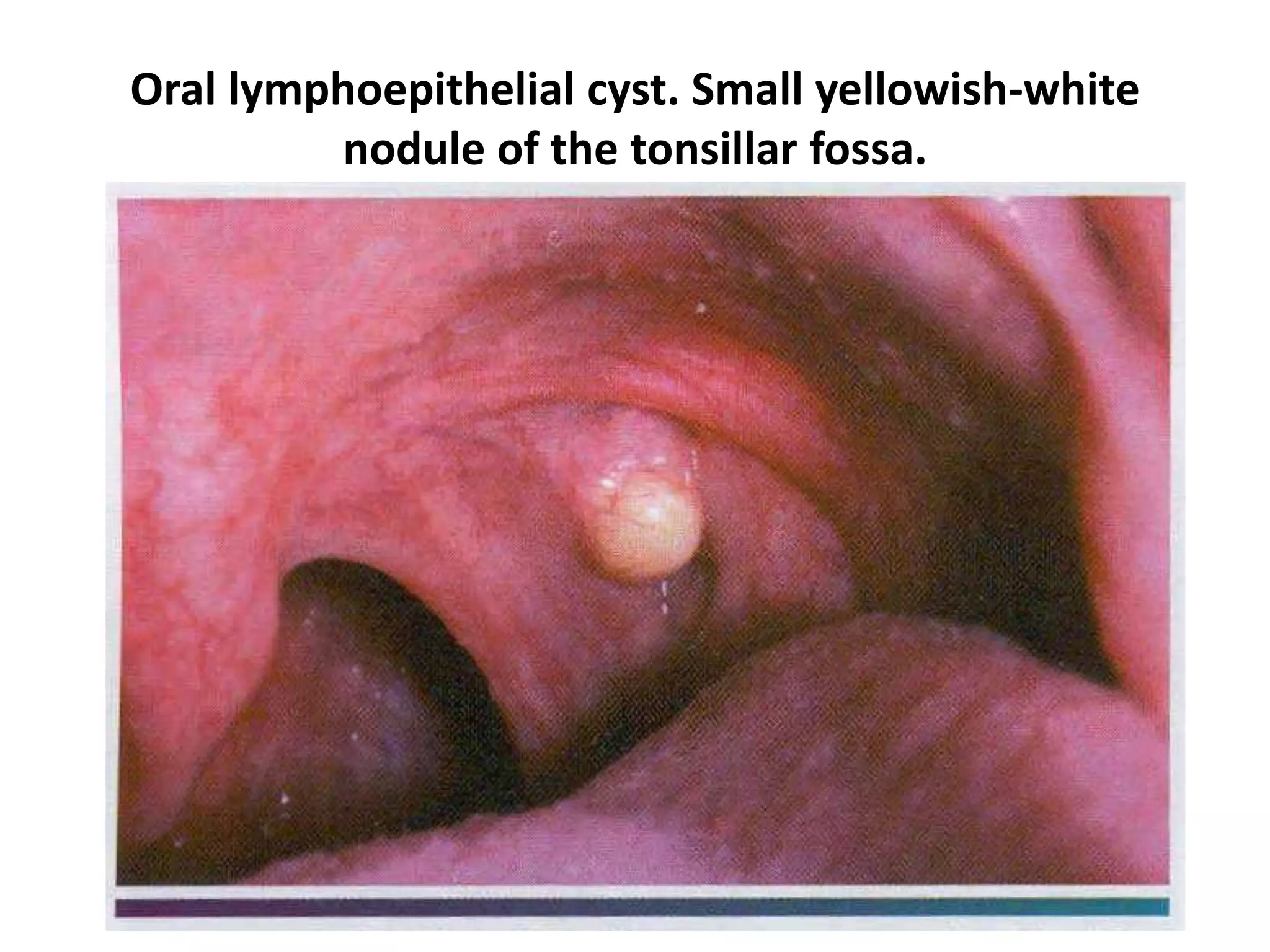
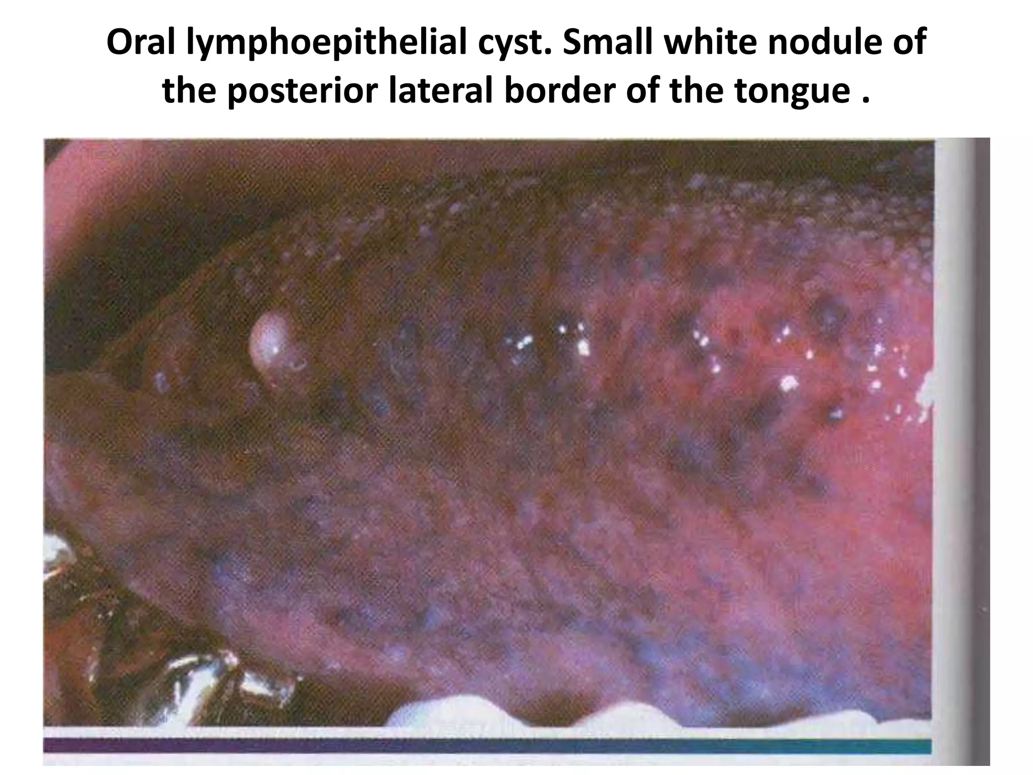
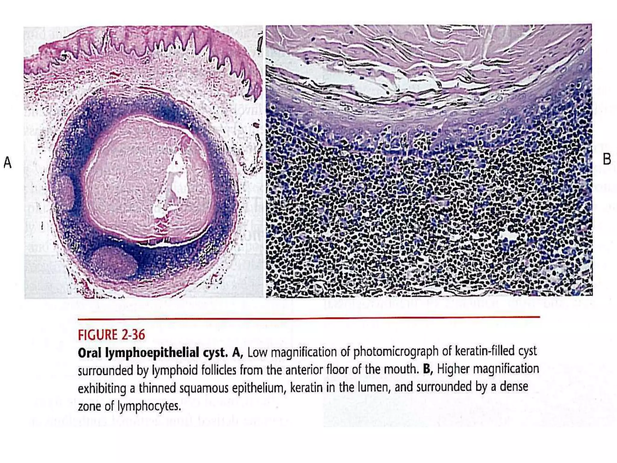
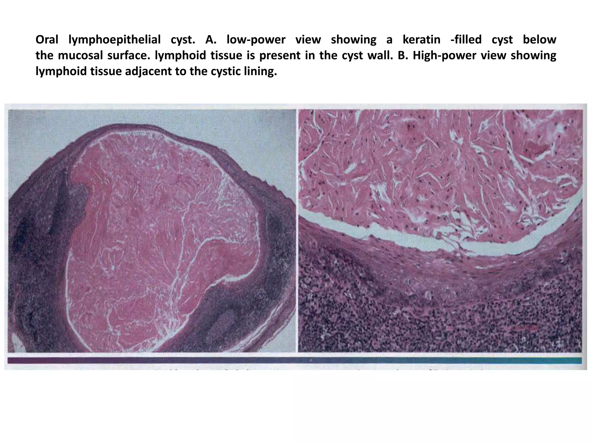
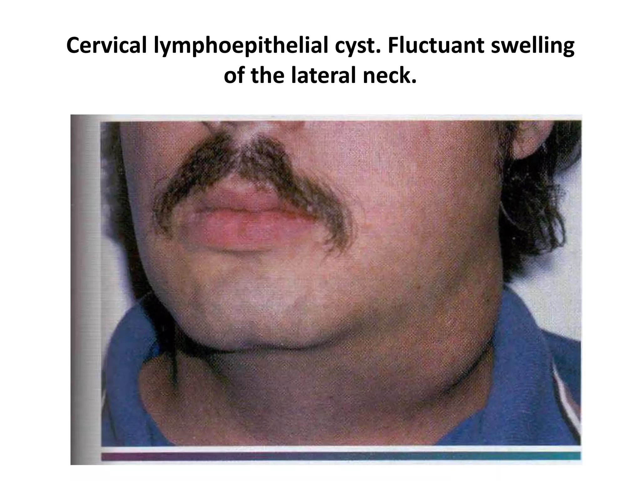
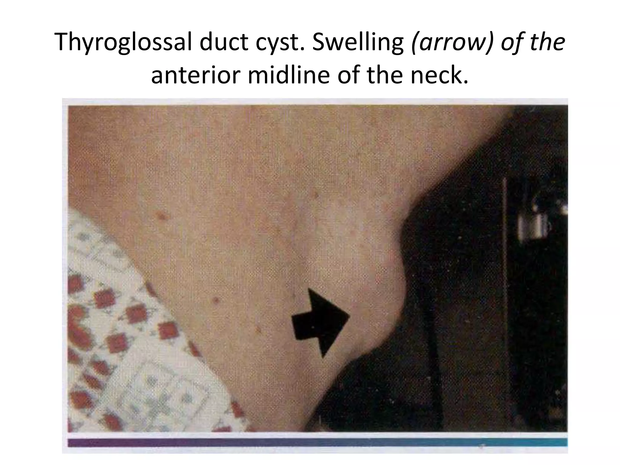
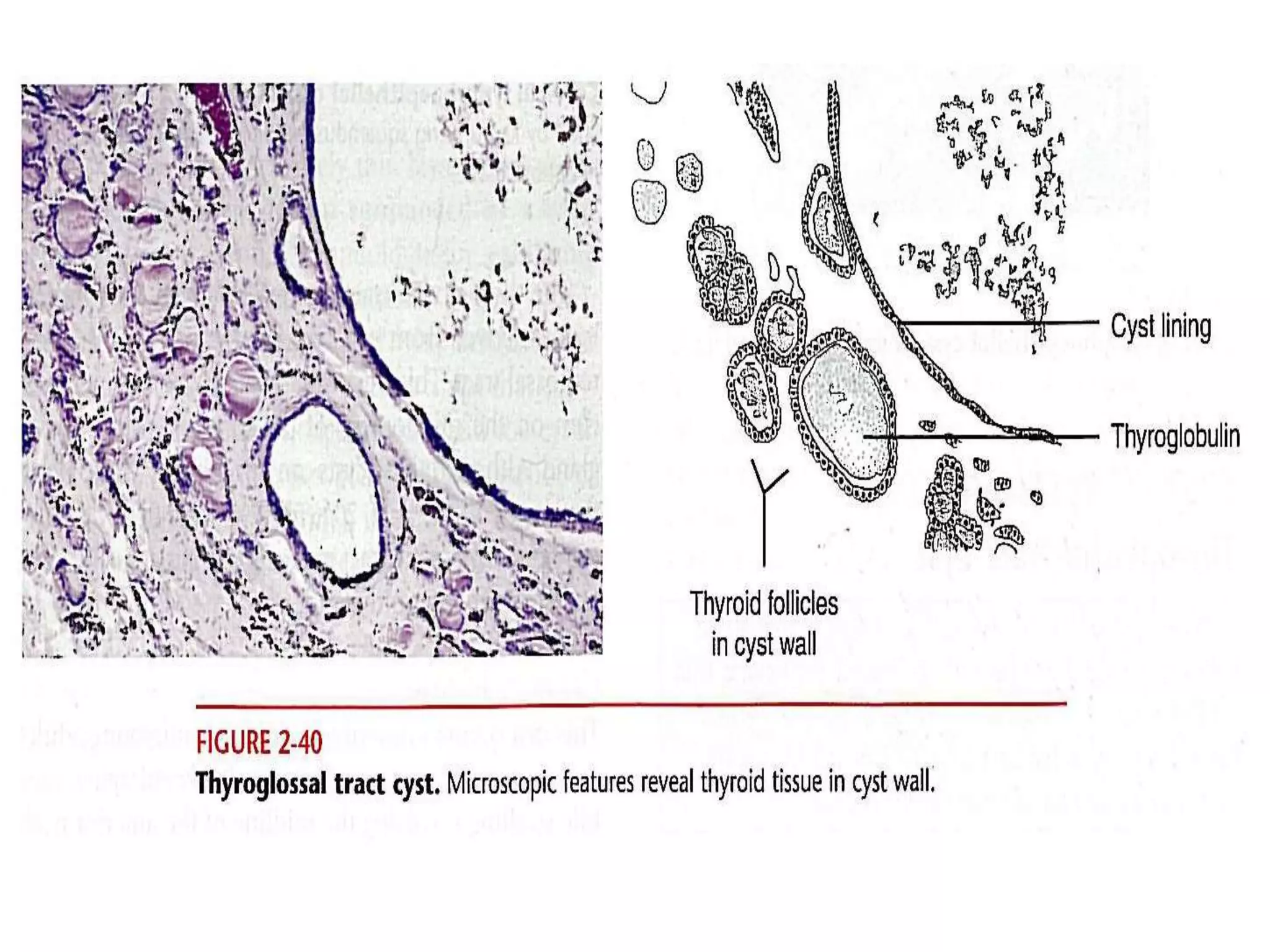
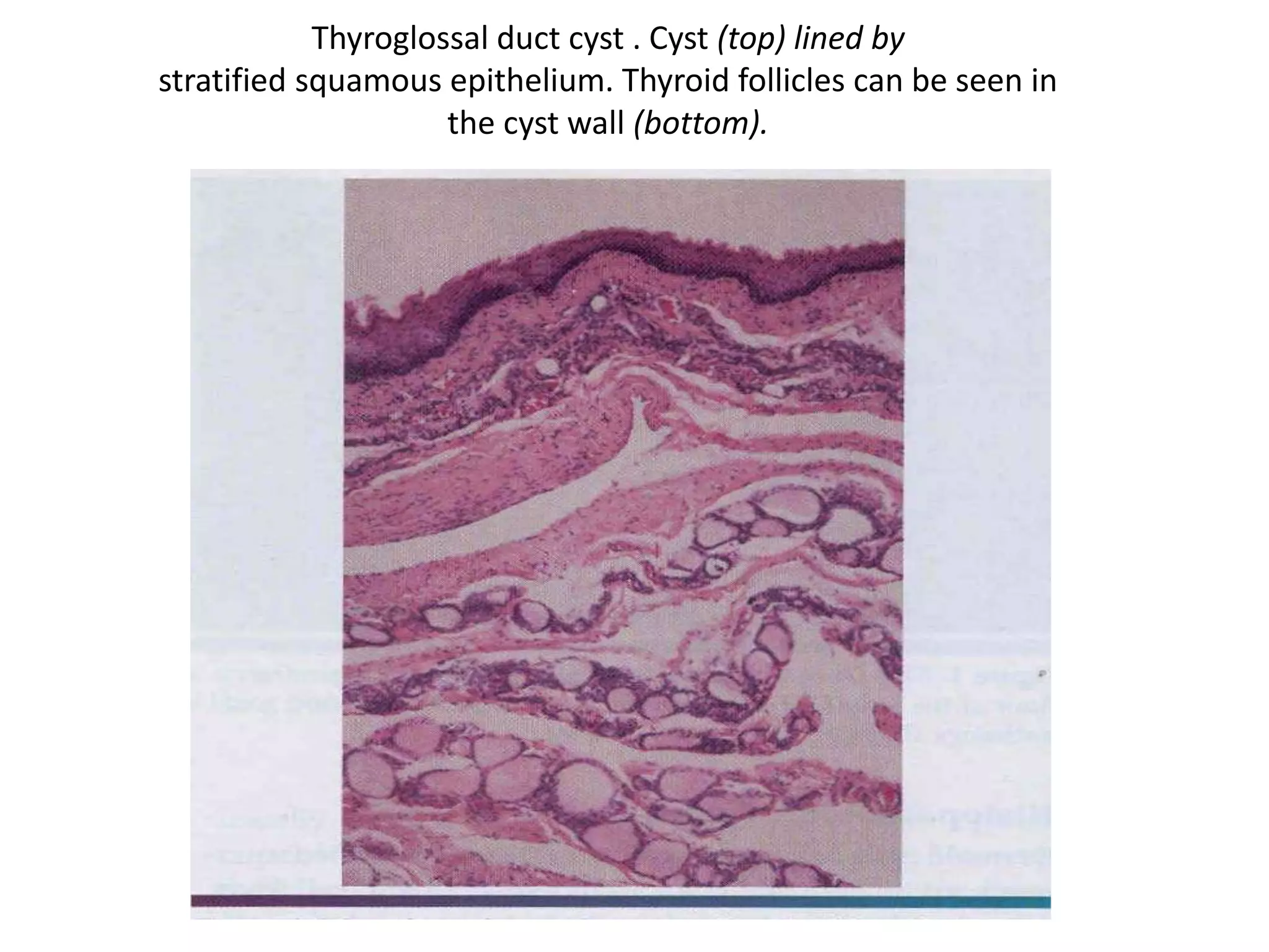
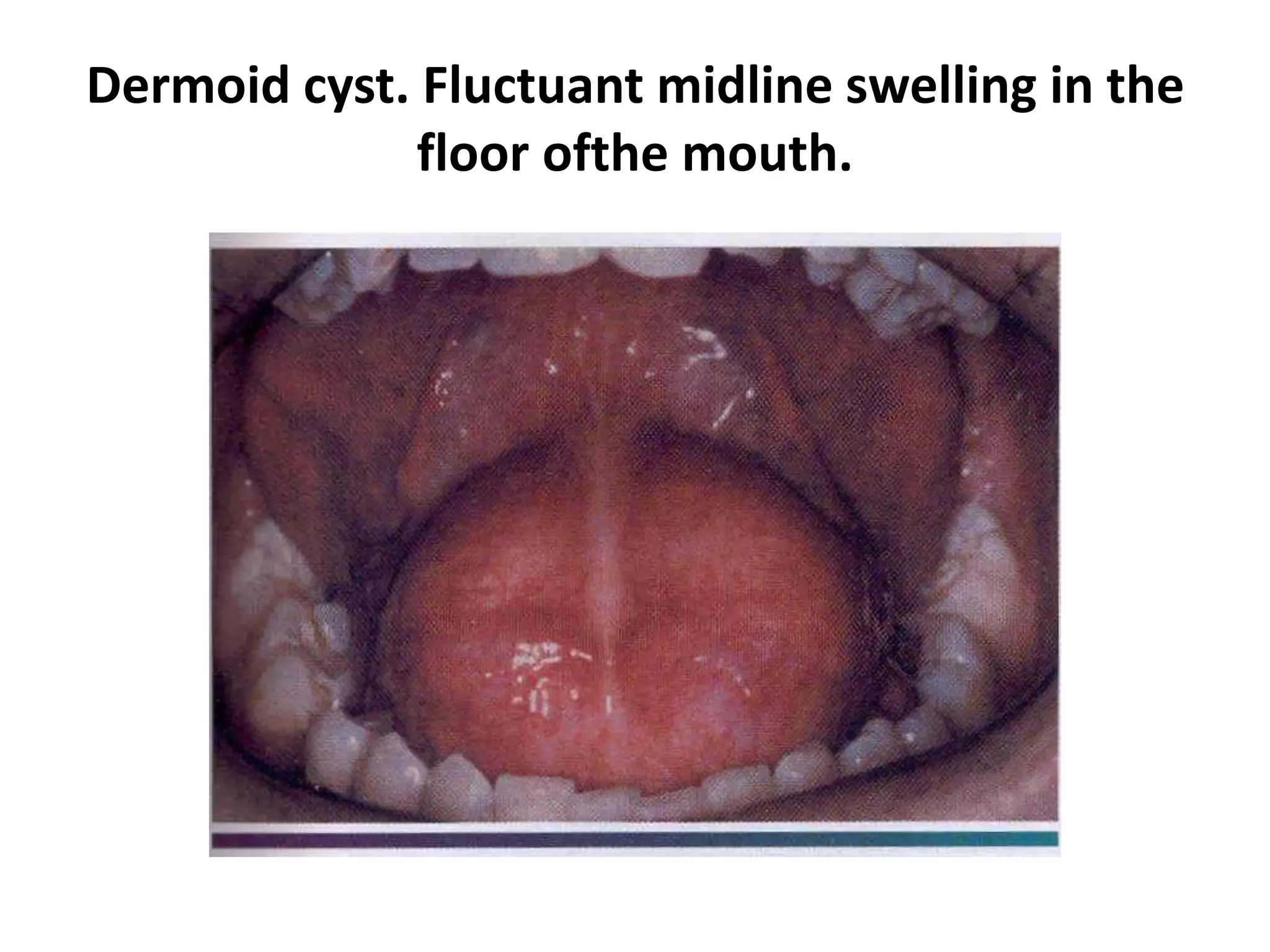
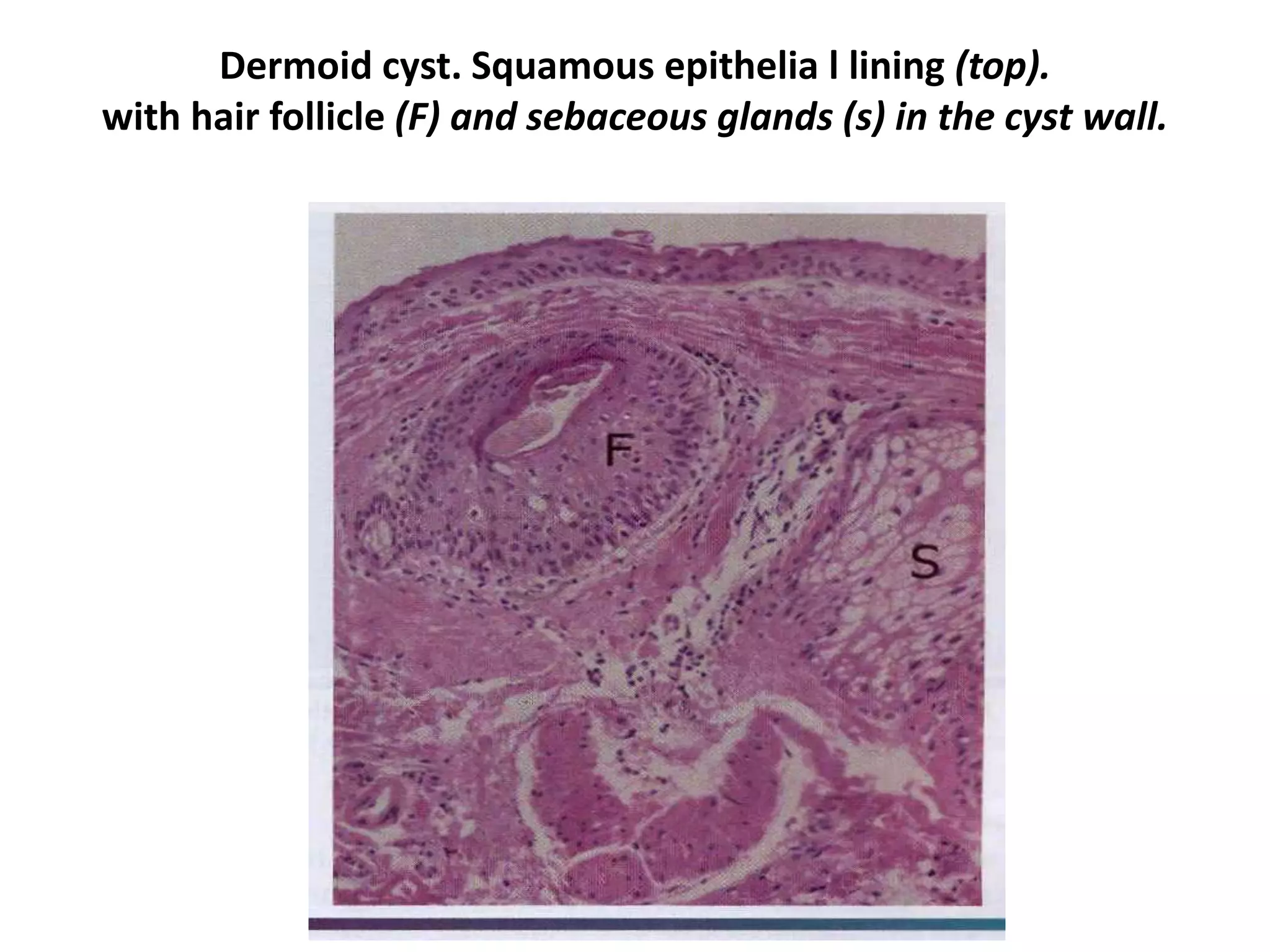
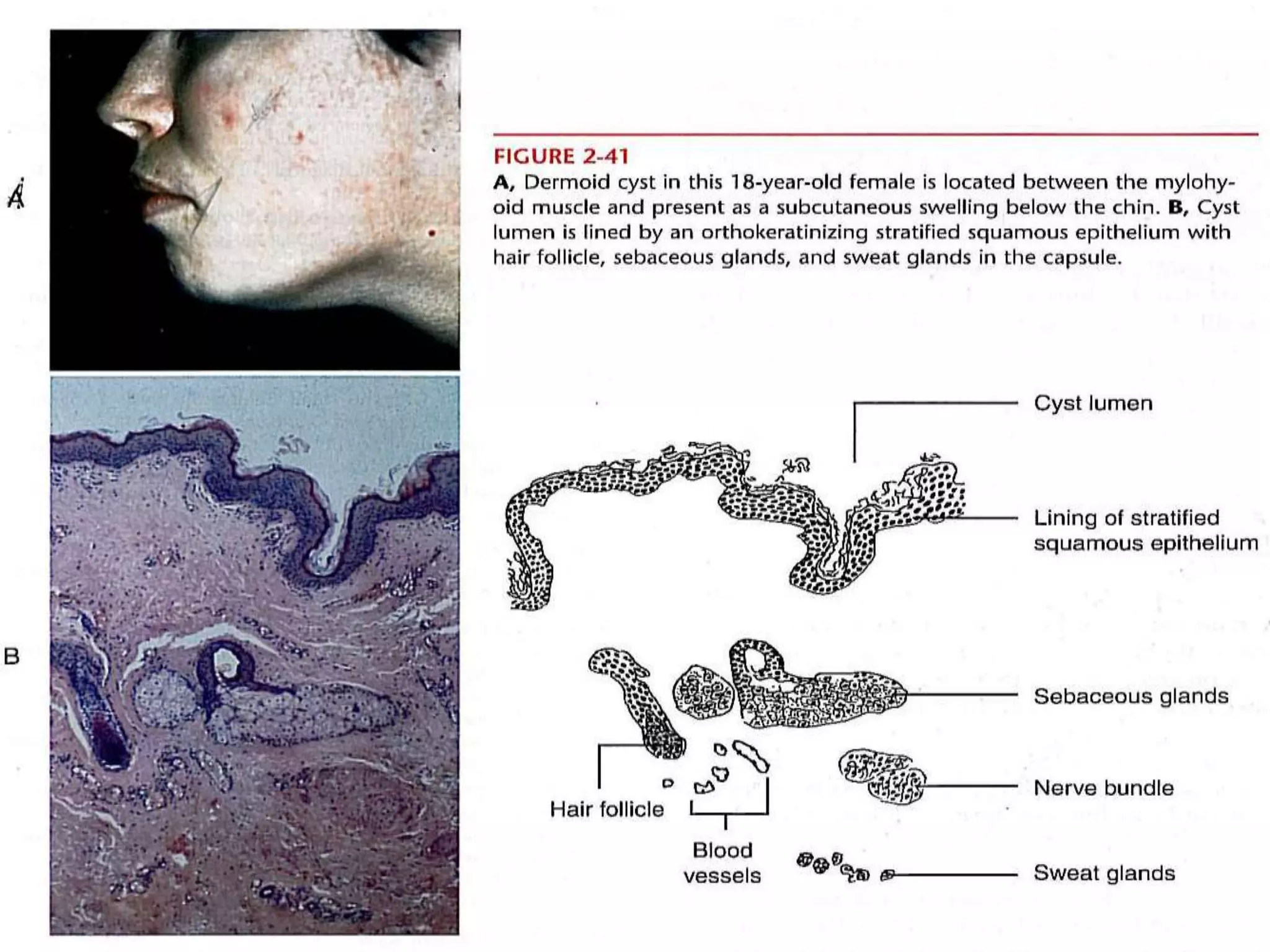
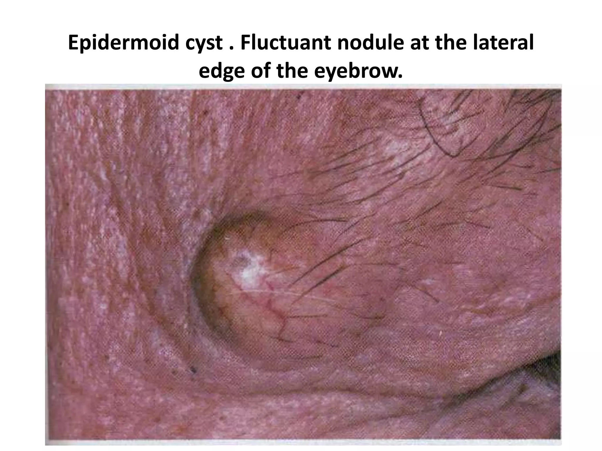
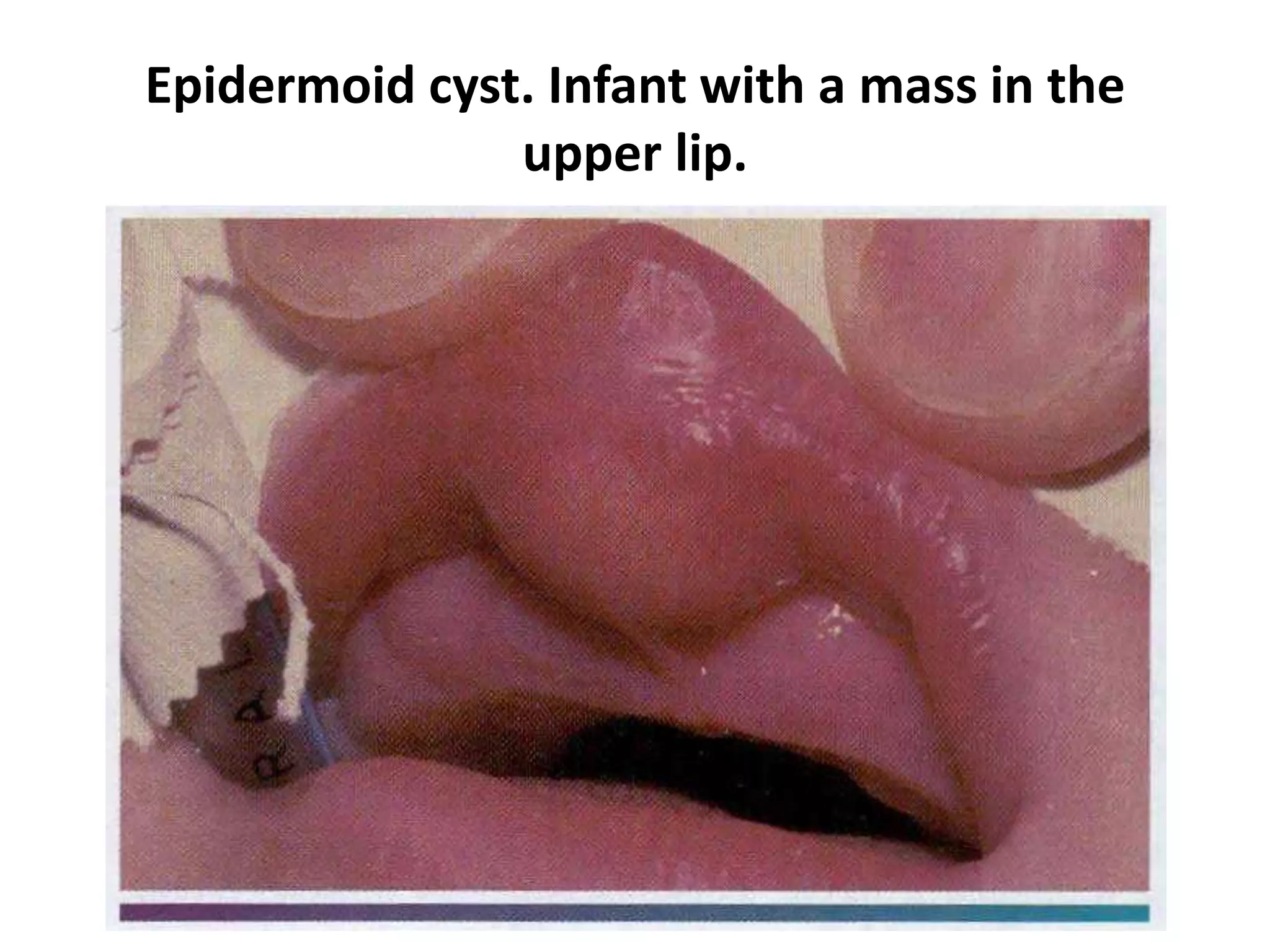

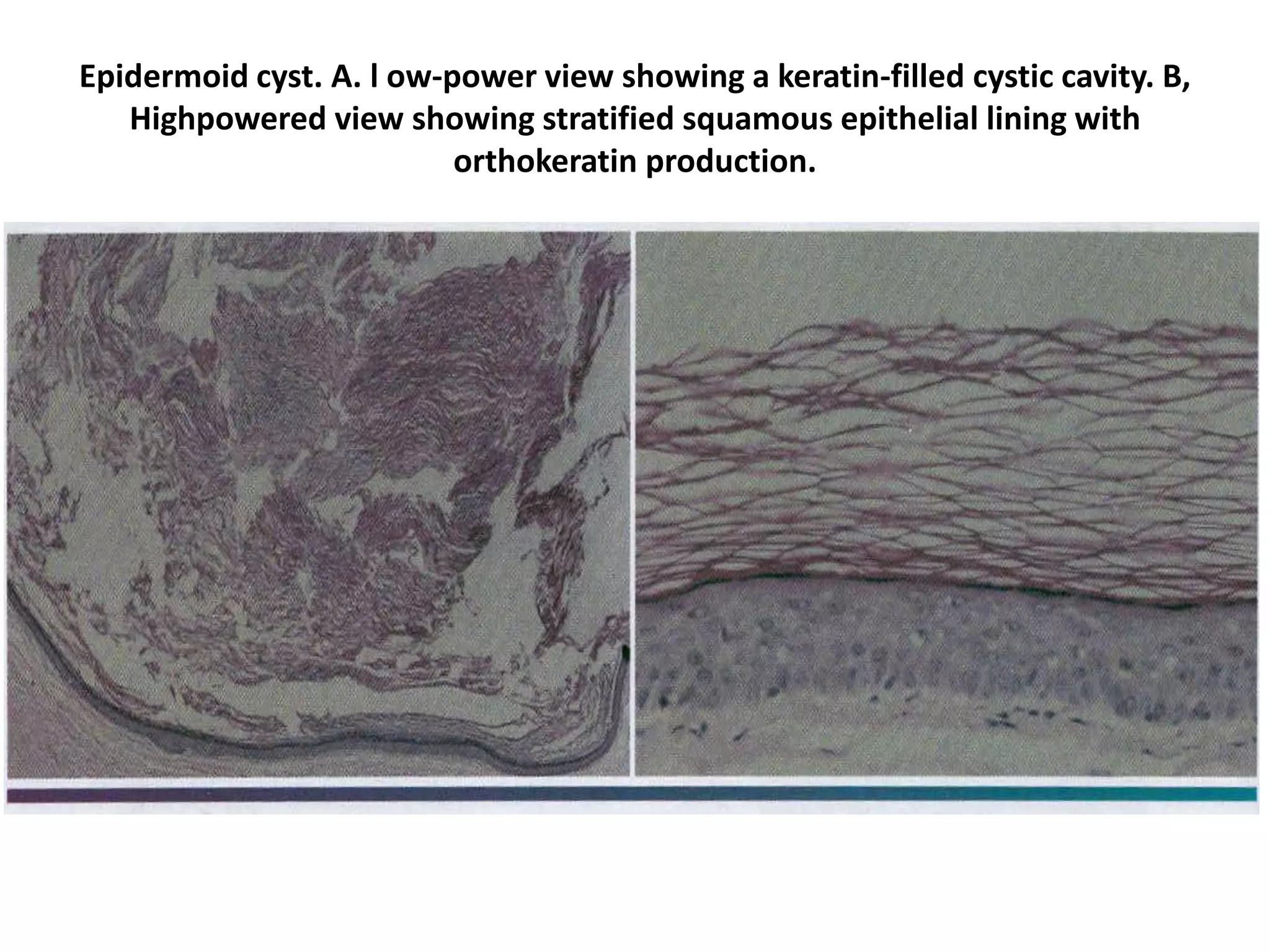

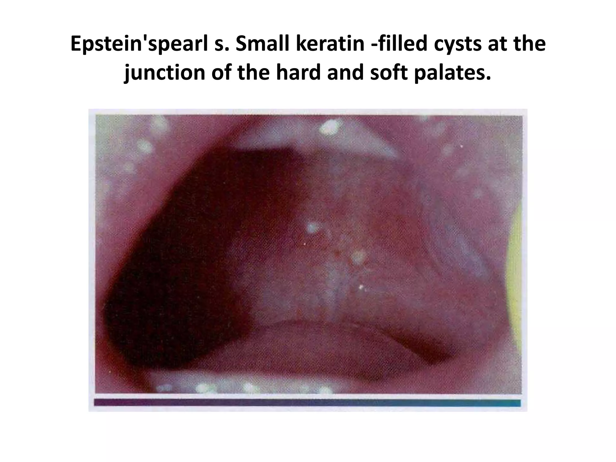


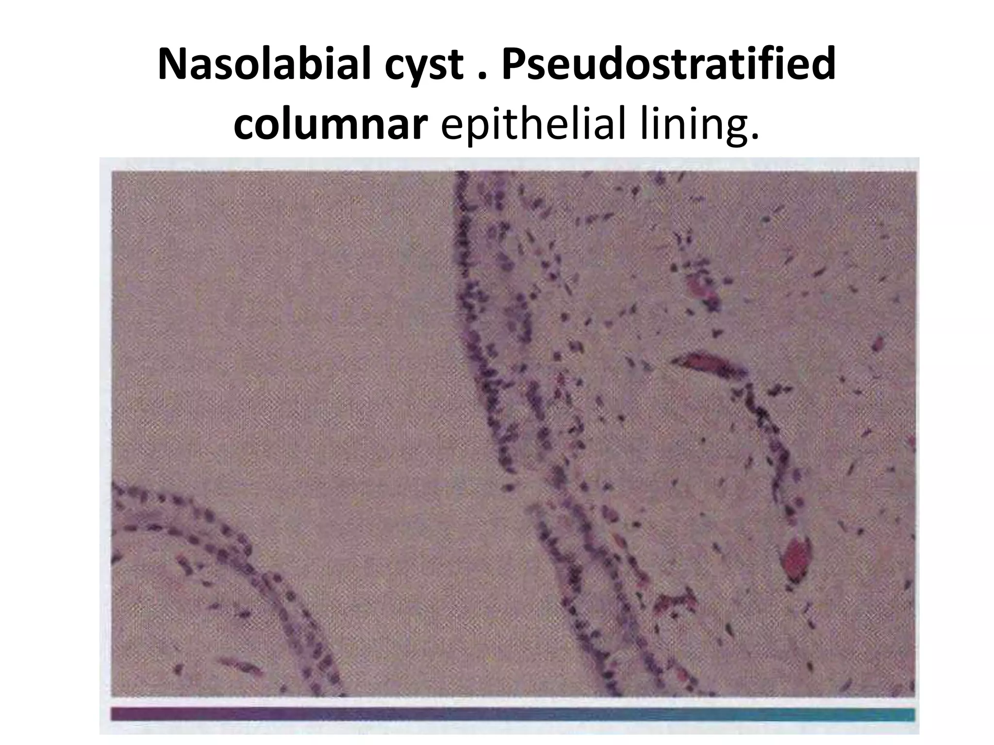
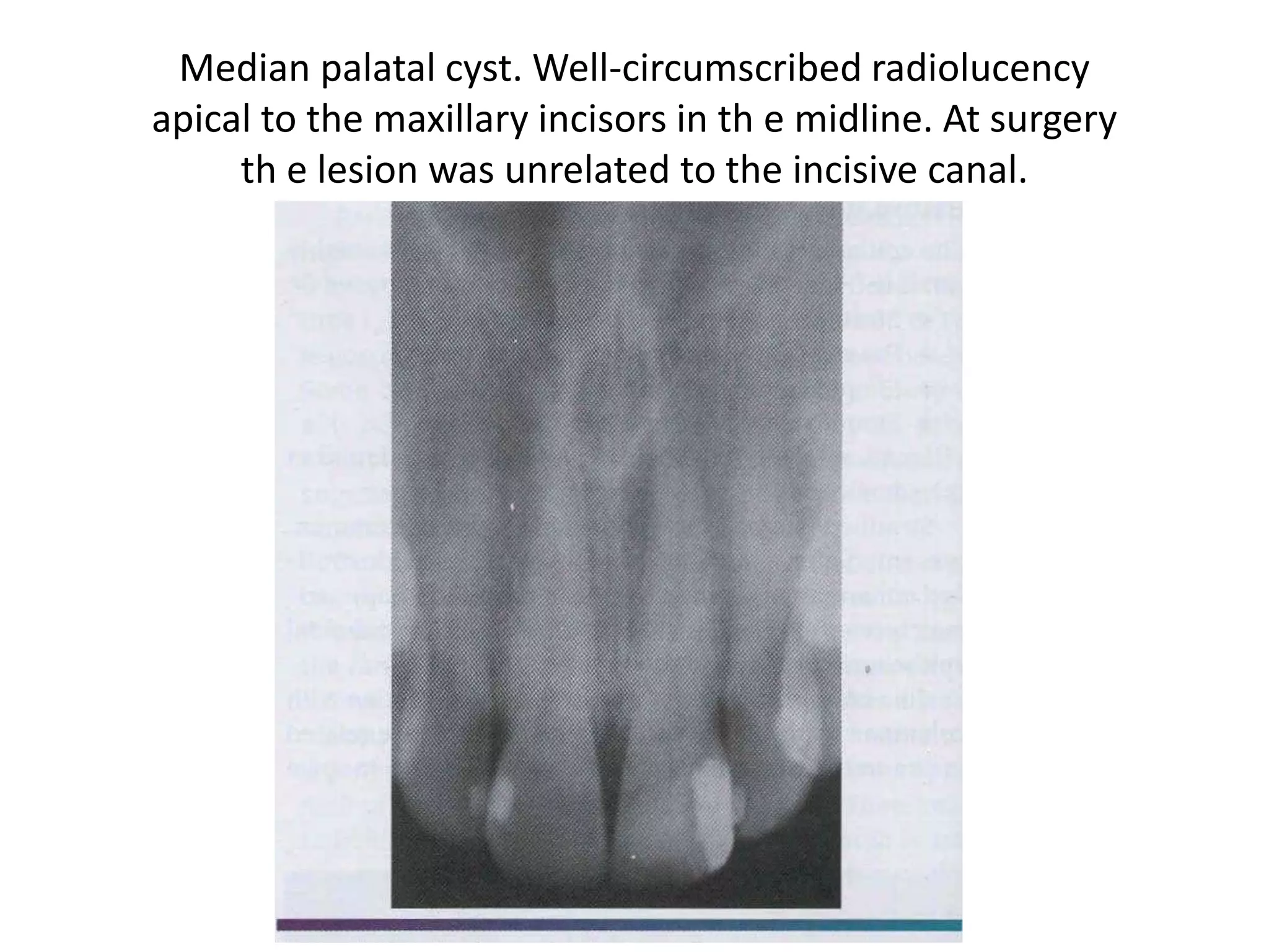
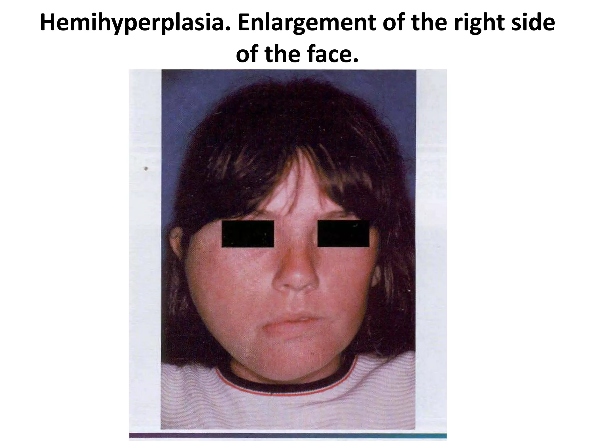
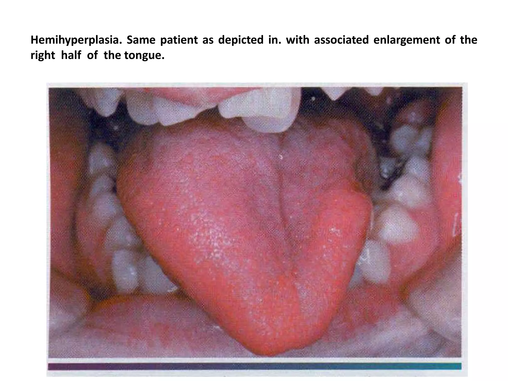
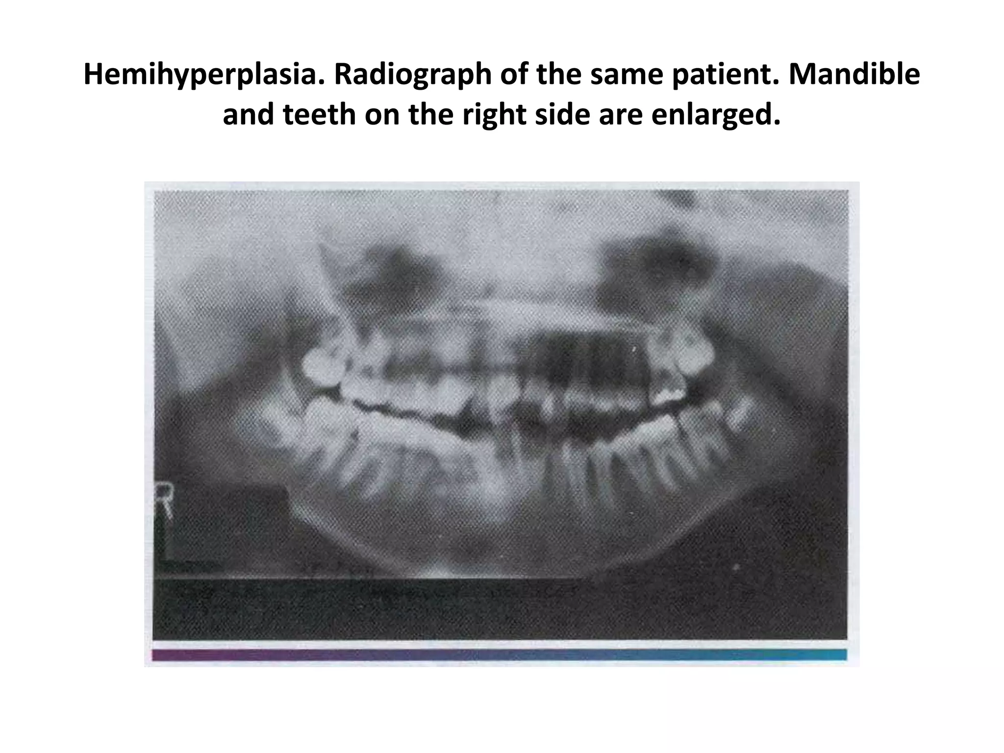
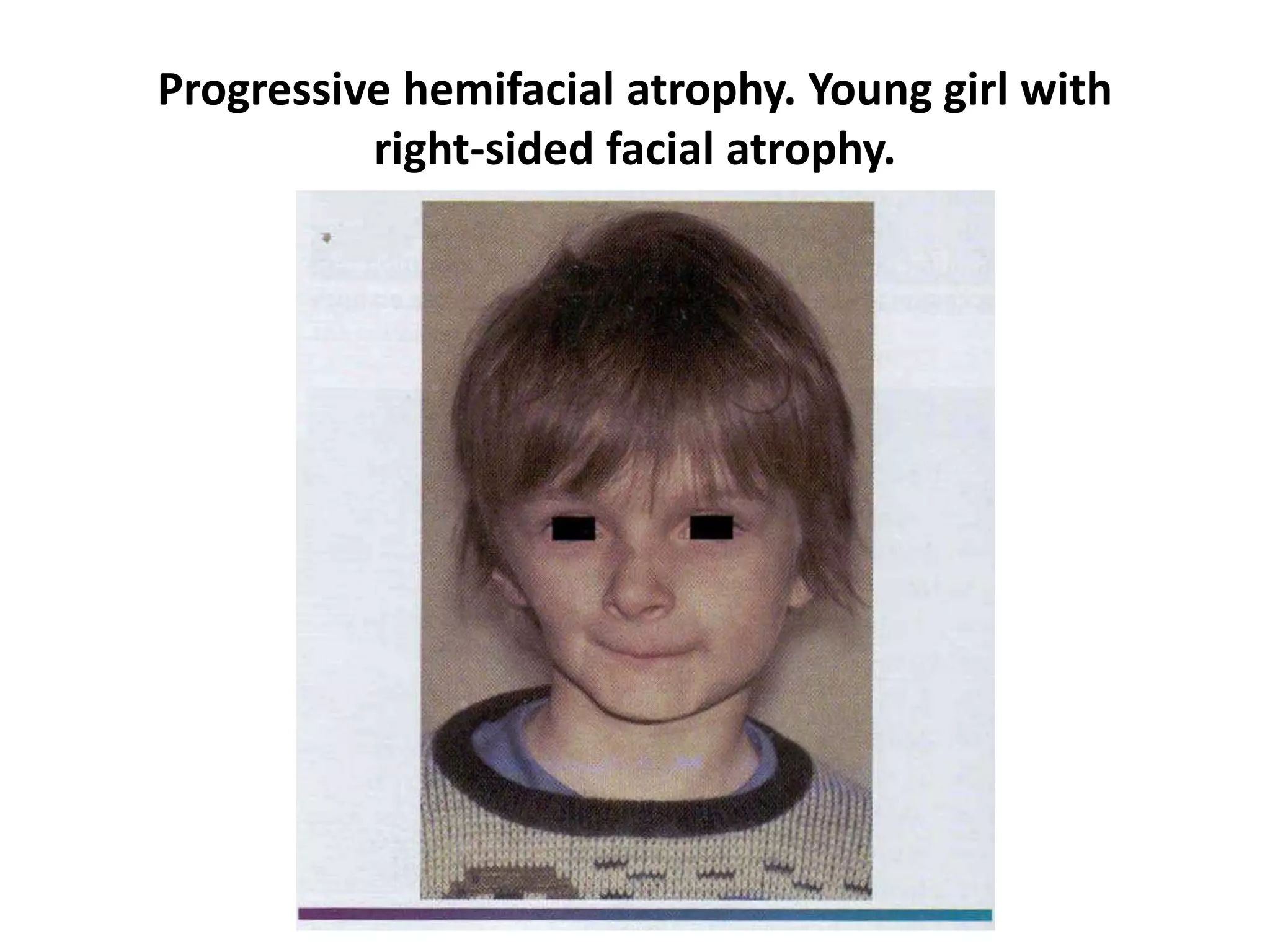
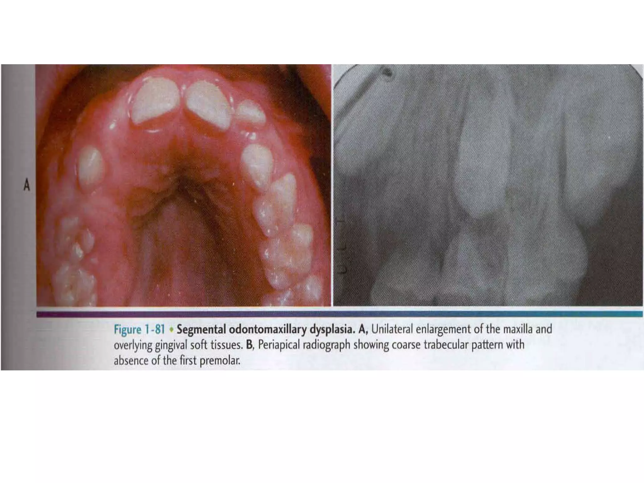
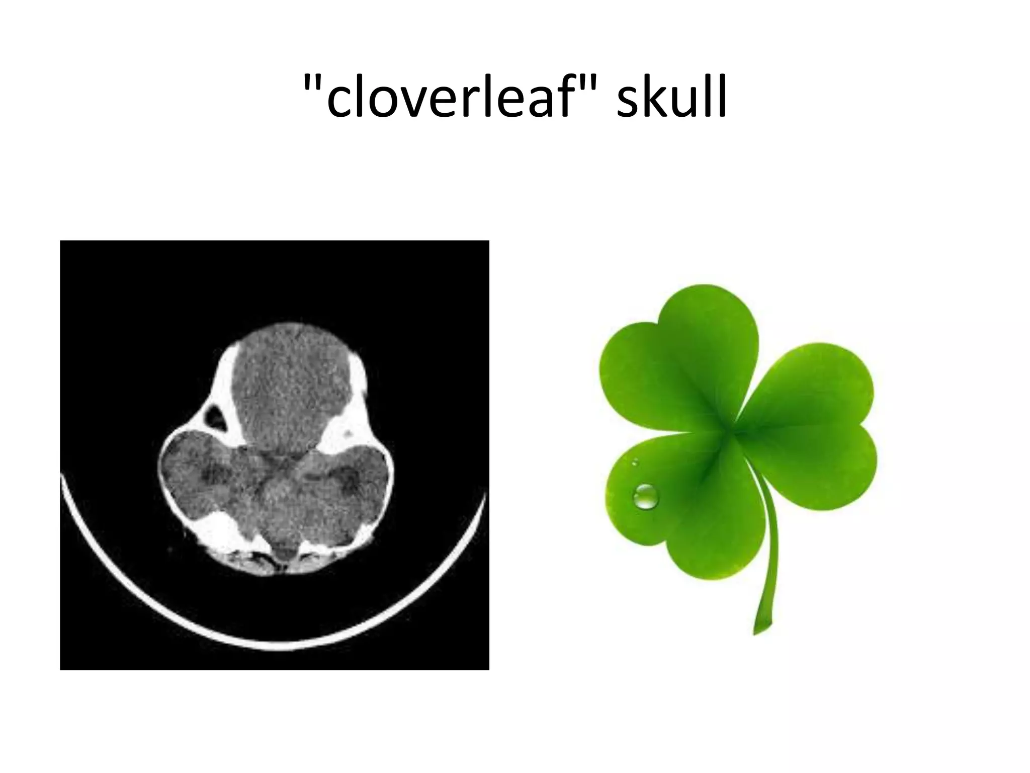
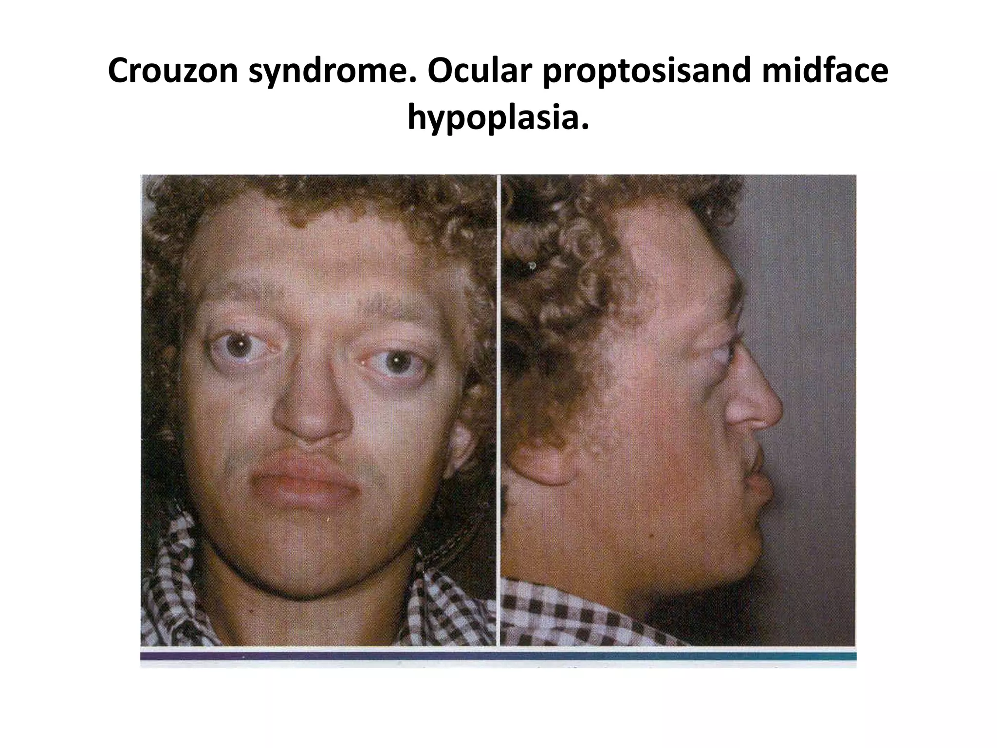

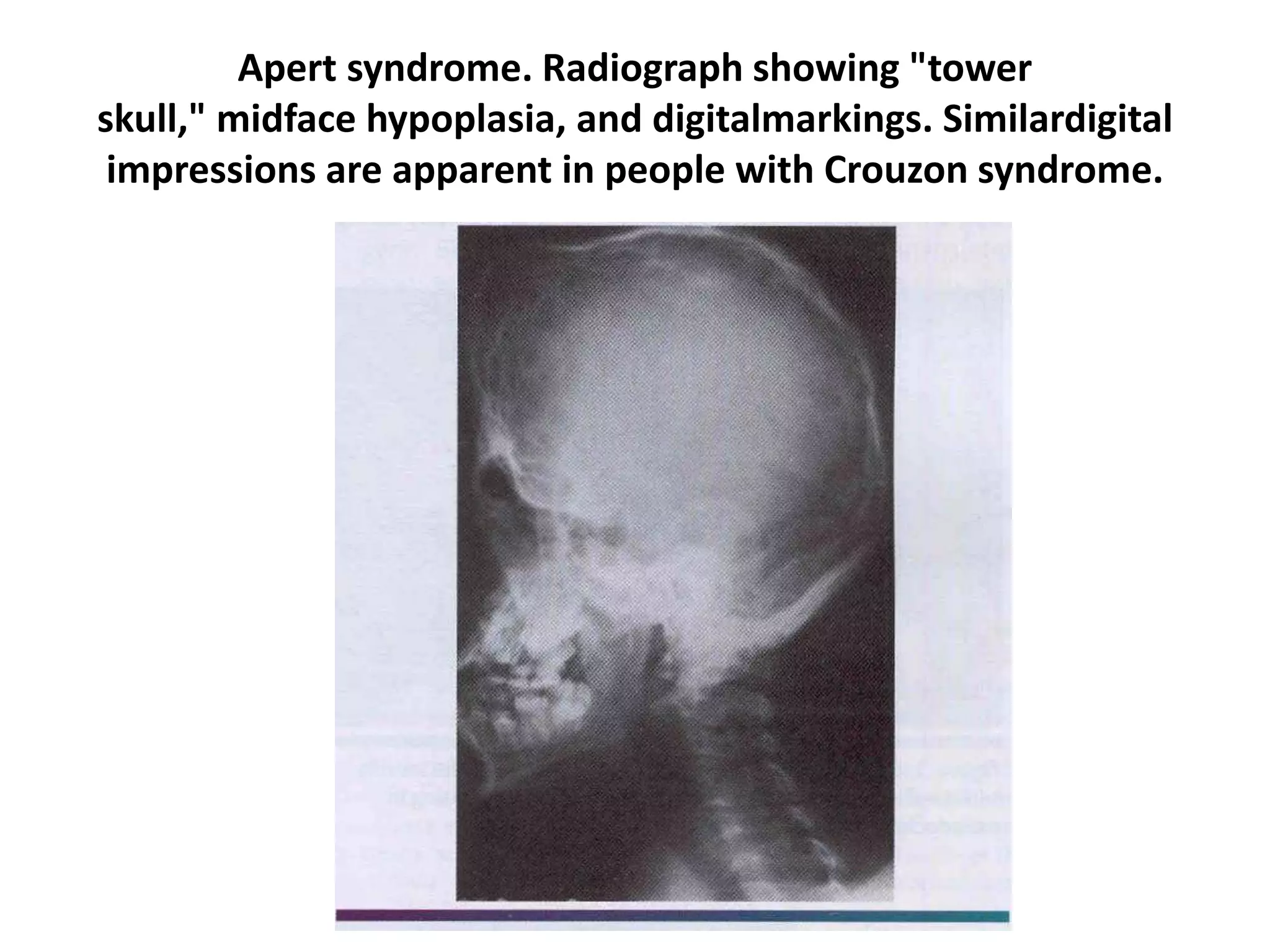

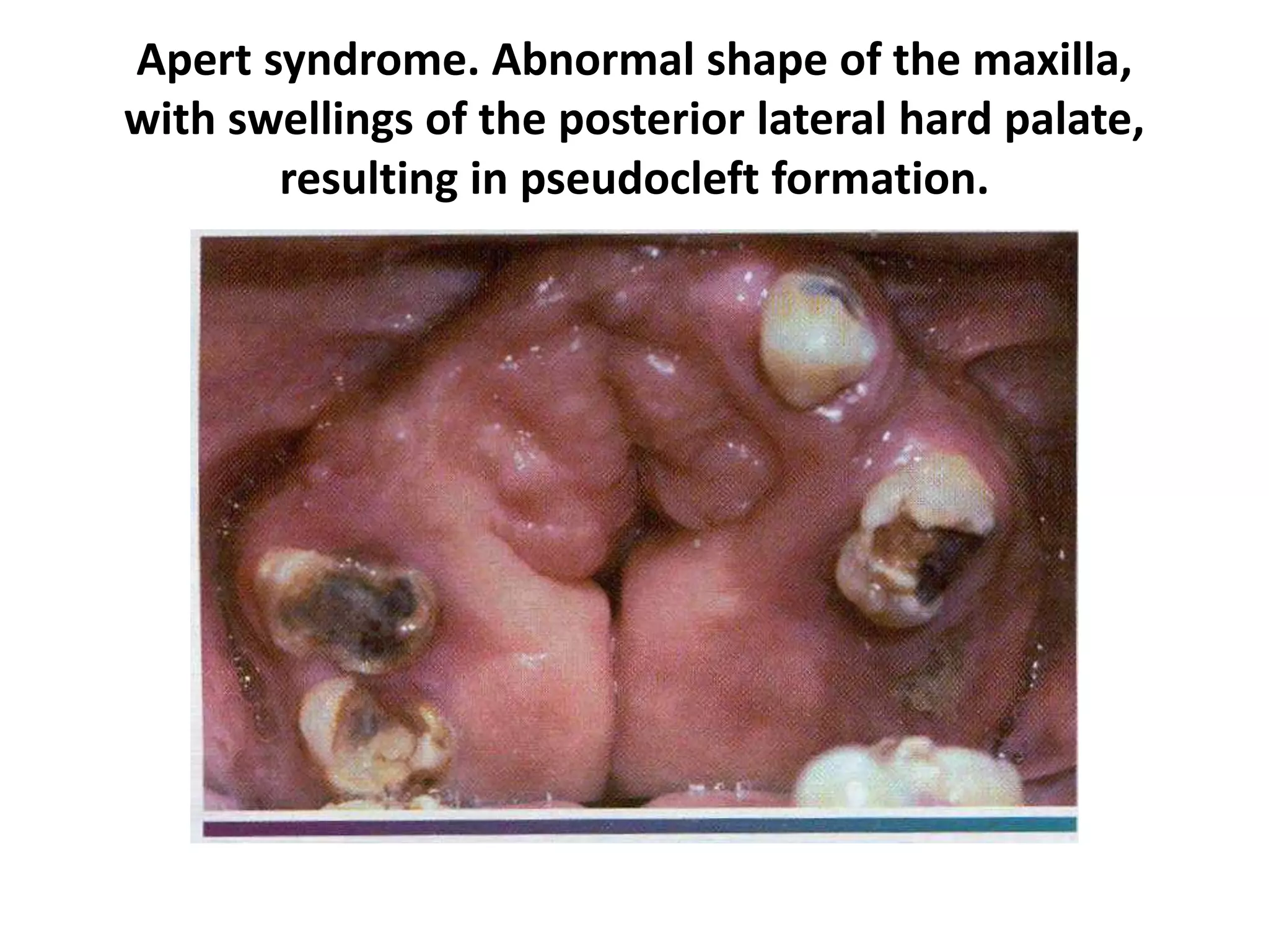
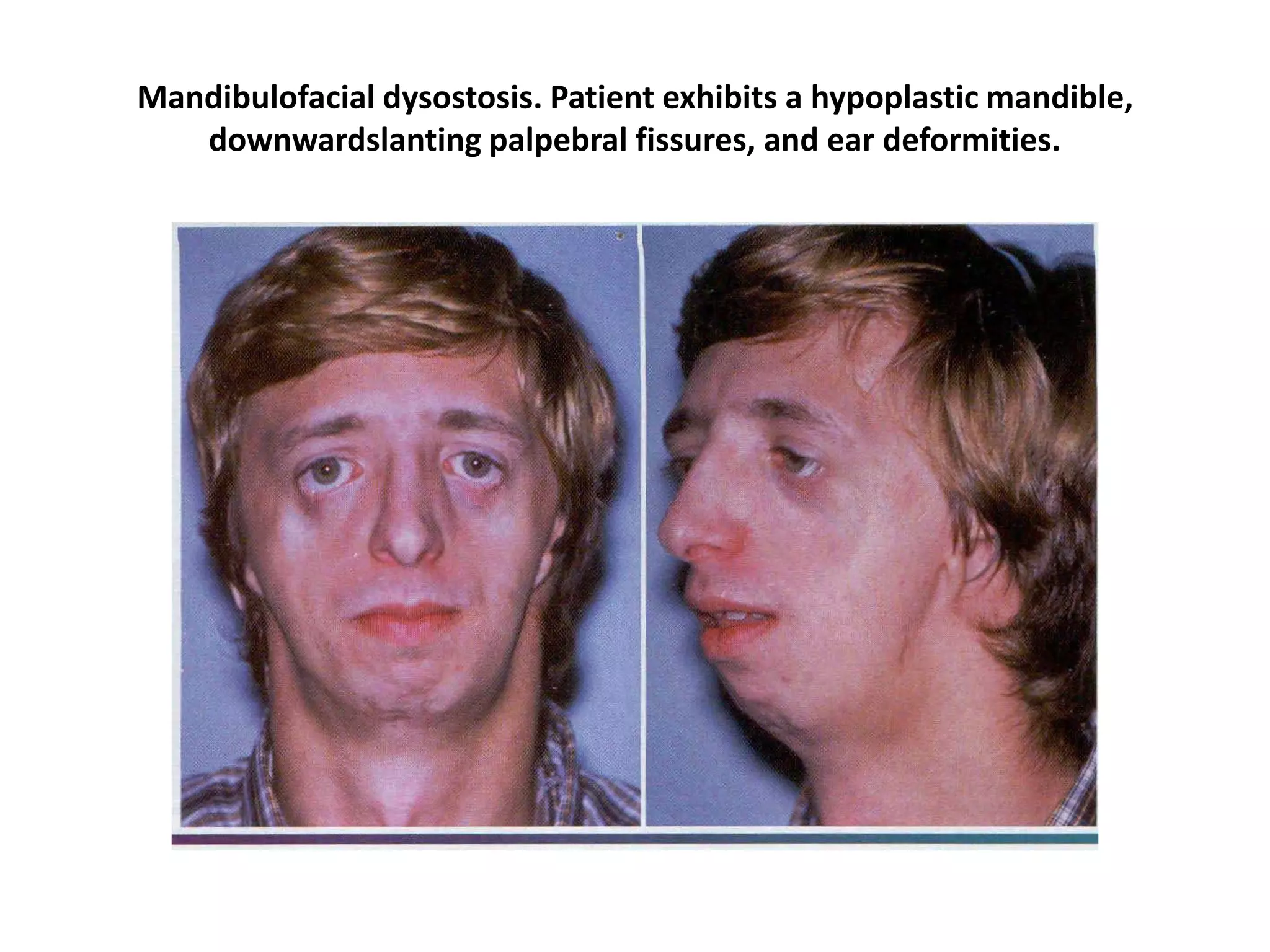

This document discusses several different types of cysts that can occur in the head and neck region, including nasopalatine duct cysts, oral lymphoepithelial cysts, dermoid cysts, epidermoid cysts, and globulomaxillary cysts. It provides details on the clinical and radiographic presentation of each cyst type as well as microscopic findings. The document also examines several congenital syndromes that affect craniofacial development, such as Apert syndrome, Crouzon syndrome, hemifacial hyperplasia, and mandibulofacial dysostosis.