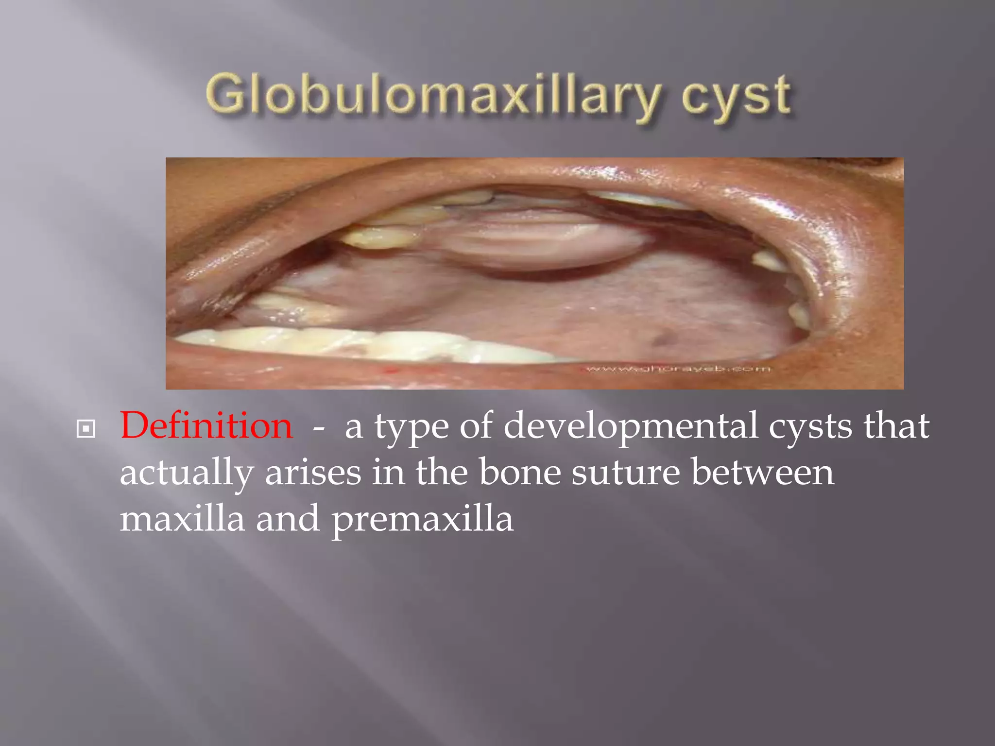
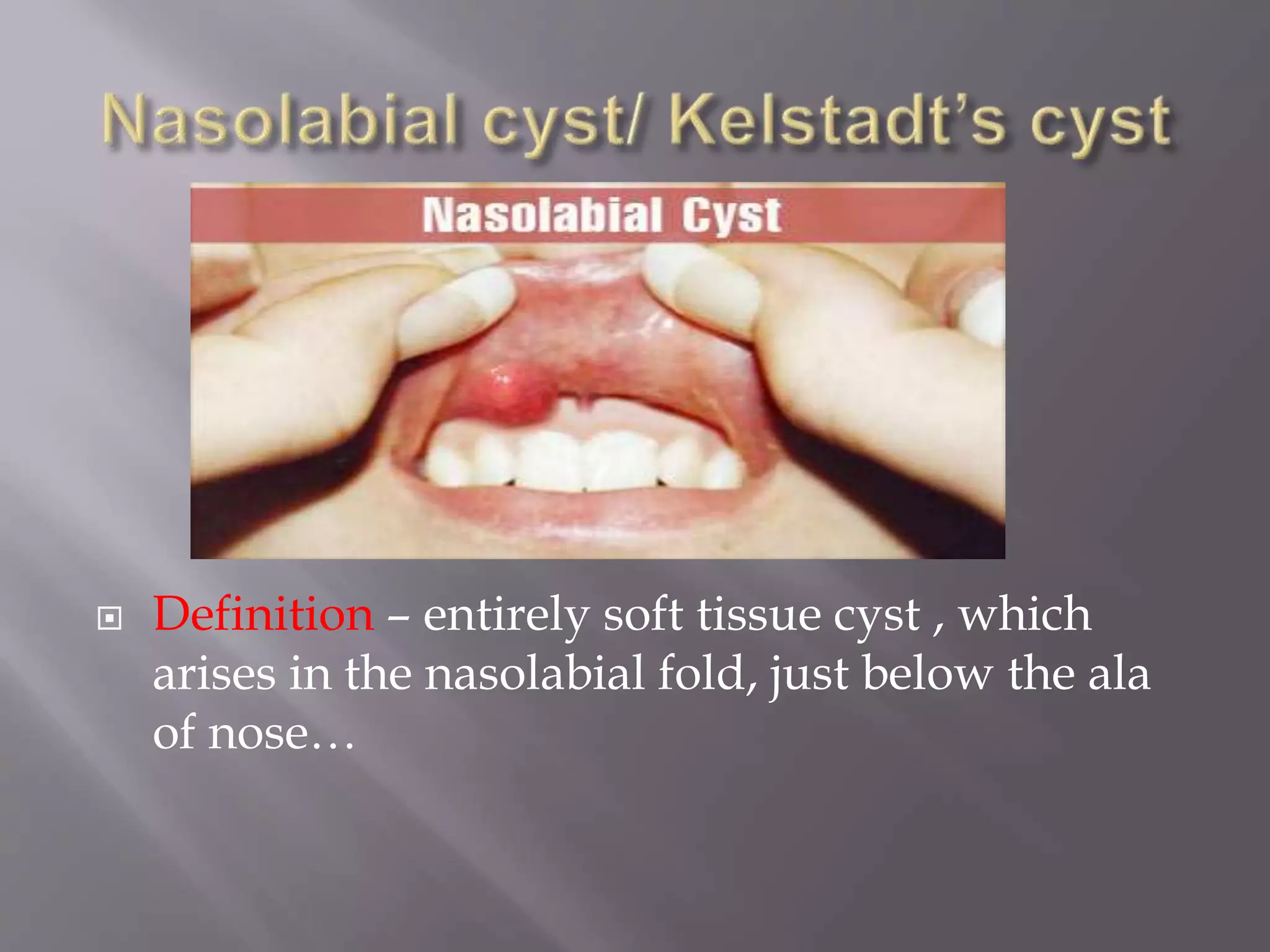
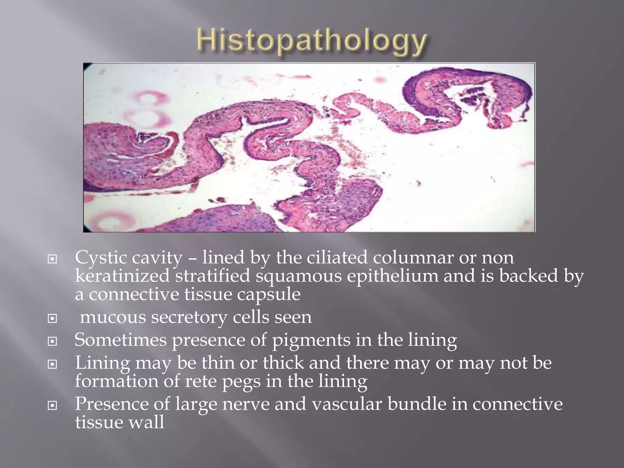
Manish deo
Roll no..10
Bds 3rd yr
 A cyst is a pathological cavity having fluid,
semifluid or gaseous contents and which is not
created by the accumulation of pus. Most cysts, but
not all, are lined by epithelium..
( Kramer 1974)
Odontogenic cyst -- Is a cyst in which lining of
lumen is derived from epthelium produced
during tooth development
Non odontogenic cyst – a cyst that arise from
epithelial inlusion or entrapments in the lines of
closure of developing facial process during
embryonic period of life…
Types of fissural cysts:
 Nasolabial cysts (nasoalveolar cysts
(located in soft tissue, not in the jaw!)
 Nasopalatine duct cysts
 Globulomaxillary cyst
 Definition - a type of developmental cysts that
actually arises in the bone suture between
maxilla and premaxilla
Earlier concept -- develops from as a result of
proliferation of the epithelium , entrapped along
the line of fusion between maxilla and premaxilla
 Nowdays --- acc. To christ
 variant of primordial cyst or lateral
periodontal cyst
Site – between lateral incisir and canine teeth
-- usually asymptomatic
-- cause pain and discomfort only when it is
secondarily infected.
--lateral maxillary and cupsid teeth will be found
to be tilted coronally with root divergence
-- vitality test is normal for both teeth
-- small swelling in between upper lateral incisor
and canine with elevation of lip is rare case
 Well circumscribed unilocular radiolucency (
inverted pear between apices of teeth)
 Roots reveals divergence
 Cystic cavity – lined by either by a
stratified/pseudostratified ciliated columnar
epithelium or thin squamous epithelium
 chronic inflammatory cell inflitration present
in capsule
 The clinical and pathohistological evidence
overwhelmingly shows that a "cyst" in the so-
called globulomaxillary region may be one of the
following lesions : Radicular cyst
 Lateral periodontal cyst
 Keratocystic odontogenic tumour
 Odontogenic tumour
 Ameloblastoma
 Central giant cell granuloma
 Apical infection
 Careful enucleation without damage to
adjoining roots ot the teeth, followed by
primary closure
 Definition – entirely soft tissue cyst , which
arises in the nasolabial fold, just below the ala
of nose…
 From remnant of nasolacrimal duct/ epithelial
lining of FOM
 Age – peak incidence in 4th and 5th decade of life
 Sex– more in female
 Site – soft tissue of anterior maxillary vestibule
below the ala of nose and deep in nasolabial area
 Usually unilateral
 Commonest complain – slowly growing and
occassionally, pain and difficult in nasal breathing
 Intraorally – bulge in labial sulcus
 Extraorally – filling out of nasolabial fold and may
lift ala nasi
 Flutuant lesion
 Seen as localized increase lucency of alveolar
process above apices of incisors ( appears as
spherical , kidney shaped lesion)
 Lucency results from presure resorption on
labial surfaces of maxilla
 Cyst lined by non ciliated pseudo stratified columnar
epithelium
 Goblet cells seen in some cases
 Occasionally, part of lining may be cuboidal / flat
sqamous
 Connective tissue wall is fibrous , relatively acellular
with fibers arranged loosely or compactly
- Submucous odontogenic abscess
- Furuncle of the nose
 Although the nasolabial cyst are extra-osseous
they lie subperiosteally and careful surgical
enucleation should be done
 Definition -- Nasopalatine duct cysts (NPDC) are
cysts that originate from epithelial remnants of
the nasopalatine duct, which become entrapped
during fusion of the palate plates
 This lesion is considered as a true
developmental cyst and it arises usually due to
the proliferation and subsequent cystic
degeneration of the epithelial remnants
remaining after closure of the embryonic
nasopalatine duct.. The initiating factors to the
development of the cyst may be trauma,
inflamMation and bacterial infection, etc.
 Age – 4th , 5th and 6th decade
 Sex – male > female ( 4:1)
 Site – usually in anterior region of mid palate near
opening of the incisive foramen
 Small, painful swelling
 often extends onto the labial aspect of upper alveolar
ridge
 Causes pressure sensation on the floor of the nose and
displacement of roots of upper central incisors
 Occasionally, there can be purulent or salty discharge
frpm the lesion
 Some patient complain of episodic swelling in the soft
tissue between the upper central incisor
 Regional teeth are always vital
 A sharply demarcated symmetrical radiolucency
in the midline of anterior maxilla
 Small round or heart shaped between the roots of
the upper central incisor in the midline with
cortical border
 Displacement of roots of the upper central incisor
are commonly seen
 Cystic cavity – lined by the ciliated columnar or non
keratinized stratified squamous epithelium and is backed by
a connective tissue capsule
 mucous secretory cells seen
 Sometimes presence of pigments in the lining
 Lining may be thin or thick and there may or may not be
formation of rete pegs in the lining
 Presence of large nerve and vascular bundle in connective
tissue wall
 Radicular cyst , if it is associated with a
pulpally involved tooth
 Large incisive canal
 Surgical excision
 Shafer, et al: A text book of oral pathology, 3rd
edition
 Essential of oral pathology - swapan kumar
purkait– 2nd edition
 Google.com
 Neville, et al:oral and maxillofacial pathology
3rd edition
- And various websites
Non odontogenic cyst

Non odontogenic cyst

  • 1.
  • 2.
     A cystis a pathological cavity having fluid, semifluid or gaseous contents and which is not created by the accumulation of pus. Most cysts, but not all, are lined by epithelium.. ( Kramer 1974)
  • 3.
    Odontogenic cyst --Is a cyst in which lining of lumen is derived from epthelium produced during tooth development Non odontogenic cyst – a cyst that arise from epithelial inlusion or entrapments in the lines of closure of developing facial process during embryonic period of life…
  • 4.
    Types of fissuralcysts:  Nasolabial cysts (nasoalveolar cysts (located in soft tissue, not in the jaw!)  Nasopalatine duct cysts  Globulomaxillary cyst
  • 5.
     Definition -a type of developmental cysts that actually arises in the bone suture between maxilla and premaxilla
  • 6.
    Earlier concept --develops from as a result of proliferation of the epithelium , entrapped along the line of fusion between maxilla and premaxilla  Nowdays --- acc. To christ  variant of primordial cyst or lateral periodontal cyst
  • 7.
    Site – betweenlateral incisir and canine teeth -- usually asymptomatic -- cause pain and discomfort only when it is secondarily infected. --lateral maxillary and cupsid teeth will be found to be tilted coronally with root divergence -- vitality test is normal for both teeth -- small swelling in between upper lateral incisor and canine with elevation of lip is rare case
  • 8.
     Well circumscribedunilocular radiolucency ( inverted pear between apices of teeth)  Roots reveals divergence
  • 9.
     Cystic cavity– lined by either by a stratified/pseudostratified ciliated columnar epithelium or thin squamous epithelium  chronic inflammatory cell inflitration present in capsule
  • 10.
     The clinicaland pathohistological evidence overwhelmingly shows that a "cyst" in the so- called globulomaxillary region may be one of the following lesions : Radicular cyst  Lateral periodontal cyst  Keratocystic odontogenic tumour  Odontogenic tumour  Ameloblastoma  Central giant cell granuloma  Apical infection
  • 11.
     Careful enucleationwithout damage to adjoining roots ot the teeth, followed by primary closure
  • 12.
     Definition –entirely soft tissue cyst , which arises in the nasolabial fold, just below the ala of nose…
  • 13.
     From remnantof nasolacrimal duct/ epithelial lining of FOM
  • 14.
     Age –peak incidence in 4th and 5th decade of life  Sex– more in female  Site – soft tissue of anterior maxillary vestibule below the ala of nose and deep in nasolabial area  Usually unilateral  Commonest complain – slowly growing and occassionally, pain and difficult in nasal breathing  Intraorally – bulge in labial sulcus  Extraorally – filling out of nasolabial fold and may lift ala nasi  Flutuant lesion
  • 15.
     Seen aslocalized increase lucency of alveolar process above apices of incisors ( appears as spherical , kidney shaped lesion)  Lucency results from presure resorption on labial surfaces of maxilla
  • 16.
     Cyst linedby non ciliated pseudo stratified columnar epithelium  Goblet cells seen in some cases  Occasionally, part of lining may be cuboidal / flat sqamous  Connective tissue wall is fibrous , relatively acellular with fibers arranged loosely or compactly
  • 17.
    - Submucous odontogenicabscess - Furuncle of the nose
  • 18.
     Although thenasolabial cyst are extra-osseous they lie subperiosteally and careful surgical enucleation should be done
  • 19.
     Definition --Nasopalatine duct cysts (NPDC) are cysts that originate from epithelial remnants of the nasopalatine duct, which become entrapped during fusion of the palate plates
  • 20.
     This lesionis considered as a true developmental cyst and it arises usually due to the proliferation and subsequent cystic degeneration of the epithelial remnants remaining after closure of the embryonic nasopalatine duct.. The initiating factors to the development of the cyst may be trauma, inflamMation and bacterial infection, etc.
  • 21.
     Age –4th , 5th and 6th decade  Sex – male > female ( 4:1)  Site – usually in anterior region of mid palate near opening of the incisive foramen  Small, painful swelling  often extends onto the labial aspect of upper alveolar ridge  Causes pressure sensation on the floor of the nose and displacement of roots of upper central incisors  Occasionally, there can be purulent or salty discharge frpm the lesion  Some patient complain of episodic swelling in the soft tissue between the upper central incisor  Regional teeth are always vital
  • 22.
     A sharplydemarcated symmetrical radiolucency in the midline of anterior maxilla  Small round or heart shaped between the roots of the upper central incisor in the midline with cortical border  Displacement of roots of the upper central incisor are commonly seen
  • 23.
     Cystic cavity– lined by the ciliated columnar or non keratinized stratified squamous epithelium and is backed by a connective tissue capsule  mucous secretory cells seen  Sometimes presence of pigments in the lining  Lining may be thin or thick and there may or may not be formation of rete pegs in the lining  Presence of large nerve and vascular bundle in connective tissue wall
  • 24.
     Radicular cyst, if it is associated with a pulpally involved tooth  Large incisive canal
  • 25.
  • 26.
     Shafer, etal: A text book of oral pathology, 3rd edition  Essential of oral pathology - swapan kumar purkait– 2nd edition  Google.com  Neville, et al:oral and maxillofacial pathology 3rd edition - And various websites