Downloaded 176 times

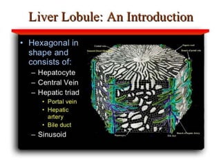
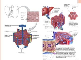
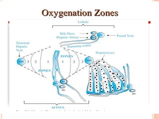
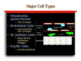
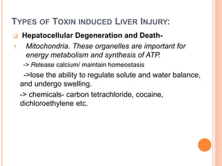
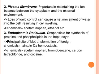
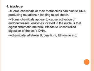
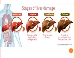
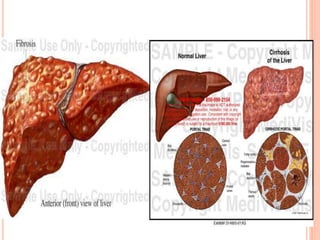
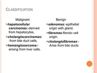
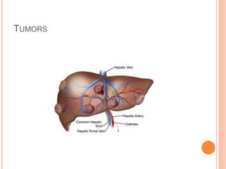
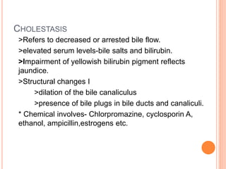
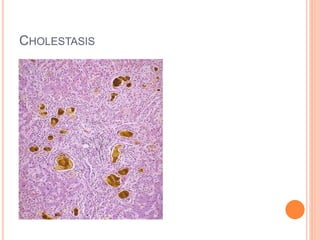
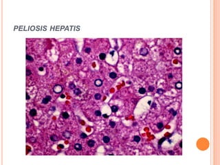
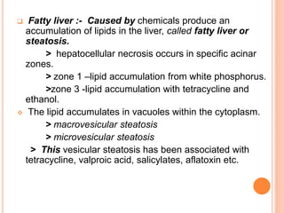
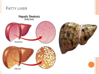
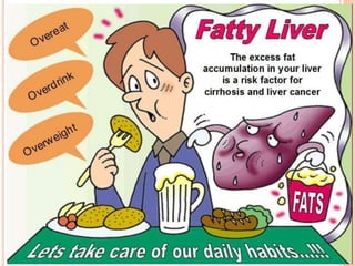


The document summarizes the different types of liver injury and conditions that can result from toxic effects on the liver. It discusses how chemicals can cause hepatocellular degeneration through damaging mitochondria, plasma membranes, the endoplasmic reticulum, or nucleus of liver cells. This can lead to fibrosis, cirrhosis, or liver tumors over time. Other toxic effects include cholestasis (decreased bile flow), sinusoidal damage, peliosis hepatis (blood-filled cysts), and fatty liver caused by lipid accumulation within liver cells. The document provides examples of chemicals that can induce each type of toxic liver effect.


