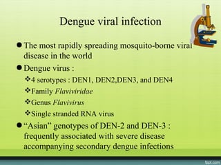
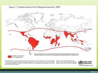
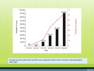
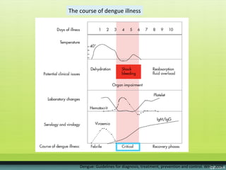
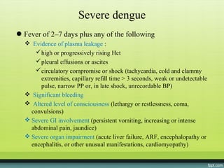
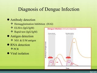
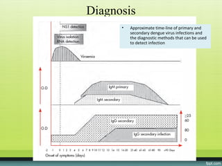
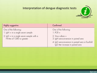
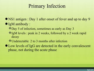
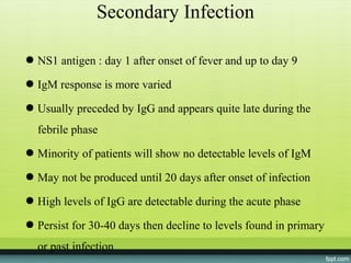
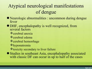
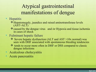
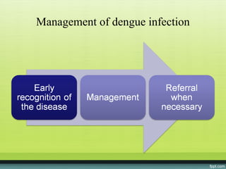
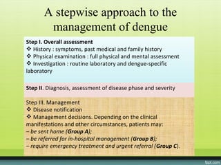
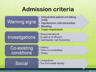
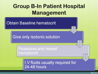
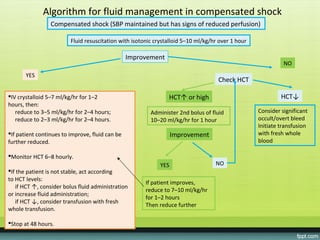
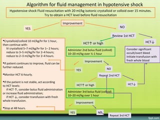
Dengue infection
  ReviseD Who
 guiDelines 2009
Dengue viral infection
● The most rapidly spreading mosquito-borne viral
  disease in the world
● Dengue virus :
  4 serotypes : DEN1, DEN2,DEN3, and DEN4
  Family Flaviviridae
  Genus Flavivirus
  Single stranded RNA virus
● “Asian” genotypes of DEN-2 and DEN-3 :
  frequently associated with severe disease
  accompanying secondary dengue infections
Average annual number of DF and DHF cases reported to WHO, and of countries reporting dengue,
1955–2007
Dengue Virus Infection
● Immunity : long lasting in same serotype, partial and
  transient to other serotypes
● Primary infection, secondary infection
● Greater risk of serious symptoms in secondary
  infection
• Plasma leakage distinguishes dengue fever from
  dengue hemorrhagic fever
• Plasma leakage,hemoconcentration and abnormalities
  in homeostasis characterize severe dengue
Dengue Virus Infection:
            Clinical Syndromes
● Undifferentiated fever
● Dengue fever: fever, headache, muscle pain,
  nausea/vomiting, rash
● DHF
● DSS


                                                Gr I, II
                         Plasma leakage

                                                Gr III, IV
Definition of Dengue Hemorrhagic
                Fever
             4 necessary criteria:
• Fever, or recent history of acute fever
• Hemorrhagic manifestations
● Low platelet count (100,000/mm3 or less)
● Objective evidence of “leaky capillaries”
    elevated hematocrit (20% or more over baseline)
    pleural or other effusions
    Fall in hematocrit >20% after I.V Fluids
Definition of Dengue Shock Syndrone


4 criteria for DHF
● Evidence of circulatory failure manifested indirectly
  by all of the following:
    Rapid and weak pulse
    Narrow pulse pressure (< 20 mm Hg) OR hypotension
     for age
    Cold, clammy skin and altered mental status
● Frank shock is direct evidence of circulatory failure
WHO classification

● Undifferentiated fever
● Dengue fever (DF)
● Dengue haemorrhagic fever (DHF)
  4 severity grades
  grades III and IV : dengue shock syndrome (DSS)
● Currently the classification into DF/DHF/DSS
  continues to be widely used
WHO classification
● patients with non-severe dengue
  patients with warning signs
  patients without warning signs
● “dengue is one disease entity with different clinical
  presentations and often with unpredictable clinical
  evolution and outcome”
Vectors

● Aedes aegypti


                Stegomyia aegypti
             (formerly Aedes aegypti)
Dengue
● incubation period of 4-10 days
● wide spectrum of illness (most, asymptomatic or
  subclinical)
● Primary infection : induce lifelong protective
  immunity to the infecting serotype
● Individuals suffering an infection are protected
  from clinical illness with a different serotype
  within 2-3 months of the primary infection
● no long-term cross-protective immunity
Risk factors (determine the severity of disease
            and secondary infection)
● Age
● Ethnicity
● Possibly chronic diseases (bronchial asthma, sickle
  cell anemia and DM)
● Young children (less able to compensate for capillary
  leakage and are consequently at greater risk of
  dengue shock)
● Secondary heterotypic infection as risk factor for
  severe dengue
The course of dengue illness
Febrile phase
● High-grade fever, 2–7 days

● facial flushing, skin erythema, body ache, myalgia, arthralgia,
  headache and N/V
● Indistinguishable between severe and non-severe dengue cases

● Monitoring for warning signs

● Mild hemorrhagic manifestations : petechiae and mucosal
  membrane bleeding (e.g. nose and gums)
● Liver often enlarged and tender after a few days of fever

● The earliest abnormality in CBC : progressive decrease in
  WBC
The course of dengue illness


    Dengue: Guidelines for diagnosis, treatment, prevention and control. WHO 2009
Critical phase
● Day 3–7 of illness

● Progressive leukopenia  rapid ↓platelet count plasma
  leakage

● Patients without ↑capillary permeability will improve
● Patients with ↑capillary permeability : worse, lose plasma
  volume

● The degree of increase above the baseline HCT reflects
  severity of plasma leakage

• Shock-WBC may increase in patients with severe bleeding
The course of dengue illness


    Dengue: Guidelines for diagnosis, treatment, prevention and control. WHO 2009
Recovery phase

● Gradual reabsorption of extravascular compartment fluid
  takes place in the following 48–72 hours
● Well-being improves, appetite returns, GI symptoms abate,
  hemodynamic status stabilizes and diuresis ensues
● Rash : “isles of white in the sea of red” Some may
  experience generalized pruritus
● Hct stabilizes or lower due to the dilutional effect of
  reabsorbed fluid
● WBC usually rise soon after defervescence but the recovery
  of platelet count is typically later than that of WBC
Febrile, critical and recovery phases in
                       dengue

Febrile phase           Dehydration; high fever may cause
                        neurological disturbances and febrile
                        seizures in young children

Critical phase          Shock from plasma leakage; severe
                        hemorrhage; organ impairment

Recovery phase          Hypervolemia (only if iv fluid therapy
                        has been excessive and/or
                        has extended into this period)
Severe dengue
● Fever of 2–7 days plus any of the following
   Evidence of plasma leakage :
      high or progressively rising Hct
      pleural effusions or ascites
      circulatory compromise or shock (tachycardia, cold and clammy
       extremities, capillary refill time > 3 seconds, weak or undetectable
       pulse, narrow PP or, in late shock, unrecordable BP)
   Significant bleeding
   Altered level of consciousness (lethargy or restlessness, coma,
    convulsions)
   Severe GI involvement (persistent vomiting, increasing or intense
    abdominal pain, jaundice)
   Severe organ impairment (acute liver failure, ARF, encephalopathy or
    encephalitis, or other unusual manifestations, cardiomyopathy)
Diagnosis of Dengue Infection
● Antibody detection
   Hemagglutination Inhibition (HAI)
   ELISA (IgG/IgM)
   Rapid test (IgG/IgM)
● Antigen detection
   NS1 & E/M antigen
● RNA detection
   PCR
● Viral isolation
Diagnosis
      •   Approximate time-line of primary and
          secondary dengue virus infections and
          the diagnostic methods that can be used
          to detect infection
Interpretation of dengue diagnostic tests
Primary Infection

● NS1 antigen : Day 1 after onset of fever and up to day 9
● IgM antibody :
  Day 5 of infection, sometimes as early as Day 3
  IgM levels : peak in 2 weeks, followed by a 2 week rapid
   decay
  Undetectable 2 to 3 months after infection
● Low levels of IgG are detected in the early convalescent
  phase, not during the acute phase
Secondary Infection

● NS1 antigen : day 1 after onset of fever and up to day 9

● IgM response is more varied

● Usually preceded by IgG and appears quite late during the
  febrile phase
● Minority of patients will show no detectable levels of IgM

● May not be produced until 20 days after onset of infection

● High levels of IgG are detectable during the acute phase

● Persist for 30-40 days then decline to levels found in primary
  or past infection
Atypical neurological manifestations
              of dengue
● Neurologic abnormalities : uncommon during dengue
  fever
● DHF, encephalopathy is well recognized, from
  several factors
  cerebral anoxia
  cerebral edema
  cerebral hemorrhage
  hyponatremia
  toxicity secondary to liver failure
● Studies in southeast Asia, encephalopathy associated
  with classic DF can occur in up to half of the cases
Atypical gastrointestinal
           manifestations of dengue
• Hepatitis
    Hepatomegaly, jaundice and raised aminotransferase levels
     (AST>ALT)
    caused by the dengue virus and ⁄or Hypoxia and tissue ischemia
     in cases of shock
• Fulminant hepatic failure
    Severe hepatic dysfunction (ALT and AST >10x normal) was
    seen with DHF associated with spontaneous bleeding tendencies
    tends to occur more often in DHF or DSS compared to classic
    dengue infections
• Acalculous cholecystitis
• Acute pancreatitis
Atypical cardiovascular manifestations
           of dengue fever
● uncommon
● Cardiac rhythm disorders :
  atrioventricular blocks
  atrial fibrillation
  sinus node dysfunction
  ectopic ventricular beats
● Most are asymptomatic, benign self limiting course with
  resolution of infection
● Attributed to viral myocarditis
Atypical respiratory manifestations of
                  dengue
● ARDS
● Pulmonary hemorrhage : thrombocytopenia,
  changes in vascular permeability, platelet
  dysfunction
Dengue myositis

● Dengue fever : break bone fever, severe muscle, joint
  and bone pain
● Acute benign myositis : elevated SGOT, SGPT, and
  CPK
● Dengue virus infection may also cause persisting,
  severe, myositis for weeks
Lymphoreticular complications of
              dengue
● Dengue virus antigen is found predominantly in cells
  of the spleen, thymus and lymph nodes
● DHF, lymphadenopathy is observed in half of the
  cases
● Splenomegaly is rarely observed
● Splenic rupture and lymph node infarction in DHF
  are rare
Management of dengue infection
A stepwise approach to the
        management of dengue
Step I. Overall assessment
 History : symptoms, past medical and family history
 Physical examination : full physical and mental assessment
 Investigation : routine laboratory and dengue-specific
laboratory

Step II. Diagnosis, assessment of disease phase and severity

Step III. Management
 Disease notification
 Management decisions. Depending on the clinical
manifestations and other circumstances, patients may:
– be sent home (Group A);
– be referred for in-hospital management (Group B);
– require emergency treatment and urgent referral (Group C).
Groups A-Patients who are sent
            home
• Encourage plenty of oral fluids

• Inform about the warning signs

• Paracetamol for high fever. Never aspirin,ibuprofen or
  other NSAIDS
Admission criteria
Group B-In Patient Hospital
      Management
Algorithm for fluid management in compensated shock
                 Compensated shock (SBP maintained but has signs of reduced perfusion)

                         Fluid resuscitation with isotonic crystalloid 5–10 ml/kg/hr over 1 hour

                                                 Improvement
                                                                                                             NO
       YES
                                                                                    Check HCT

IV crystalloid 5–7 ml/kg/hr for 1–2                            HCT↑ or high                                HCT↓
hours, then:
   reduce to 3–5 ml/kg/hr for 2–4 hours;                 Administer 2nd bolus of fluid             Consider significant
   reduce to 2–3 ml/kg/hr for 2–4 hours.                 10–20 ml/kg/hr for 1 hour                 occult/overt bleed
                                                                                                   Initiate transfusion
If patient continues to improve, fluid can be                     Improvement                     with fresh whole
further reduced.                                                                                   blood

Monitor HCT 6–8 hourly.
                                                             YES                   NO
If the patient is not stable, act according
to HCT levels:
                                                       If patient improves,
   if HCT ↑, consider bolus fluid administration
                                                       reduce to 7–10 ml/kg/hr
or increase fluid administration;
                                                       for 1–2 hours
   if HCT ↓, consider transfusion with fresh
                                                       Then reduce further
whole transfusion.

Stop at 48 hours.
Algorithm for fluid management in hypotensive shock
     Hypotensive shock Fluid resuscitation with 20 ml/kg isotonic crystalloid or colloid over 15 minutes.
                            Try to obtain a HCT level before fluid resuscitation

                                                   Improvement
                  YES                                                                           NO

                                                                                            Review 1st HCT
Crystalloid/colloid 10 ml/kg/hr for 1 hour,                                                                        HCT↓
then continue with:                                                HCT↑ or high
   IV crystalloid 5–7 ml/kg/hr for 1– 2 hours;       Administer 2nd bolus fluid (colloid)            Consider significant
   reduce to 3–5 ml/kg/hr for 2–4 hours;             10–20 ml/kg over ½-1 hour                       occult/overt bleed
   reduce to 2–3 ml/kg/hr for 2–4 hours.                                                             Initiate transfusion with
                                                                   Improvement                       fresh whole blood
If patient continues to improve, fluid can be
further reduced.
                                                             YES                     NO
Monitor HCT 6-hourly.                                                        Repeat 2nd HCT

If the patient is not stable, act according
                                                             HCT↑ or high                             HCT↓
to HCT levels:
   if HCT ↑, consider bolus fluid administration
                                                     Administer 3rd bolus fluid (colloid)
or increase fluid administration;
                                                     10–20 ml/kg over 1 hour
   if HCT ↓, consider transfusion with fresh
whole transfusion.
                                                             Improvement
Stop at 48 hours.
                                                       YES                   NO
                                                                                       Repeat 3rd HCT
Treatment of hemorrhagic
               complications
● Mucosal bleeding :
   if patient remains stable with fluid resuscitation/replacement,
   considered as minor


● Bleeding improves rapidly during recovery phase

● Patients with profound thrombocytopenia :
   strict bed rest and protect from trauma
   not give i.m injections (avoid hematoma)
   prophylactic platelet transfusions for severe thrombocytopaenia in
   hemodynamically stable patients not shown to be effective and not
   necessary
Management of Dengue Infection

● No hemorrhagic manifestations and patient is well-
  hydrated:
   home treatment


● Hemorrhagic manifestations or hydration borderline:
   outpatient observation center or hospitalization


● Warning signs (even without profound shock) or
  DSS:
   hospitalize
Treatment of Dengue Fever

● Fluids
● Rest
● Antipyretics (avoid aspirin and NSAIDs)
● Monitor blood pressure, hematocrit, platelet
  count, level of consciousness
Dengue with who guidelines

Dengue with who guidelines

  • 1.
    Dengue infection ReviseD Who guiDelines 2009
  • 2.
    Dengue viral infection ●The most rapidly spreading mosquito-borne viral disease in the world ● Dengue virus : 4 serotypes : DEN1, DEN2,DEN3, and DEN4 Family Flaviviridae Genus Flavivirus Single stranded RNA virus ● “Asian” genotypes of DEN-2 and DEN-3 : frequently associated with severe disease accompanying secondary dengue infections
  • 4.
    Average annual numberof DF and DHF cases reported to WHO, and of countries reporting dengue, 1955–2007
  • 5.
    Dengue Virus Infection ●Immunity : long lasting in same serotype, partial and transient to other serotypes ● Primary infection, secondary infection ● Greater risk of serious symptoms in secondary infection • Plasma leakage distinguishes dengue fever from dengue hemorrhagic fever • Plasma leakage,hemoconcentration and abnormalities in homeostasis characterize severe dengue
  • 6.
    Dengue Virus Infection: Clinical Syndromes ● Undifferentiated fever ● Dengue fever: fever, headache, muscle pain, nausea/vomiting, rash ● DHF ● DSS Gr I, II Plasma leakage Gr III, IV
  • 7.
    Definition of DengueHemorrhagic Fever 4 necessary criteria: • Fever, or recent history of acute fever • Hemorrhagic manifestations ● Low platelet count (100,000/mm3 or less) ● Objective evidence of “leaky capillaries”  elevated hematocrit (20% or more over baseline)  pleural or other effusions  Fall in hematocrit >20% after I.V Fluids
  • 8.
    Definition of DengueShock Syndrone 4 criteria for DHF ● Evidence of circulatory failure manifested indirectly by all of the following:  Rapid and weak pulse  Narrow pulse pressure (< 20 mm Hg) OR hypotension for age  Cold, clammy skin and altered mental status ● Frank shock is direct evidence of circulatory failure
  • 9.
    WHO classification ● Undifferentiatedfever ● Dengue fever (DF) ● Dengue haemorrhagic fever (DHF) 4 severity grades grades III and IV : dengue shock syndrome (DSS) ● Currently the classification into DF/DHF/DSS continues to be widely used
  • 10.
  • 11.
    ● patients withnon-severe dengue patients with warning signs patients without warning signs ● “dengue is one disease entity with different clinical presentations and often with unpredictable clinical evolution and outcome”
  • 13.
    Vectors ● Aedes aegypti Stegomyia aegypti (formerly Aedes aegypti)
  • 14.
    Dengue ● incubation periodof 4-10 days ● wide spectrum of illness (most, asymptomatic or subclinical) ● Primary infection : induce lifelong protective immunity to the infecting serotype ● Individuals suffering an infection are protected from clinical illness with a different serotype within 2-3 months of the primary infection ● no long-term cross-protective immunity
  • 15.
    Risk factors (determinethe severity of disease and secondary infection) ● Age ● Ethnicity ● Possibly chronic diseases (bronchial asthma, sickle cell anemia and DM) ● Young children (less able to compensate for capillary leakage and are consequently at greater risk of dengue shock) ● Secondary heterotypic infection as risk factor for severe dengue
  • 16.
    The course ofdengue illness
  • 17.
    Febrile phase ● High-gradefever, 2–7 days ● facial flushing, skin erythema, body ache, myalgia, arthralgia, headache and N/V ● Indistinguishable between severe and non-severe dengue cases ● Monitoring for warning signs ● Mild hemorrhagic manifestations : petechiae and mucosal membrane bleeding (e.g. nose and gums) ● Liver often enlarged and tender after a few days of fever ● The earliest abnormality in CBC : progressive decrease in WBC
  • 18.
    The course ofdengue illness Dengue: Guidelines for diagnosis, treatment, prevention and control. WHO 2009
  • 19.
    Critical phase ● Day3–7 of illness ● Progressive leukopenia  rapid ↓platelet count plasma leakage ● Patients without ↑capillary permeability will improve ● Patients with ↑capillary permeability : worse, lose plasma volume ● The degree of increase above the baseline HCT reflects severity of plasma leakage • Shock-WBC may increase in patients with severe bleeding
  • 20.
    The course ofdengue illness Dengue: Guidelines for diagnosis, treatment, prevention and control. WHO 2009
  • 21.
    Recovery phase ● Gradualreabsorption of extravascular compartment fluid takes place in the following 48–72 hours ● Well-being improves, appetite returns, GI symptoms abate, hemodynamic status stabilizes and diuresis ensues ● Rash : “isles of white in the sea of red” Some may experience generalized pruritus ● Hct stabilizes or lower due to the dilutional effect of reabsorbed fluid ● WBC usually rise soon after defervescence but the recovery of platelet count is typically later than that of WBC
  • 22.
    Febrile, critical andrecovery phases in dengue Febrile phase Dehydration; high fever may cause neurological disturbances and febrile seizures in young children Critical phase Shock from plasma leakage; severe hemorrhage; organ impairment Recovery phase Hypervolemia (only if iv fluid therapy has been excessive and/or has extended into this period)
  • 23.
    Severe dengue ● Feverof 2–7 days plus any of the following  Evidence of plasma leakage :  high or progressively rising Hct  pleural effusions or ascites  circulatory compromise or shock (tachycardia, cold and clammy extremities, capillary refill time > 3 seconds, weak or undetectable pulse, narrow PP or, in late shock, unrecordable BP)  Significant bleeding  Altered level of consciousness (lethargy or restlessness, coma, convulsions)  Severe GI involvement (persistent vomiting, increasing or intense abdominal pain, jaundice)  Severe organ impairment (acute liver failure, ARF, encephalopathy or encephalitis, or other unusual manifestations, cardiomyopathy)
  • 24.
    Diagnosis of DengueInfection ● Antibody detection  Hemagglutination Inhibition (HAI)  ELISA (IgG/IgM)  Rapid test (IgG/IgM) ● Antigen detection  NS1 & E/M antigen ● RNA detection  PCR ● Viral isolation
  • 25.
    Diagnosis • Approximate time-line of primary and secondary dengue virus infections and the diagnostic methods that can be used to detect infection
  • 26.
    Interpretation of denguediagnostic tests
  • 27.
    Primary Infection ● NS1antigen : Day 1 after onset of fever and up to day 9 ● IgM antibody : Day 5 of infection, sometimes as early as Day 3 IgM levels : peak in 2 weeks, followed by a 2 week rapid decay Undetectable 2 to 3 months after infection ● Low levels of IgG are detected in the early convalescent phase, not during the acute phase
  • 28.
    Secondary Infection ● NS1antigen : day 1 after onset of fever and up to day 9 ● IgM response is more varied ● Usually preceded by IgG and appears quite late during the febrile phase ● Minority of patients will show no detectable levels of IgM ● May not be produced until 20 days after onset of infection ● High levels of IgG are detectable during the acute phase ● Persist for 30-40 days then decline to levels found in primary or past infection
  • 29.
    Atypical neurological manifestations of dengue ● Neurologic abnormalities : uncommon during dengue fever ● DHF, encephalopathy is well recognized, from several factors cerebral anoxia cerebral edema cerebral hemorrhage hyponatremia toxicity secondary to liver failure ● Studies in southeast Asia, encephalopathy associated with classic DF can occur in up to half of the cases
  • 30.
    Atypical gastrointestinal manifestations of dengue • Hepatitis  Hepatomegaly, jaundice and raised aminotransferase levels (AST>ALT)  caused by the dengue virus and ⁄or Hypoxia and tissue ischemia in cases of shock • Fulminant hepatic failure  Severe hepatic dysfunction (ALT and AST >10x normal) was seen with DHF associated with spontaneous bleeding tendencies  tends to occur more often in DHF or DSS compared to classic dengue infections • Acalculous cholecystitis • Acute pancreatitis
  • 31.
    Atypical cardiovascular manifestations of dengue fever ● uncommon ● Cardiac rhythm disorders : atrioventricular blocks atrial fibrillation sinus node dysfunction ectopic ventricular beats ● Most are asymptomatic, benign self limiting course with resolution of infection ● Attributed to viral myocarditis
  • 32.
    Atypical respiratory manifestationsof dengue ● ARDS ● Pulmonary hemorrhage : thrombocytopenia, changes in vascular permeability, platelet dysfunction
  • 33.
    Dengue myositis ● Denguefever : break bone fever, severe muscle, joint and bone pain ● Acute benign myositis : elevated SGOT, SGPT, and CPK ● Dengue virus infection may also cause persisting, severe, myositis for weeks
  • 34.
    Lymphoreticular complications of dengue ● Dengue virus antigen is found predominantly in cells of the spleen, thymus and lymph nodes ● DHF, lymphadenopathy is observed in half of the cases ● Splenomegaly is rarely observed ● Splenic rupture and lymph node infarction in DHF are rare
  • 35.
  • 36.
    A stepwise approachto the management of dengue Step I. Overall assessment  History : symptoms, past medical and family history  Physical examination : full physical and mental assessment  Investigation : routine laboratory and dengue-specific laboratory Step II. Diagnosis, assessment of disease phase and severity Step III. Management  Disease notification  Management decisions. Depending on the clinical manifestations and other circumstances, patients may: – be sent home (Group A); – be referred for in-hospital management (Group B); – require emergency treatment and urgent referral (Group C).
  • 38.
    Groups A-Patients whoare sent home • Encourage plenty of oral fluids • Inform about the warning signs • Paracetamol for high fever. Never aspirin,ibuprofen or other NSAIDS
  • 39.
  • 40.
    Group B-In PatientHospital Management
  • 41.
    Algorithm for fluidmanagement in compensated shock Compensated shock (SBP maintained but has signs of reduced perfusion) Fluid resuscitation with isotonic crystalloid 5–10 ml/kg/hr over 1 hour Improvement NO YES Check HCT IV crystalloid 5–7 ml/kg/hr for 1–2 HCT↑ or high HCT↓ hours, then: reduce to 3–5 ml/kg/hr for 2–4 hours; Administer 2nd bolus of fluid Consider significant reduce to 2–3 ml/kg/hr for 2–4 hours. 10–20 ml/kg/hr for 1 hour occult/overt bleed Initiate transfusion If patient continues to improve, fluid can be Improvement with fresh whole further reduced. blood Monitor HCT 6–8 hourly. YES NO If the patient is not stable, act according to HCT levels: If patient improves, if HCT ↑, consider bolus fluid administration reduce to 7–10 ml/kg/hr or increase fluid administration; for 1–2 hours if HCT ↓, consider transfusion with fresh Then reduce further whole transfusion. Stop at 48 hours.
  • 42.
    Algorithm for fluidmanagement in hypotensive shock Hypotensive shock Fluid resuscitation with 20 ml/kg isotonic crystalloid or colloid over 15 minutes. Try to obtain a HCT level before fluid resuscitation Improvement YES NO Review 1st HCT Crystalloid/colloid 10 ml/kg/hr for 1 hour, HCT↓ then continue with: HCT↑ or high IV crystalloid 5–7 ml/kg/hr for 1– 2 hours; Administer 2nd bolus fluid (colloid) Consider significant reduce to 3–5 ml/kg/hr for 2–4 hours; 10–20 ml/kg over ½-1 hour occult/overt bleed reduce to 2–3 ml/kg/hr for 2–4 hours. Initiate transfusion with Improvement fresh whole blood If patient continues to improve, fluid can be further reduced. YES NO Monitor HCT 6-hourly. Repeat 2nd HCT If the patient is not stable, act according HCT↑ or high HCT↓ to HCT levels: if HCT ↑, consider bolus fluid administration Administer 3rd bolus fluid (colloid) or increase fluid administration; 10–20 ml/kg over 1 hour if HCT ↓, consider transfusion with fresh whole transfusion. Improvement Stop at 48 hours. YES NO Repeat 3rd HCT
  • 43.
    Treatment of hemorrhagic complications ● Mucosal bleeding :  if patient remains stable with fluid resuscitation/replacement, considered as minor ● Bleeding improves rapidly during recovery phase ● Patients with profound thrombocytopenia :  strict bed rest and protect from trauma  not give i.m injections (avoid hematoma)  prophylactic platelet transfusions for severe thrombocytopaenia in hemodynamically stable patients not shown to be effective and not necessary
  • 44.
    Management of DengueInfection ● No hemorrhagic manifestations and patient is well- hydrated:  home treatment ● Hemorrhagic manifestations or hydration borderline:  outpatient observation center or hospitalization ● Warning signs (even without profound shock) or DSS:  hospitalize
  • 45.
    Treatment of DengueFever ● Fluids ● Rest ● Antipyretics (avoid aspirin and NSAIDs) ● Monitor blood pressure, hematocrit, platelet count, level of consciousness

Editor's Notes

  • #7 - Grade I     มีไข้และมีอาการร่วมอื่นๆแต่ไม่จำเพาะ แต่เมื่อทำ tourniquet test จะให้ผล positive - Grade II     อาการเหมือน grade I แต่ที่เพิ่มเติมคือ พบเลือดออกเป็นจุดเลือดใต้ผิวหนัง - Grade III     ระบบไหลเวียนโลหิตเริ่มล้มเหลวเกิดอาการช็อค ชีพจรเร็ว เบา pulse pressure แคบ ความดันโลหิตต่ำ ริมฝีปากเขียว ตัวเย็น กระสับกระส่าย - Grade IV     แสดงอาการช็อครุนแรง ความดันโลหิตและชีพจรวัดไม่ได้  
  • #8 21
  • #9 22
  • #10 - Grade I     มีไข้และมีอาการร่วมอื่นๆแต่ไม่จำเพาะ แต่เมื่อทำ tourniquet test จะให้ผล positive - Grade II     อาการเหมือน grade I แต่ที่เพิ่มเติมคือ พบเลือดออกเป็นจุดเลือดใต้ผิวหนัง - Grade III     ระบบไหลเวียนโลหิตเริ่มล้มเหลวเกิดอาการช็อค ชีพจรเร็ว เบา pulse pressure แคบ ความดันโลหิตต่ำ ริมฝีปากเขียว ตัวเย็น กระสับกระส่าย - Grade IV     แสดงอาการช็อครุนแรง ความดันโลหิตและชีพจรวัดไม่ได้
  • #27 dengue infection is defined as primary if IgM/IgG OD ratio &gt; 1.2 (using pt’s sera at 1/100 dilution) or 1.4 (using pt’s sera at 1/20 dilutions) secondary if ratio &lt;1.2 or 1.4 Ratios : vary between lab