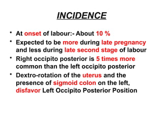
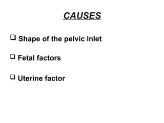
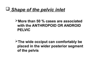
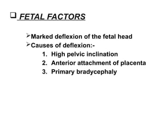
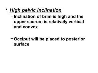
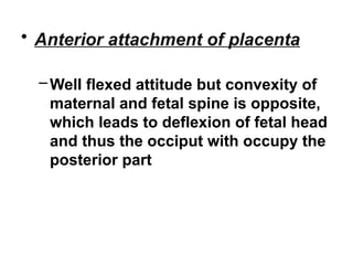
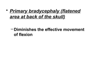
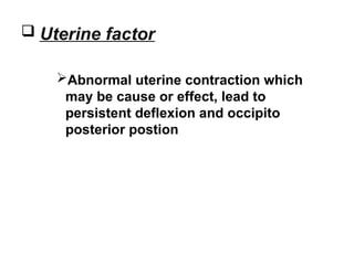
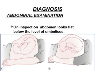
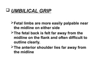
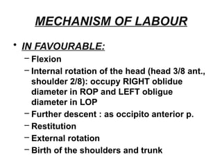
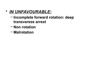
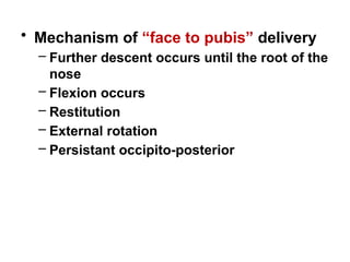
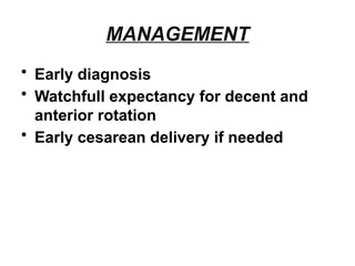
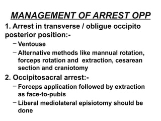
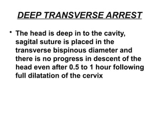
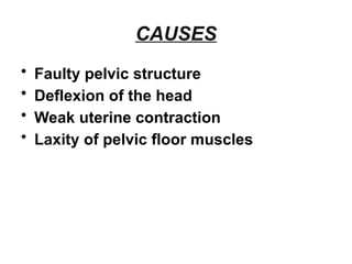
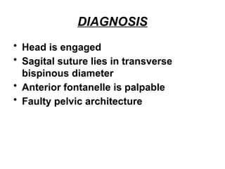
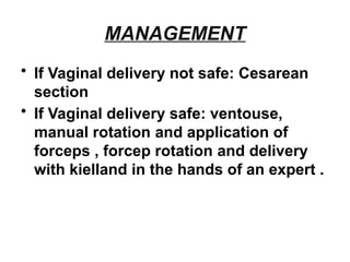
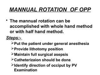
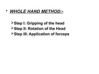
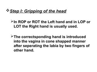
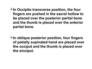
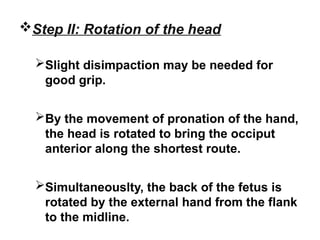
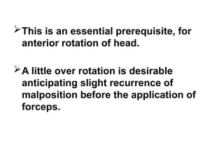
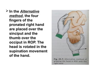
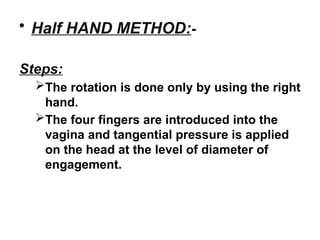
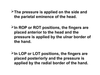
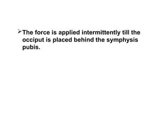
OCCIPITO POSTERIOR
POSITION
MALPOSITION
“ Malposition refers to any
position of the vertex other than the
flexed occipito-anterior one.”
OCCIPITO POSTERIOR POSITION
“In a vertex presentation where
the occiput is placed posteriorly over
the sacroiliac joint or directly over
sacrum called occipito-posterior
postion.”
• Occiput placed over:- Right sacroiliac
joint called RIGHT OCCIPITO
POSTERIOR
• Occiput placed over:- Left sacroiliac
joint called LEFT OCCIPITO
POSTERIOR
• Traditionally called 3rd and 4th position
of the vertex.
• Occiput placed over:- sacrum called
DIRECT OCCIPITO POSTERIOR
• All the three positions are Primary
(before the onset of labour ) or
Secondary ( developing after labour
starts )
• In majority of cases (90 %), ANTERIOR
ROTATION of occiput occurs and
follows the course like that of an
occipito anterior position and it is
favorable position
• But as the posterior position
occasionaly gives rise to dytocia, it is
described along with malpositions
INCIDENCE
• At onset of labour:- About 10 %
• Expected to be more during late pregnancy
and less during late second stage of labour
• Right occipito posterior is 5 times more
common than the left occipito posterior
• Dextro-rotation of the uterus and the
presence of sigmoid colon on the left,
disfavor Left Occipito Posterior Position
CAUSES
 Shape of the pelvic inlet
 Fetal factors
 Uterine factor
 Shape of the pelvic inlet
More than 50 % cases are associated
with the ANTHROPOID OR ANDROID
PELVIC
The wide occiput can comfortably be
placed in the wider posterior segment
of the pelvis
 FETAL FACTORS
Marked deflexion of the fetal head
Causes of deflexion:-
1. High pelvic inclination
2. Anterior attachment of placenta
3. Primary bradycephaly
• High pelvic inclination
–Inclination of brim is high and the
upper sacrum is relatively vertical
and convex
–Occiput will be placed to posterior
surface
• Anterior attachment of placenta
–Well flexed attitude but convexity of
maternal and fetal spine is opposite,
which leads to deflexion of fetal head
and thus the occiput with occupy the
posterior part
• Primary bradycephaly (flatened
area at back of the skull)
–Diminishes the effective movement
of flexion
 Uterine factor
Abnormal uterine contraction which
may be cause or effect, lead to
persistent deflexion and occipito
posterior postion
DIAGNOSIS
ABDOMINAL EXAMINATION
On inspection abdomen looks flat
below the level of umbelicus
 UMBILICAL GRIP
Fetal limbs are more easily palpable near
the midline on either side
The fetal back is felt far away from the
midline on the flank and often difficult to
outline clearly.
The anterior shoulder lies far away from
the midline
 PELVIC GRIP
Head is not engaged
Sinciput not felt as in well flexed occipito
posterior
 AUSCULTATION
Intensity of fetal heart sound felt on the
flank and often difficult to locate
 VAGINAL EXAMINATION
Elongated bag of membranes which is
likely to rupture during examination
Sagital suture occupies any oblique
diameters of the pelvis
Posterior fontanelle felt near the sacroiliac
joint
Anterior fontanelle felt more easily
because of deflexion of the head, lower
than posterior fontanelle
MECHANISM OF LABOUR
• IN FAVOURABLE:
– Flexion
– Internal rotation of the head (head 3/8 ant.,
shoulder 2/8): occupy RIGHT oblidue
diameter in ROP and LEFT obligue
diameter in LOP
– Further descent : as occipito anterior p.
– Restitution
– External rotation
– Birth of the shoulders and trunk
• IN UNFAVOURABLE:
– Incomplete forward rotation: deep
transverese arrest
– Non rotation
– Malrotation
• Mechanism of “face to pubis” delivery
– Further descent occurs until the root of the
nose
– Flexion occurs
– Restitution
– External rotation
– Persistant occipito-posterior
MANAGEMENT
• Early diagnosis
• Watchfull expectancy for decent and
anterior rotation
• Early cesarean delivery if needed
MANAGEMENT OF ARREST OPP
1. Arrest in transverse / obligue occipito
posterior position:-
– Ventouse
– Alternative methods like mannual rotation,
forceps rotation and extraction, cesarean
section and craniotomy
2. Occipitosacral arrest:-
– Forceps application followed by extraction
as face-to-pubis
– Liberal mediolateral episiotomy should be
done
DEEP TRANSVERSE ARREST
• The head is deep in to the cavity,
sagital suture is placed in the
transverse bispinous diameter and
there is no progress in descent of the
head even after 0.5 to 1 hour following
full dilatation of the cervix
CAUSES
• Faulty pelvic structure
• Deflexion of the head
• Weak uterine contraction
• Laxity of pelvic floor muscles
DIAGNOSIS
• Head is engaged
• Sagital suture lies in transverse
bispinous diameter
• Anterior fontanelle is palpable
• Faulty pelvic architecture
MANAGEMENT
• If Vaginal delivery not safe: Cesarean
section
• If Vaginal delivery safe: ventouse,
manual rotation and application of
forceps , forcep rotation and delivery
with kielland in the hands of an expert .
MANNUAL ROTATION OF OPP
• The mannual rotation can be
accomplished with whole hand method
or with half hand method.
Steps:-
Put the patient under general anesthesia
Provide lithotomy position
Maintain full surgical asepsis
Catheterizaion should be done
Identify direction of occiput by PV
Examination
• WHOLE HAND METHOD:-
Step I: Gripping of the head
Step II: Rotation of the Head
Step III: Application of forceps
Step I: Gripping of the head
In ROP or ROT the Left hand and in LOP or
LOT the Right hand is usually used.
The correctsponding hand is introduced
into the vagina in cone shapped manner
after seperating the labia by two fingers of
other hand.
In Occipito transverse position, the four
fingers are pushed in the sacral hollow to
be placed over the posterior parital bone
and the thumb is placed over the anterior
parital bone.
In oblique posterior position, four fingers
of patially supinated hand are placed over
the occiput and the thumb is placed over
the sinciput.
Step II: Rotation of the head
Slight disimpaction may be needed for
good grip.
By the movement of pronation of the hand,
the head is rotated to bring the occiput
anterior along the shortest route.
Simultaneouslty, the back of the fetus is
rotated by the external hand from the flank
to the midline.
This is an essential prerequisite, for
anterior rotation of head.
A little over rotation is desirable
anticipating slight recurrence of
malposition before the application of
forceps.
 In the Alternative
method, the four
fingers of the
pronated right hand
are placed over the
sinciput and the
thumb over the
occiput in ROP. The
head is rotated in the
supination movement
of the hand.
Step III: Application of the forceps
 Following Rotation, when the right hand is
placed over the left side of the pelvis, left
blade of the forcep is introduced.
When the left hand is used, it is placed on
the right side of the pelvis after rotation, as
such the right blade is to be introduced
first and the left blade is then to be
introduced underneath the right blade.
While introducing the blades, it is
preferable that an assistant fixes the head
by suprapubic pressure in a manner of
first pelvic grip.
As it is a mid forceps application, axis
traction device should be used.
DIFFICULTIES:-
Failure to grip the head adequetly due to
lack of space
Failure to dislodge the head from the
impacted position
Inadequate anesthesia
Wrong case selection
DANGERS-
Accidental slipping of the head above the
pelvic brim and prolapse of the cord
 It is better to be perform cesarean section
in such a situation.
• Half HAND METHOD:-
Steps:
The rotation is done only by using the right
hand.
The four fingers are introduced into the
vagina and tangential pressure is applied
on the head at the level of diameter of
engagement.
The pressure is applied on the side and
the parietal eminence of the head.
In ROP or ROT positions, the fingers are
placed anterior to the head and the
pressure is applied by the ulnar border of
the hand.
In LOP or LOT positions, the fingers are
placed posteriorly and the pressure is
applied by the radial border of the hand.
The force is applied intermittently till the
occiput is placed behind the symphysis
pubis.
THANK YOU

DEEP TRANSVERSE ARREST .pptx in obstetrics

  • 1.
  • 2.
    MALPOSITION “ Malposition refersto any position of the vertex other than the flexed occipito-anterior one.” OCCIPITO POSTERIOR POSITION “In a vertex presentation where the occiput is placed posteriorly over the sacroiliac joint or directly over sacrum called occipito-posterior postion.”
  • 3.
    • Occiput placedover:- Right sacroiliac joint called RIGHT OCCIPITO POSTERIOR • Occiput placed over:- Left sacroiliac joint called LEFT OCCIPITO POSTERIOR • Traditionally called 3rd and 4th position of the vertex.
  • 4.
    • Occiput placedover:- sacrum called DIRECT OCCIPITO POSTERIOR • All the three positions are Primary (before the onset of labour ) or Secondary ( developing after labour starts )
  • 5.
    • In majorityof cases (90 %), ANTERIOR ROTATION of occiput occurs and follows the course like that of an occipito anterior position and it is favorable position • But as the posterior position occasionaly gives rise to dytocia, it is described along with malpositions
  • 7.
    INCIDENCE • At onsetof labour:- About 10 % • Expected to be more during late pregnancy and less during late second stage of labour • Right occipito posterior is 5 times more common than the left occipito posterior • Dextro-rotation of the uterus and the presence of sigmoid colon on the left, disfavor Left Occipito Posterior Position
  • 8.
    CAUSES  Shape ofthe pelvic inlet  Fetal factors  Uterine factor
  • 9.
     Shape ofthe pelvic inlet More than 50 % cases are associated with the ANTHROPOID OR ANDROID PELVIC The wide occiput can comfortably be placed in the wider posterior segment of the pelvis
  • 11.
     FETAL FACTORS Markeddeflexion of the fetal head Causes of deflexion:- 1. High pelvic inclination 2. Anterior attachment of placenta 3. Primary bradycephaly
  • 12.
    • High pelvicinclination –Inclination of brim is high and the upper sacrum is relatively vertical and convex –Occiput will be placed to posterior surface
  • 13.
    • Anterior attachmentof placenta –Well flexed attitude but convexity of maternal and fetal spine is opposite, which leads to deflexion of fetal head and thus the occiput with occupy the posterior part
  • 14.
    • Primary bradycephaly(flatened area at back of the skull) –Diminishes the effective movement of flexion
  • 16.
     Uterine factor Abnormaluterine contraction which may be cause or effect, lead to persistent deflexion and occipito posterior postion
  • 17.
    DIAGNOSIS ABDOMINAL EXAMINATION On inspectionabdomen looks flat below the level of umbelicus
  • 18.
     UMBILICAL GRIP Fetallimbs are more easily palpable near the midline on either side The fetal back is felt far away from the midline on the flank and often difficult to outline clearly. The anterior shoulder lies far away from the midline
  • 20.
     PELVIC GRIP Headis not engaged Sinciput not felt as in well flexed occipito posterior  AUSCULTATION Intensity of fetal heart sound felt on the flank and often difficult to locate
  • 21.
     VAGINAL EXAMINATION Elongatedbag of membranes which is likely to rupture during examination Sagital suture occupies any oblique diameters of the pelvis Posterior fontanelle felt near the sacroiliac joint Anterior fontanelle felt more easily because of deflexion of the head, lower than posterior fontanelle
  • 23.
    MECHANISM OF LABOUR •IN FAVOURABLE: – Flexion – Internal rotation of the head (head 3/8 ant., shoulder 2/8): occupy RIGHT oblidue diameter in ROP and LEFT obligue diameter in LOP – Further descent : as occipito anterior p. – Restitution – External rotation – Birth of the shoulders and trunk
  • 24.
    • IN UNFAVOURABLE: –Incomplete forward rotation: deep transverese arrest – Non rotation – Malrotation
  • 26.
    • Mechanism of“face to pubis” delivery – Further descent occurs until the root of the nose – Flexion occurs – Restitution – External rotation – Persistant occipito-posterior
  • 27.
    MANAGEMENT • Early diagnosis •Watchfull expectancy for decent and anterior rotation • Early cesarean delivery if needed
  • 28.
    MANAGEMENT OF ARRESTOPP 1. Arrest in transverse / obligue occipito posterior position:- – Ventouse – Alternative methods like mannual rotation, forceps rotation and extraction, cesarean section and craniotomy 2. Occipitosacral arrest:- – Forceps application followed by extraction as face-to-pubis – Liberal mediolateral episiotomy should be done
  • 29.
    DEEP TRANSVERSE ARREST •The head is deep in to the cavity, sagital suture is placed in the transverse bispinous diameter and there is no progress in descent of the head even after 0.5 to 1 hour following full dilatation of the cervix
  • 30.
    CAUSES • Faulty pelvicstructure • Deflexion of the head • Weak uterine contraction • Laxity of pelvic floor muscles
  • 31.
    DIAGNOSIS • Head isengaged • Sagital suture lies in transverse bispinous diameter • Anterior fontanelle is palpable • Faulty pelvic architecture
  • 32.
    MANAGEMENT • If Vaginaldelivery not safe: Cesarean section • If Vaginal delivery safe: ventouse, manual rotation and application of forceps , forcep rotation and delivery with kielland in the hands of an expert .
  • 33.
    MANNUAL ROTATION OFOPP • The mannual rotation can be accomplished with whole hand method or with half hand method. Steps:- Put the patient under general anesthesia Provide lithotomy position Maintain full surgical asepsis Catheterizaion should be done Identify direction of occiput by PV Examination
  • 34.
    • WHOLE HANDMETHOD:- Step I: Gripping of the head Step II: Rotation of the Head Step III: Application of forceps
  • 35.
    Step I: Grippingof the head In ROP or ROT the Left hand and in LOP or LOT the Right hand is usually used. The correctsponding hand is introduced into the vagina in cone shapped manner after seperating the labia by two fingers of other hand.
  • 36.
    In Occipito transverseposition, the four fingers are pushed in the sacral hollow to be placed over the posterior parital bone and the thumb is placed over the anterior parital bone. In oblique posterior position, four fingers of patially supinated hand are placed over the occiput and the thumb is placed over the sinciput.
  • 37.
    Step II: Rotationof the head Slight disimpaction may be needed for good grip. By the movement of pronation of the hand, the head is rotated to bring the occiput anterior along the shortest route. Simultaneouslty, the back of the fetus is rotated by the external hand from the flank to the midline.
  • 38.
    This is anessential prerequisite, for anterior rotation of head. A little over rotation is desirable anticipating slight recurrence of malposition before the application of forceps.
  • 39.
     In theAlternative method, the four fingers of the pronated right hand are placed over the sinciput and the thumb over the occiput in ROP. The head is rotated in the supination movement of the hand.
  • 40.
    Step III: Applicationof the forceps  Following Rotation, when the right hand is placed over the left side of the pelvis, left blade of the forcep is introduced. When the left hand is used, it is placed on the right side of the pelvis after rotation, as such the right blade is to be introduced first and the left blade is then to be introduced underneath the right blade.
  • 41.
    While introducing theblades, it is preferable that an assistant fixes the head by suprapubic pressure in a manner of first pelvic grip. As it is a mid forceps application, axis traction device should be used.
  • 42.
    DIFFICULTIES:- Failure to gripthe head adequetly due to lack of space Failure to dislodge the head from the impacted position Inadequate anesthesia Wrong case selection
  • 43.
    DANGERS- Accidental slipping ofthe head above the pelvic brim and prolapse of the cord  It is better to be perform cesarean section in such a situation.
  • 44.
    • Half HANDMETHOD:- Steps: The rotation is done only by using the right hand. The four fingers are introduced into the vagina and tangential pressure is applied on the head at the level of diameter of engagement.
  • 45.
    The pressure isapplied on the side and the parietal eminence of the head. In ROP or ROT positions, the fingers are placed anterior to the head and the pressure is applied by the ulnar border of the hand. In LOP or LOT positions, the fingers are placed posteriorly and the pressure is applied by the radial border of the hand.
  • 46.
    The force isapplied intermittently till the occiput is placed behind the symphysis pubis.
  • 47.