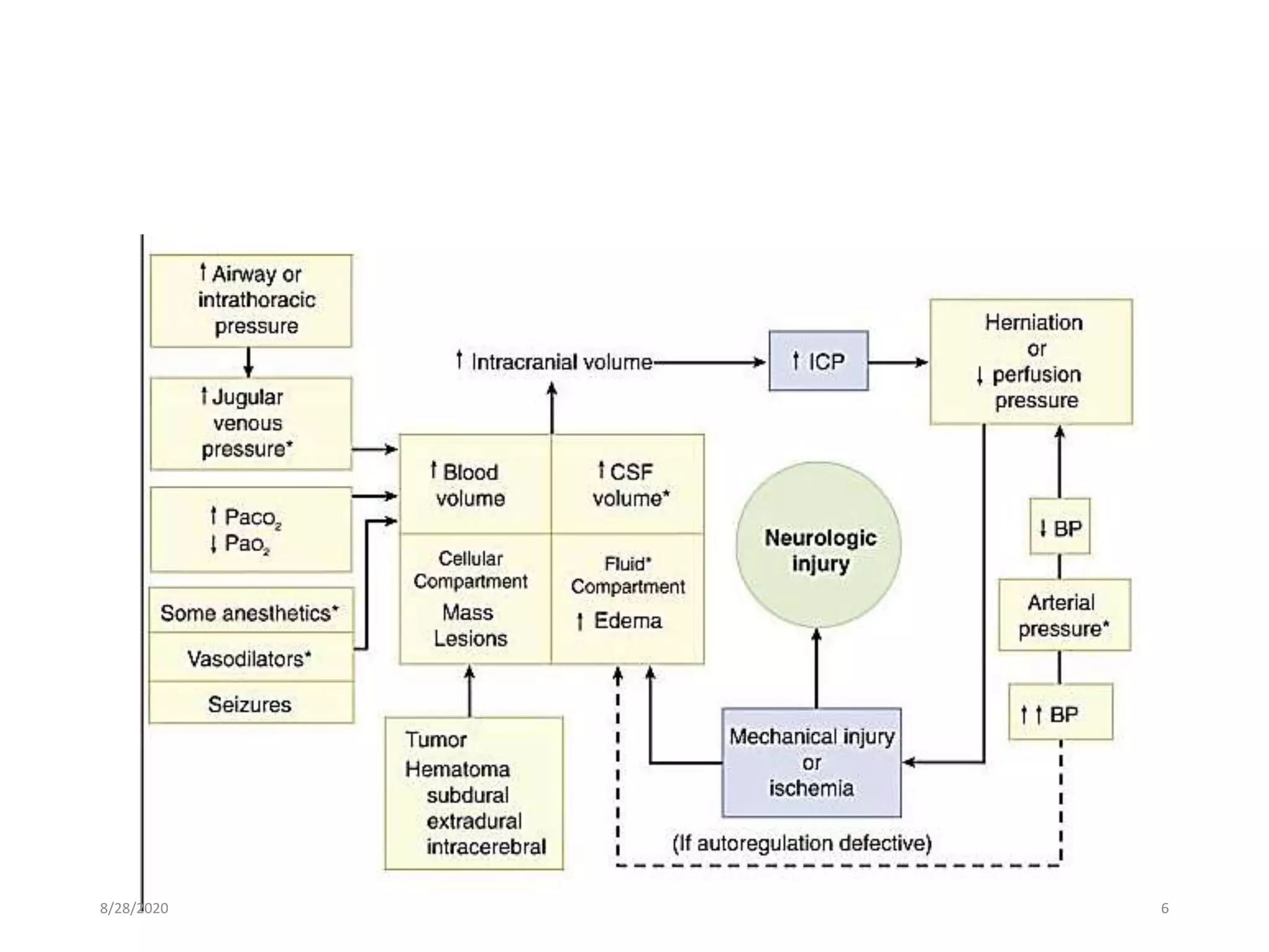
Anaesthesia and craniotomy
for mass lesion
Dr. ZIKRULLAH
8/28/2020 1
Introduction
• Anaesthesia for neurosurgical procedures requires understanding of
the normal anatomy and physiology of the CNS
• Changes that occur in response to the presence of space occupying
lesions, trauma or infection.
• Balanced anaesthesia with smooth induction and emergence is
advocated.
• Maintenance of an adequate cerebral perfusion pressure (CPP),
avoidance of intracranial hypertension
• Provision of optimal surgical conditions to avoid further progression
of neurological insult
8/28/2020 2
Space occupying lesion
• Intracranial masses may be congenital, neoplastic , infectious
(abscess or cyst), or vascular (hematoma or arteriovenous
malformation).
• Craniotomy is commonly undertaken for primary and metastatic
neoplasms of the brain.
• Primary tumors usually arise from glial cells (astrocytoma,
oligodendroglioma, or glioblastoma), ependymal cells
(ependymoma), or supporting tissues (meningioma, schwannoma, or
choroidal papilloma).
• Childhood tumors include medulloblastoma, neuroblastoma, and
chordoma.
• About 80% of the tumors are located in the supratentorial
compartment and about 20% in the posterior fossa.
8/28/2020 3
Sign and symptoms
• Regardless of the cause, intracranial masses present according to
growth rate, location, and ICP.
• Slowly growing masses are frequently asymptomatic for long periods,
whereas rapidly growing ones usually present acutely.
• Common presentations include headache, seizures, a general decline
in cognitive or specific neurological functions, and focal neurological
deficits.
• Supratentorial masses typically present as seizures, hemiplegia, or
aphasia.
• Infratentorial masses more commonly present as cerebellar
dysfunction (ataxia, nystagmus, and dysarthria) or brain stem
compression (cranial nerve palsies, altered consciousness, or
abnormal respiration).
• As ICP increases, signs of intracranial hypertension also develop.
8/28/2020 4
PATHOPHYSIOLOGIC CONSIDERATIONS
• Normal intracranial pressure (ICP) is about 10-15 mmHg.
• Intracranial hypertension generally results from the
omass lesion itself,
oDepressed skull fracture,
oInterference with normal absorption of cerebrospinal fluid (CSF),
oExcessive cerebral blood volume (CBV), or
oSystemic disturbances promoting brain edema.
8/28/2020 5
8/28/2020 6
• Natural mechanisms such as displacement of intracranial blood
volume and cerebrospinal fluid (CSF) and increased reabsorption of
CSF tend to limit the increase in ICP as the tumor increases in size.
• As these mechanisms are exhausted, ICP increases steeply.
• Such steep increase in ICP leads to rapid neurological deterioration.
8/28/2020 7
8/28/2020 8
• The important consequences of raised ICP are: Cerebral ischemia due
to reduction of cerebral perfusion pressure (CPP) and Brain shifts.
• If ICP exceeds 30 mmHg, CBF progressively decreases and vicious
circle is established: ischaemia causes brain oedema, which increases
ICP hence more ischaemia
• Significant gradients of ICP within various compartments of
intracranial cavity lead to herniation of brain structures.
• The most common forms of herniation are herniation of the
uncus of the temporal lobe through the hiatus in the tentorium cerebelli,
cingulate gyrus under the falx cerebri, and
cerebellar tonsils through formen magnum.
8/28/2020 9
Various herniation pathways: (1) subfalcine, (2) uncal (transtentorial), (3) cerebellar,
and (4) transcalvarial. (From Fishman RA: Brain edema. N Engl J Med 293:706-711,
1975.)8/28/2020 10
Cerebral Oedema
• Increase in brain water content- produced by several mechanisms:
• Vasogenic : Most common, d/t disruption of BBB. Loss of cerebral
autoregulation which allows entry of plasma-like fluid into the brain.
• Causes : Mechanical trauma, inflammatory lesion, tumours, hypertension &
infarction.
• Cytotoxic :Following metabolic insults hypoxaemia or ischaemia,
results in failure of brain cells to actively extrude sodium &
progressive cellular swelling.
• The various clinical indicators of increased ICP include headache
(awakens the patient at night), nausea and vomiting, HTN,
bradycardia, blurred vision, somnolence, and papilledema or
• Late signs: deteriorating GCS, Cushings reflex, dilated pupils,
decorticate then decerebrate posturing and coma.
8/28/2020 11
• Suggestive findings of CT include midline shift, obliteration of the
basal cisterns, loss of sulci, ventricular effacement and edema.
• Edema appears on a CT scan as a region of hypodensity.
• The basal cisterns appear on CT as a black (fluid) halo around the
upper end of the brainstem.
• They include the interpeduncular cistern, which lies between the two
cerebral peduncles; the quadrigeminal cistern, which overlies the
four colliculi; and the ambient cisterns, which lie lateral to the
cerebral peduncles.
8/28/2020 12
CT scan depicting normal (left) and compressed (right) basal cisterns, in a
patient with diffuse cerebral swelling ,the cisterns have been obliterated8/28/2020 13
Treatment
• Directed at underlying cause.
• Metabolic disturbances are corrected & operative intervention
undertaken whenever possible.
• Vasogenic oedema (tumours) responds to steroids (dexamethasone).
• A single 10 mg dose can significantly increase blood glucose
concentrations in non-diabetic patients. (Pasternak J et al. Effect of single dose
dexamethasone on blood glucose concentration in patients undergoing craniotomy. J
Neurosurg Anesthesiol 2005; 16: 122–5)
• There is evidence to support tight glycaemic control in critically ill,
neurologically impaired patients
8/28/2020 14
• Fluid restriction, osmotic agents & loop diuretics usually effective in
temporarily decreasing oedema.
• Hyperventilation
• Normal pts may tolerate pCO2 upto 25 mmHg without ischaemia
but even moderate hyperventilation (PaCO2= 30-33) – may aggravate
ischaemia in patients with focal ischaemia
• Mannitol 0.5-1gm/kg effective in rapid reduction in ICP
8/28/2020 15
Anaesthetic Consideration
8/28/2020 16
Aims of anaesthesia
• Optimal operating conditions
• Maintenance of stable ICP
• Stable haemodynamics, oxygenation and ventilation parameters
• Appropriate CPP while minimising CMRO2 to protection against
ischaemia
• Early detection & prompt management of intra-op complications
such as VAE in post fossa surgery, intracranial bleed during cerebral
aneurysm rupture
• Controlled but rapid emergence to enable early assessment &
monitoring of neurological status.
8/28/2020 17
Preanesthetic evaluation
• Confirm diagnosis
• Routine pre-op assessment
• Airway, CVS and respiratory system
• Investigations appropriate for age, general status of patient and type
of surgery
• The preanesthetic evaluation should attempt to establish the
presence or absence of intracranial hypertension.
• Examination should include a neurological assessment documenting
mental status/ signs of raised ICT and any existing sensory or motor
deficits.
• Computed tomographic (CT) and magnetic resonance imaging (MRI)
scans should be reviewed for evidence of brain edema, ventricular
size and midline shift greater than 0.5 cm.
8/28/2020 18
• Observe respiratory effort in terms of tachypnoea, laboured
breathing or Cheyne-Stokes pattern of breathing
• Assess the presence of cough/gag reflex if bulbar involvement is
suspected
• Medications should be reviewed with special reference to
corticosteroid, diuretic, and anticonvulsant therapy.
• Laboratory evaluation should rule out corticosteroid-induced
hyperglycemia and electrolyte disturbances due to diuretics or
abnormalities in secretion of antidiuretic hormone
• Anticonvulsant levels should be measured, particularly when seizures
are not well controlled.
8/28/2020 19
• Based on overall assessment, identify patients who would requires
post-op ventilation in ICU
• GCS ≤ 6
• Evidence of raised ICP
• Large or deep seated tumour
• Presence of midline shift and/or significant cerebral oedema
8/28/2020 20
Premedication
• Opioid premedication often avoided when intracranial hypertension
is suspected due to secondary hypercarbia leading to further increase
in ICT.
• For pts. with normal ICT, benzodiazepines such as midazolam/
diazepam may be given.
• Dexamethasone 10 mg IV/PO followed by 10 mg x 6 hrly is generally
given 48 hrs prior (may be given 24 hrs before also) to surgery.
• corticosteroids, anticonvulsants should be continued until the time of
surgery.
• Fasting instruction for the patient
8/28/2020 21
Monitoring
• Standard ASA monitoring devices
• Direct intra-arterial BP monitoring and bladder catheterization.
• End-tidal CO2 measurements alone cannot be relied upon, arterial
blood gas are necessary to closely regulate PaCO2.
• CVP monitoring should be done for pts requiring vasopressors.
• Neuromuscular function should be monitored
• Monitoring visual evoked potentials may be useful in preventing
optic nerve damage during resections of large pituitary tumors.
8/28/2020 22
Cont….
• Monitoring for air embolism:includes
• Precordial doppler device,
• Transoesophageal echocardiography
• Et CO2 / pulmonary artery pressure / end tidal nitrogen (shows increase)
• Brain monitoring
• Electroencephalogram,
• Evoked potentials,
• Jugular venous bulb oxygen saturation (Sjo2),
• Flow velocity measured by transcranial Doppler (TCD),
• Brain tissue Po2 (btPo2),
• ICP
8/28/2020 23
• Management of patients with intracranial hypertension is greatly
facilitated by monitoring ICP perioperatively.
• A ventriculostomy or subdural bolt is commonly employed and is
usually placed by the neurosurgeon preoperatively under local
anesthesia.
• A ventriculostomy has the added advantage of allowing removal of
CSF to decrease ICP.
• Electronic monitoring of ICP is possible utilizing saline-filled tubing
with a pressure transducer. The transducer should be zeroed to the
same reference level as the arterial pressure transducer (usually the
external auditory meatus).
8/28/2020 24
INDUCTION
• Induction of anesthesia and tracheal intubation are critical periods
for patients with compromised intracranial elastance or an already
elevated ICP.
• Intracranial elastance can be improved by osmotic diuresis, steroids,
or removal of CSF via a ventriculostomy drain immediately prior to
induction.
• Smooth induction &intubation prevents increase in ICP/ decrease
CPP and consequent herniation.
• Arterial hypotension during induction also detrimental.
8/28/2020 25
Technique
• Thiopental or propofol together with hyperventilation is a preferred
technique.
• Intravenous opioid—eg, fentanyl, 2.5-5 µg/kg—just prior to
thiopental blunts the sympathetic response, Esmolol: 0.5–1.0 mg/kg,
is effective in preventing tachycardia
• Propofol has the added benefit of a very short recovery time and
reduced CBF, as measured by PET, more than sevoflurane at
equipotent concentrations. (Maksimow A et al. Correlation of EEG spectral entropy
with regional cerebral blood flow during sevoflurane and propofol anaesthesia.
Anaesthesia 2005; 60: 862–9)
• substitution of etomidate for thiopental may provide greater
protection against circulatory depression.
8/28/2020 26
• Patients with reactive airways (bronchospastic disease), the
combination of incremental doses of thiopental and low-dose
isoflurane with hyperventilation may be preferable.
• A nondepolarizing tneuromuscular blocking agent (NMBA) is given to
facilitate ventilation and prevent straining or coughing
• Succinylcholine may increase ICP, particularly if intubation is
attempted prior to the establishment of deep thiopental anesthesia
and hyperventilation.
• However, still may be the agent of choice in patients at increased risk
for aspiration or with a potentially difficult airway
8/28/2020 27
• Hypertension during induction should be treated with esmolol or by
deepening the anesthetic with additional thiopental or propofol, or
by hyperventilation with low doses (< 1 MAC) of isoflurane.
• Potentially deleterious effect on cerebral blood volume and ICP-
vasodilators (such as nitroprusside, nitroglycerin, calcium channel
blockers, and hydralazine) should generally be avoided until the dura
is opened.
• Transient hypotension should generally be treated with incremental
doses of vasopressors (ephedrine or phenylephrine) rather than
intravenous fluids.
8/28/2020 28
Positioning
• Pressure points should be padded
• Prevent pressure and traction on nerves
• Prevention of thromboembolic complications-compression devices
• Frontal, temporal, and parietooccipital craniotomies are performed in
the supine position.
• avoid extreme flexion or rotation of the neck as it impedes jugular
venous drainage and can increase ICP
• The head is elevated 15°–30° to facilitate venous and CSF drainage.
• Head is maintained flat in chronic SDH (discourage reaccumulation)
and just after CSF shunting (to avoid rapid collapse of ventricles)
8/28/2020 29
• Prone position can lead to retinal ischemia, blindness, macroglossia
• Secure your lines before draping
• Re-check placement of ETT after positioning. During positioning, the
tracheal tube should be well secured and all breathing circuit
connections checked.
• Risk of unrecognized disconnection as the patient's airway is not
easily accessible.
• Head is often secured in place using Mayfield3-point fixator
• An additional dose of Fentanyl before the pins inserted helps to
prevent marked hypertension and tachycardia in cases of intracranial
HTN
• Poor positioning can lead to problems during the entire case -
CAREFUL ATTENTION IS REQUIRED
8/28/2020 30
Maintainance
• Anesthesia is usually maintained with:
• nitrous oxide–opioid–NMBA technique:70% nitrous, 30% oxygen, narcotics
(fentanyl 2-5 mcg/kg/min), pancuronium 0.02-0.05 mg/kg/hr
• balance anesthesia with volatile agent
• Persistent hypertension requires the use balanced anesthesia with
50-70% nitrous, muscle relaxant, volatile agent usually isoflurane 0.5-
1 MAC or a TIVA (propofol, NMB, narcs, dex) may be used.
• Sevoflurane gives smooth induction, rapid onset and offset of action
• In a study comparing desflurane, isoflurane, and sevoflurane in a
porcine model of intracranial hypertension, at equipotent doses and
normocapnia, CBF and ICP were greatest with desflurane and least
with sevoflurane. (Holmstrom A et al, J Neurosurg Anesthesiol 2004; 16: 136)
8/28/2020 31
• Nitrous oxide causes cerebral vasodilatation, increased CBV and ICP.
Also contribute to development of pneumoencephalocele.
• Should be avoided:-in patient with cerebral ischaemia, reduced
intracranial compliance, Surgery with significant risk of VAE (posterior
fossa surgery)
• Neuromuscular blockade is recommended—unless electromyography
is used—to prevent straining, bucking, or movement.
• Analgesia maintained with intermittent boluses of Fentanyl or
infusion of Remifentanyl
• Increased anesthetics are required during stimulating periods:
laryngoscopy–intubation, skin incision, dural opening, periosteal
manipulations, and closure.
• ICP should be reduced by administering mannitol 0.5-1g/kg and/or
furosemide 0.5mg/kg.
8/28/2020 32
• Maintain PaO2>100mmHg and PaCO2 between 30-35 mmHg.
• Avoid overventilation since hypocarbia may result in cerebral
vasoconstriction and reduce cerebral perfusion
• Positive end-expiratory pressure (PEEP) and ventilatory patterns
resulting in high mean airway pressures (a low rate with large tidal
volumes) should be avoided because of a potentially adverse effect
on ICP by increasing central venous pressure.
• Hypoxic patients may require PEEP and higher mean airway
pressures; in such patients, the effect of PEEP on ICP is variable.
8/28/2020 33
Fluid management
• Goals with respect to fluids in neuro: keep them dry but maintain
CPP.
• IV fluid used judiciously and be sufficient to maintain IV volume and
hemodynamic stability
• Dextrose-containing solutions should be avoided unless indicated
• Hypo-osmolar causing fluid shift
• Hyperglycemia can cause impaired neurological recovery
• Ringers lactate is also hypo-osmolar and can cause increase plasma
glucose via lactate metabolism
• 0.9% saline is the preferred crystalloid but may cause
hyperchloraemic acidosis when large doses are infused
8/28/2020 34
• Colloid solutions should generally be used to restore intravascular
volume deficits, whereas isotonic crystalloid solutions are used for
maintenance fluid requirements.
• Intraoperative fluid replacement should be below calculated
maintenance requirements for patients with severe brain edema or
increased ICP.
• Neurosurgical procedures result in minimal redistributive fluid losses
but are often associated with "occult" blood loss.
• Medical judgment should be used for making decisions on blood
transfusions
8/28/2020 35
Temperature control
• Permissive hypothermia 33-35 deg celcius decreases CMRO2 and may
increase the period of ischaemia tolerated intra-op. e.g aneurysm surgery
• However, d/ t lack of demonstrated efficacy routine use NOT advocated
• Normothermia should be achieved before patient awakens to avoid
shivering which markedly increases O2 demand
Thromboembolic prophylaxis
• Neurosurgical patients are at risk for DVT and PE
• Heparin should NOT be used because of risk of bleeding in confined cavity
• Mechanical means-graduated compression stockings and intermittent
pneumatic leg compression
8/28/2020 36
Emergence
• Like induction, emergence must be slow and controlled
• Aims during emergence from anaesthesia are maintenance of stable
blood pressure and ICP without coughing or straining
• Extubation is important to allow for neurological examination of
patient
• Extubate patients if normal criteria are met and if High ICP is not out
of control, patients who remain intubated should be sedated if
agitation is a problem.
• The patient should not be allowed to cough through ETT(tachycardia,
hypertension and increased ICP), it may precipitate intracranial
hemorrhage or worsen cerebral edema.
8/28/2020 37
• Administration of vasoactive agents such as Labetalol, esmolol,
enalapiril, nicardipine, diltiazem have been used successfully to
control HTN.
• The α-2 agonist dexmedetomidine has been shown to provide good
haemodynamic stability during intracranial tumour surgery,
attenuating the response to intubation and emergence. (Tanskanen PE et
al. Dexmedetomidine as an anaesthetic adjuvant in patients undergoing intracranial
tumour surgery. Br J Anaesth 2006; 97: 658–65)
• Intravenous lidocaine, 1.5 mg/kg, or a small dose of propofol (20–30
mg) or thiopental (25–50 mg), just before suctioning to suppress
coughing prior to extubation may be given.
• The increased use of remifentanil may be associated with more
postoperative hypertension--avoided with effective transitional
analgesia
8/28/2020 38
• As the skin is being closed, attempts should be made to have the
patient breathe spontaneously. After the head dressing is applied and
full access to the patient is regained, anesthetic gases are completely
discontinued, and the NMBA is reversed (morgan)
• Emergence should be timed to coincide with conclusion of dressing
rather than final suture. Early discontinuation of volatile agents with
supplementation of residual N2O with propofol in the terminal
stages of craniotomy.(miller)
8/28/2020 39
• The advantages of early versus delayed extubation are a subject of
debate. The potential benefits of early awakening are feasibility of
early neurological examination, and low cost.
• Advantages of delaying the extubation by a few hours have been
reported recently. The potential benefits of delaying extubation are
reduced risk of hypoxemia, better respiratory and haemodynamic
control, and lower incidence of postoperative haematoma formation.
• Delayed awakening may be seen following opioid overdose or
prolonged administration of the volatile agent.
• Opioid overdosing is manifested by small pupils and slow respirations
(< 12/min)---naloxone can be given in 0.04-mg increments in titrated
manner
• Most patients are taken to the intensive care unit postoperatively for
close monitoring of neurological function.
8/28/2020 40
Causes of delayed emergence:
• Unplanned delayed emergence is not an infrequent event after
surgery for brain tumors.
• Some of the potential causes for such delay are seizure, intracranial
haematoma, brain oedema or swelling, and tension
pneumocephalus.
• Hypothermia, metabolic acidosis and hyponatremia may also
contribute
8/28/2020 41
PACU MANAGEMENT
• Regular neurological observations
• Any neurological deterioration should raise suspicion of ICB/
oedema. Urgent CT should be considered.
• Other aspects:
• Hemodynamic should be closely monitored to maintain adequate
CPP.
• Electrolyte imbalance (esp sodium)
• U/o should be monitored(diabetes insipidus)
8/28/2020 42
Post operative pain:
• more than 50% craniotomy patients experience postoperative pain
of moderate or severe intensity
• Craniotomy pain generally less severe than pain of extracranial
surgery
• Intermediate - long duration opioids most commonly used:
-Meperidine, 10–20 mg
(0.25–0.5 mg/kg in children),
-Hydromorphone 0.25–0.5 mg
(0.015–0.02 mg/kg in children
-Morphine 2–4 mg
(0.025–0.05 mg/kg in children
8/28/2020 43
Cont..
• When the patient is fully awake, patient-controlled analgesia (PCA)
can be instituted
• Commonly used opioids are
• Morphine
Adult bolus=0.5-2.5 mg
Pediatric =0.01-0.03 mg/kg (max 0.15 mg/kg/hr)
Infusion = 0.01-0.03 mg/kg/hr
• Fentanyl (0.01 mg/ml)
Adult= 10-20 µg
Pediatric =0.5-1 µg/kg (max 4µg/kg/hr)
Infusion = 0.5-1 µg/kg/hr
• Pentazocine (0.3-0.5 mg/kg)
8/28/2020 44
Contd..
• Other alternatives:
- Alfentanil
- Methadone
- Buprenorphine
- Tramadol (caution in seizures or increased ICP).
NURSE-ADMINISTERED INTERMITTENT ANALGESIA
• Single-Dose Analgesics Providing >50% Relief of Moderate to Severe
Postoperative Pain
Diclofenac (100 mg PO)
Rofecoxib (50 mg PO)
Codeine (60 mg) + acetaminophen (1000 mg PO)
Ketorolac
8/28/2020 45
PONV:
• very common after craniotomy despite the widespread use of
dexamethasone.
• An important cause is post op pain.
• Infratentorial surgery - higher risk than supratentorial surgery.
• Awake craniotomy - lower risk than GA.
• Decreased incidence with Propofol anesthesia.
8/28/2020 46
• Treatment / prevention:
• Ondansetron 4 mg (0.1 mg/kg in children)
• Granisetron 0.01–0.04 mg/kg
• Dolasetron 12.5 (0.035 mg/kg in children )
• Refractory cases
• Dexamethasone 4–10 mg (0.10 mg/kg in children), combined with another
antiemetic.
• Nonpharmacological prophylaxis
• adequate hydration (20 mL/kg) after fasting.
• stimulation of the P6 acupuncture point (wrist).
8/28/2020 47
8/28/2020 48
Post-op ventilatory support
• Patients with poor pre-op neurological status
• Intra-op events (duration and complexity of surgery, hemodynamic
instability, complications)
• Lower cranial nerve dysfunction
• Brain swelling is either marked during operation or expected to occur
postoperatively.
• Patients who have sustained multiple traumatic injuries.
8/28/2020 49
INTENSIVE CARE UNIT MANAGEMENT
• Aim is to achieve optimum cerebral perfusion
• Maintain CPP at 60-70 mm Hg
• maintaining ICP at 20-25 Hg of water
• maintain MAP by fluid/vassopresor
Cerebral blood flow:
• Xenon-enhanced CT scan/Nitrous oxide clearance/positron
emission tomographyscan/SPECT (single photon emission
computed tomography/Thermal diffusion probe/Laser
Doppler probe/Transcranial Doppler sonogram
.
8/28/2020 50
Intracranial Pressure measurement-
• fiberoptic based intra parencymal or intra ventricular devices, or
plain intraventricular catheters are used.
• It does not allow CSF drainage for immediate ICP management,
Coupling it with an intraventricular drain overcomes this
disadvantage.
• External ventricular drainage via an intraventricular catheter offers
the gold standard for ICP measurement and immediate treatment for
ICP elevations
8/28/2020 51
Cont..
Measure to reduce ICP:
• External ventricular drainage via an intraventricular catheter
• Hyperosmolar Therapy
• Mannitol @ 0.1 g/kg/min or 1 g/kg as 20% solution delivered over 10
minutes or more is recommended.
• Careful monitoring of urine output with aggressive replacement of this fluid
loss is also recommended to prevent hypotension associated with the use of
mannitol.
• hypertonic saline in concentrations of 3%, 7.2%, 7.5%, 10%, and 23.4%
• hypertonic saline is given as an infusion, the goal of which is to elevate serum
sodium to 155-160 mEq/L.
8/28/2020 52
Cont..
Hyperventilation:
Acceptable modality in the presence of impending herniation
for short periods of time or in the presence of
elevated ICP refractory to sedation, paralysis, CSF drainage, or
osmotic diuresis.
Did not always work to reduce ICP and precipitate cerebral
ischemia.
Cerebral metabolic suppresant( Barbiturate Therapy):
used as a third-tier therapy for elevated ICP when other more
standard therapies have failed
Hemodynamic instability
8/28/2020 53
8/28/2020 54
Cont..
fluid therapy:
• Guided by clinical &laboratory assessment of volume status by invasive
hemodynamic monitor.
• Generally 30-40 ml/kg/day maintenance fluid
• Avoid hypertonic and dextrose containing fluid
• Maintain serum osmolarity 290-320 mosm/l
Optimum Hemoglobin and Hematocrit:
• Hematocrit of 30-35% and a haemoglobin of 10 mg/dl current
recommendations.
8/28/2020 55
Cont..
Sedation &analgesia
• To control ICP.
• Decrease cerebral metabolism
• Facilitate mechanical ventilation.
• Provide amnesia
• Morphine :0.05-0.07mg/kg/hrs
• Midazolam :20-200mcg/kg/hrs
• fentanyl :1-10mcg/kg/hrs
• Propofol : 3-5mg/kg/hrs
8/28/2020 56
Cont..
Propofol infusion syndrome:
• occur when propofol is used in doses greater than 5 mg/kg/hour or
for more than 48 hrs
• Present with hyperkalemia, hepatomegaly, lipidemia, metabolic
acidosis, myocardial failure, and rhabdomyolysis.
Neuromuscular blockade
• Should be PNS guided
• To avoid coughing and bucking on tube
• To synchronize ventilation
• Minimize increase in ICP
• but prolonged use can cause neuromyopathy
• Atracurium infusion (0.4-0.6mg/kg/hrs) is not associated with myopathy
8/28/2020 57
Cont…
Nutrition/Feeding :
• Energy requirement is high.
• Early feeding within 24 hrs
• Enteral feeding- lower incidence of hyperglycemia and protect against gastric
ulceration.
• Total parenteral nutrition
• Strict blood sugar control and gastric ulceration prophylaxis is required.
8/28/2020 58
Specific procedures
8/28/2020 59
Posterior Fossa Surgery
• Anatomy:Lies between tentorium cerebelli and foramen
magnum
• Contains cerebellum and brainstem
• Cranial nerve IX (glossopharyngeal), X(vagus),
XI(accessory), XII(hypoglossal)
• Emissary veins (valveless veins that drain external veins of
skulls into dural venous sinuses)
8/28/2020 60
Indications:
• Resection or biopsy of tumours (glioma, astrocytoma,
meningioma, medulloblastoma, acoustic neuroma,
hemangioblastoma)
• Resection of vascular lesion (aneurysm, angioma, AVM)
• Abscess, haematoma, congenital lesions (Arnold-chiari
malformation
8/28/2020 61
• Three main problems exist here
a. Unusual Positioning
b. Potential for brainstem injury
c. Obstructive hydrocephalus
• Special problems : Confined space-not much room for
oedema/ bleeding which if uncontrolled can cause coning
through foramen magnum
8/28/2020 62
Position:
• Prone, lateral or semiprone (park-bench), sitting is rarely adopted.
• Sitting position may complicate to VAE, pneumocephalus,
quadriplegia or macroglossia
• Extreme care must be taken while turning the patient
• Avoid extreme neck flexion which may cause
• venous and lymphatic obstruction (can cause upper airway oedema)
• Cord hypoperfusion (resulting in quadriparesis) esp in elderly
Hydrocephalus:
• Obstruction to CSF flow at the aqueduct/ fourth ventricle results in
hydrocephalus
8/28/2020 63
Brainstem injury:
• Main motor and sensory pathways are in close proximity to operative
site (lower cranial nerve nuclei and vital centres controlling
respiratory and CVS functions in brainstem)
• Irritation of lower pons & upper medulla- floor of 4th ventricle
• Injury to 5th nerve occur near cerebello-pontine-angle
• CVS response may include brady with hypotention, Tachy with HTN,
or brady with HTN and ventricular dysrhythmias.
• Precipitous decrease in HR often signifies brainstem ischaemia and
should be notified to the surgeon
• Resolves spontaneously when surgical retraction is removed
• Atropine is required in severe bradyarrythmias
8/28/2020 64
• Injury take the form of ischemia/infarction with possibility of
lower cranial nerve dysfunction(IX, X,XII) and bulbar paresis.
• Clinical sequel include: Loss of airway reflexes leading to
aspiration (Insert ryles tubes) and postop apnea.
• nitrous should be avoided:Increase CMRO2 and CBF,
aggravate VAE or pneumocephalus
• TIVA is preferred
8/28/2020 65
• Post-op ICU with mechanical ventilation is often indicated:
In patients with low GCS
There is evidence of airway oedema or bulbar paresis
The surgical resection is extensive or complicated
There are intra-op complications
8/28/2020 66
Awake Craniotomy
• Awake craniotomy is gaining popularity worldwide
• Used for the excision of tumours located in the functional cortex,
namely the motor strip, speech areas and short term memory area.
• Intra-op testing allows optimal tumour resection while preserving
functional tissue
• low cost, minimal post-op neurological dysfunction, early discharge
• The enthusiasm for awake craniotomy is such that it has even been
suggested that it could become routine for supratentorial tumours
irrespective of functional cortex.
8/28/2020 67
• In a prospective trial of 200 patients, the procedure was well
tolerated with reduced intensive care time and hospital stay.
(Taylor MD et al. Awake craniotomy with brain mapping as the routine surgical approach to
treating patients with supratentorial intraaxial tumors.J Neurosurg 1999; 90: 35– 41)
• Contraindications
• Patient refusal
• Communication difficulties, confused or extreme anxiety
• Obese or those with oesophageal reflux & large vascular tumour are
best excluded
8/28/2020 68
Presurgical preparation
• Before resection, pts must undergo:
I. Wada test ,or
II. Video telemetry
Wada test:
• Done to localize the hemisphere that controls speech or to confirm
that there is bilateral representation for short-term memory, or both.
• involves selectively anesthetizing the cerebral hemispheres, usually
by injection of sodium amytal into the carotid artery.
• Speech is an issue when the posterolateral portions of the temporal
lobe are involved, and memory is the concern when the involvement
is medial.
Video telemetry:
• performed to localize the seizure focus with the use of continuous EEG
8/28/2020 69
Anaesthetic Techniques
• Pt must may be explained about the procedure and limitations of
his/her movement.
• Minimize pt discomfort associated with pain.
• Ensure pt responsiveness and compliance during evaluation.
• Various techniques are:
Sedation only
Asleep-wake-asleep
8/28/2020 70
Sedation only
• Local anaesthesia combined with appropriate sedation and MAC
• Generally accepted technique is propofol-remifentanyl combination
• Dexmedetomidine provides sedation and analgesia without
respiratory depression and has been used as a sole agent, an adjunct,
and a rescue drug for awake craniotomy.
• A loading dose of 0.5–1.0 µg /kg over 20 min is then followed by an
infusion rate of 0.2–0.7 µg /kg/hr depending on the level of sedation
required
8/28/2020 71
Asleep-awake-asleep using GA
• Important to maintain airway & ventilatory control
• It makes use of TIVA with target –controlled infusion of propofol &
remifentanil.
• Propofol is the most frequently used drug for both sedation and
general anaesthesia.
• provides titratable sedation and a rapid smooth recovery, decreases
the incidence of seizures and, when stopped for awakening,
minimizes interference with electrocorticographic recordings.
• Controlled ventilation is maintained via LMA or proseal.
• Infusion rates are adjusted in response to changes in haemodynamics
& surgical stimulation- guided by BIS
8/28/2020 72
• When tumour is exposed, remifentanil infusion is reduced until
spontaneous respiration resumes.
• Propofol infusion is stopped & LMA removed as patient awakens.
• Background infusion of remifentanil 0.005-0.01mcg/kg/min is used to
provide additional analgesia during awake period.
• When tumour is resected, pt is reanaethetised & LMA inserted.
• LMA is superior to others because it minimizes the risk of coughing or
straining & subsequent vomiting during lightening of anaesthesia.
• Controlled ventilation via LMA obviates problems of apnea,
hypoventilation or airway obstruction.
8/28/2020 73
Thank You
8/28/2020 74

Craniotomy for brain tumour

  • 1.
    Anaesthesia and craniotomy formass lesion Dr. ZIKRULLAH 8/28/2020 1
  • 2.
    Introduction • Anaesthesia forneurosurgical procedures requires understanding of the normal anatomy and physiology of the CNS • Changes that occur in response to the presence of space occupying lesions, trauma or infection. • Balanced anaesthesia with smooth induction and emergence is advocated. • Maintenance of an adequate cerebral perfusion pressure (CPP), avoidance of intracranial hypertension • Provision of optimal surgical conditions to avoid further progression of neurological insult 8/28/2020 2
  • 3.
    Space occupying lesion •Intracranial masses may be congenital, neoplastic , infectious (abscess or cyst), or vascular (hematoma or arteriovenous malformation). • Craniotomy is commonly undertaken for primary and metastatic neoplasms of the brain. • Primary tumors usually arise from glial cells (astrocytoma, oligodendroglioma, or glioblastoma), ependymal cells (ependymoma), or supporting tissues (meningioma, schwannoma, or choroidal papilloma). • Childhood tumors include medulloblastoma, neuroblastoma, and chordoma. • About 80% of the tumors are located in the supratentorial compartment and about 20% in the posterior fossa. 8/28/2020 3
  • 4.
    Sign and symptoms •Regardless of the cause, intracranial masses present according to growth rate, location, and ICP. • Slowly growing masses are frequently asymptomatic for long periods, whereas rapidly growing ones usually present acutely. • Common presentations include headache, seizures, a general decline in cognitive or specific neurological functions, and focal neurological deficits. • Supratentorial masses typically present as seizures, hemiplegia, or aphasia. • Infratentorial masses more commonly present as cerebellar dysfunction (ataxia, nystagmus, and dysarthria) or brain stem compression (cranial nerve palsies, altered consciousness, or abnormal respiration). • As ICP increases, signs of intracranial hypertension also develop. 8/28/2020 4
  • 5.
    PATHOPHYSIOLOGIC CONSIDERATIONS • Normalintracranial pressure (ICP) is about 10-15 mmHg. • Intracranial hypertension generally results from the omass lesion itself, oDepressed skull fracture, oInterference with normal absorption of cerebrospinal fluid (CSF), oExcessive cerebral blood volume (CBV), or oSystemic disturbances promoting brain edema. 8/28/2020 5
  • 6.
  • 7.
    • Natural mechanismssuch as displacement of intracranial blood volume and cerebrospinal fluid (CSF) and increased reabsorption of CSF tend to limit the increase in ICP as the tumor increases in size. • As these mechanisms are exhausted, ICP increases steeply. • Such steep increase in ICP leads to rapid neurological deterioration. 8/28/2020 7
  • 8.
  • 9.
    • The importantconsequences of raised ICP are: Cerebral ischemia due to reduction of cerebral perfusion pressure (CPP) and Brain shifts. • If ICP exceeds 30 mmHg, CBF progressively decreases and vicious circle is established: ischaemia causes brain oedema, which increases ICP hence more ischaemia • Significant gradients of ICP within various compartments of intracranial cavity lead to herniation of brain structures. • The most common forms of herniation are herniation of the uncus of the temporal lobe through the hiatus in the tentorium cerebelli, cingulate gyrus under the falx cerebri, and cerebellar tonsils through formen magnum. 8/28/2020 9
  • 10.
    Various herniation pathways:(1) subfalcine, (2) uncal (transtentorial), (3) cerebellar, and (4) transcalvarial. (From Fishman RA: Brain edema. N Engl J Med 293:706-711, 1975.)8/28/2020 10
  • 11.
    Cerebral Oedema • Increasein brain water content- produced by several mechanisms: • Vasogenic : Most common, d/t disruption of BBB. Loss of cerebral autoregulation which allows entry of plasma-like fluid into the brain. • Causes : Mechanical trauma, inflammatory lesion, tumours, hypertension & infarction. • Cytotoxic :Following metabolic insults hypoxaemia or ischaemia, results in failure of brain cells to actively extrude sodium & progressive cellular swelling. • The various clinical indicators of increased ICP include headache (awakens the patient at night), nausea and vomiting, HTN, bradycardia, blurred vision, somnolence, and papilledema or • Late signs: deteriorating GCS, Cushings reflex, dilated pupils, decorticate then decerebrate posturing and coma. 8/28/2020 11
  • 12.
    • Suggestive findingsof CT include midline shift, obliteration of the basal cisterns, loss of sulci, ventricular effacement and edema. • Edema appears on a CT scan as a region of hypodensity. • The basal cisterns appear on CT as a black (fluid) halo around the upper end of the brainstem. • They include the interpeduncular cistern, which lies between the two cerebral peduncles; the quadrigeminal cistern, which overlies the four colliculi; and the ambient cisterns, which lie lateral to the cerebral peduncles. 8/28/2020 12
  • 13.
    CT scan depictingnormal (left) and compressed (right) basal cisterns, in a patient with diffuse cerebral swelling ,the cisterns have been obliterated8/28/2020 13
  • 14.
    Treatment • Directed atunderlying cause. • Metabolic disturbances are corrected & operative intervention undertaken whenever possible. • Vasogenic oedema (tumours) responds to steroids (dexamethasone). • A single 10 mg dose can significantly increase blood glucose concentrations in non-diabetic patients. (Pasternak J et al. Effect of single dose dexamethasone on blood glucose concentration in patients undergoing craniotomy. J Neurosurg Anesthesiol 2005; 16: 122–5) • There is evidence to support tight glycaemic control in critically ill, neurologically impaired patients 8/28/2020 14
  • 15.
    • Fluid restriction,osmotic agents & loop diuretics usually effective in temporarily decreasing oedema. • Hyperventilation • Normal pts may tolerate pCO2 upto 25 mmHg without ischaemia but even moderate hyperventilation (PaCO2= 30-33) – may aggravate ischaemia in patients with focal ischaemia • Mannitol 0.5-1gm/kg effective in rapid reduction in ICP 8/28/2020 15
  • 16.
  • 17.
    Aims of anaesthesia •Optimal operating conditions • Maintenance of stable ICP • Stable haemodynamics, oxygenation and ventilation parameters • Appropriate CPP while minimising CMRO2 to protection against ischaemia • Early detection & prompt management of intra-op complications such as VAE in post fossa surgery, intracranial bleed during cerebral aneurysm rupture • Controlled but rapid emergence to enable early assessment & monitoring of neurological status. 8/28/2020 17
  • 18.
    Preanesthetic evaluation • Confirmdiagnosis • Routine pre-op assessment • Airway, CVS and respiratory system • Investigations appropriate for age, general status of patient and type of surgery • The preanesthetic evaluation should attempt to establish the presence or absence of intracranial hypertension. • Examination should include a neurological assessment documenting mental status/ signs of raised ICT and any existing sensory or motor deficits. • Computed tomographic (CT) and magnetic resonance imaging (MRI) scans should be reviewed for evidence of brain edema, ventricular size and midline shift greater than 0.5 cm. 8/28/2020 18
  • 19.
    • Observe respiratoryeffort in terms of tachypnoea, laboured breathing or Cheyne-Stokes pattern of breathing • Assess the presence of cough/gag reflex if bulbar involvement is suspected • Medications should be reviewed with special reference to corticosteroid, diuretic, and anticonvulsant therapy. • Laboratory evaluation should rule out corticosteroid-induced hyperglycemia and electrolyte disturbances due to diuretics or abnormalities in secretion of antidiuretic hormone • Anticonvulsant levels should be measured, particularly when seizures are not well controlled. 8/28/2020 19
  • 20.
    • Based onoverall assessment, identify patients who would requires post-op ventilation in ICU • GCS ≤ 6 • Evidence of raised ICP • Large or deep seated tumour • Presence of midline shift and/or significant cerebral oedema 8/28/2020 20
  • 21.
    Premedication • Opioid premedicationoften avoided when intracranial hypertension is suspected due to secondary hypercarbia leading to further increase in ICT. • For pts. with normal ICT, benzodiazepines such as midazolam/ diazepam may be given. • Dexamethasone 10 mg IV/PO followed by 10 mg x 6 hrly is generally given 48 hrs prior (may be given 24 hrs before also) to surgery. • corticosteroids, anticonvulsants should be continued until the time of surgery. • Fasting instruction for the patient 8/28/2020 21
  • 22.
    Monitoring • Standard ASAmonitoring devices • Direct intra-arterial BP monitoring and bladder catheterization. • End-tidal CO2 measurements alone cannot be relied upon, arterial blood gas are necessary to closely regulate PaCO2. • CVP monitoring should be done for pts requiring vasopressors. • Neuromuscular function should be monitored • Monitoring visual evoked potentials may be useful in preventing optic nerve damage during resections of large pituitary tumors. 8/28/2020 22
  • 23.
    Cont…. • Monitoring forair embolism:includes • Precordial doppler device, • Transoesophageal echocardiography • Et CO2 / pulmonary artery pressure / end tidal nitrogen (shows increase) • Brain monitoring • Electroencephalogram, • Evoked potentials, • Jugular venous bulb oxygen saturation (Sjo2), • Flow velocity measured by transcranial Doppler (TCD), • Brain tissue Po2 (btPo2), • ICP 8/28/2020 23
  • 24.
    • Management ofpatients with intracranial hypertension is greatly facilitated by monitoring ICP perioperatively. • A ventriculostomy or subdural bolt is commonly employed and is usually placed by the neurosurgeon preoperatively under local anesthesia. • A ventriculostomy has the added advantage of allowing removal of CSF to decrease ICP. • Electronic monitoring of ICP is possible utilizing saline-filled tubing with a pressure transducer. The transducer should be zeroed to the same reference level as the arterial pressure transducer (usually the external auditory meatus). 8/28/2020 24
  • 25.
    INDUCTION • Induction ofanesthesia and tracheal intubation are critical periods for patients with compromised intracranial elastance or an already elevated ICP. • Intracranial elastance can be improved by osmotic diuresis, steroids, or removal of CSF via a ventriculostomy drain immediately prior to induction. • Smooth induction &intubation prevents increase in ICP/ decrease CPP and consequent herniation. • Arterial hypotension during induction also detrimental. 8/28/2020 25
  • 26.
    Technique • Thiopental orpropofol together with hyperventilation is a preferred technique. • Intravenous opioid—eg, fentanyl, 2.5-5 µg/kg—just prior to thiopental blunts the sympathetic response, Esmolol: 0.5–1.0 mg/kg, is effective in preventing tachycardia • Propofol has the added benefit of a very short recovery time and reduced CBF, as measured by PET, more than sevoflurane at equipotent concentrations. (Maksimow A et al. Correlation of EEG spectral entropy with regional cerebral blood flow during sevoflurane and propofol anaesthesia. Anaesthesia 2005; 60: 862–9) • substitution of etomidate for thiopental may provide greater protection against circulatory depression. 8/28/2020 26
  • 27.
    • Patients withreactive airways (bronchospastic disease), the combination of incremental doses of thiopental and low-dose isoflurane with hyperventilation may be preferable. • A nondepolarizing tneuromuscular blocking agent (NMBA) is given to facilitate ventilation and prevent straining or coughing • Succinylcholine may increase ICP, particularly if intubation is attempted prior to the establishment of deep thiopental anesthesia and hyperventilation. • However, still may be the agent of choice in patients at increased risk for aspiration or with a potentially difficult airway 8/28/2020 27
  • 28.
    • Hypertension duringinduction should be treated with esmolol or by deepening the anesthetic with additional thiopental or propofol, or by hyperventilation with low doses (< 1 MAC) of isoflurane. • Potentially deleterious effect on cerebral blood volume and ICP- vasodilators (such as nitroprusside, nitroglycerin, calcium channel blockers, and hydralazine) should generally be avoided until the dura is opened. • Transient hypotension should generally be treated with incremental doses of vasopressors (ephedrine or phenylephrine) rather than intravenous fluids. 8/28/2020 28
  • 29.
    Positioning • Pressure pointsshould be padded • Prevent pressure and traction on nerves • Prevention of thromboembolic complications-compression devices • Frontal, temporal, and parietooccipital craniotomies are performed in the supine position. • avoid extreme flexion or rotation of the neck as it impedes jugular venous drainage and can increase ICP • The head is elevated 15°–30° to facilitate venous and CSF drainage. • Head is maintained flat in chronic SDH (discourage reaccumulation) and just after CSF shunting (to avoid rapid collapse of ventricles) 8/28/2020 29
  • 30.
    • Prone positioncan lead to retinal ischemia, blindness, macroglossia • Secure your lines before draping • Re-check placement of ETT after positioning. During positioning, the tracheal tube should be well secured and all breathing circuit connections checked. • Risk of unrecognized disconnection as the patient's airway is not easily accessible. • Head is often secured in place using Mayfield3-point fixator • An additional dose of Fentanyl before the pins inserted helps to prevent marked hypertension and tachycardia in cases of intracranial HTN • Poor positioning can lead to problems during the entire case - CAREFUL ATTENTION IS REQUIRED 8/28/2020 30
  • 31.
    Maintainance • Anesthesia isusually maintained with: • nitrous oxide–opioid–NMBA technique:70% nitrous, 30% oxygen, narcotics (fentanyl 2-5 mcg/kg/min), pancuronium 0.02-0.05 mg/kg/hr • balance anesthesia with volatile agent • Persistent hypertension requires the use balanced anesthesia with 50-70% nitrous, muscle relaxant, volatile agent usually isoflurane 0.5- 1 MAC or a TIVA (propofol, NMB, narcs, dex) may be used. • Sevoflurane gives smooth induction, rapid onset and offset of action • In a study comparing desflurane, isoflurane, and sevoflurane in a porcine model of intracranial hypertension, at equipotent doses and normocapnia, CBF and ICP were greatest with desflurane and least with sevoflurane. (Holmstrom A et al, J Neurosurg Anesthesiol 2004; 16: 136) 8/28/2020 31
  • 32.
    • Nitrous oxidecauses cerebral vasodilatation, increased CBV and ICP. Also contribute to development of pneumoencephalocele. • Should be avoided:-in patient with cerebral ischaemia, reduced intracranial compliance, Surgery with significant risk of VAE (posterior fossa surgery) • Neuromuscular blockade is recommended—unless electromyography is used—to prevent straining, bucking, or movement. • Analgesia maintained with intermittent boluses of Fentanyl or infusion of Remifentanyl • Increased anesthetics are required during stimulating periods: laryngoscopy–intubation, skin incision, dural opening, periosteal manipulations, and closure. • ICP should be reduced by administering mannitol 0.5-1g/kg and/or furosemide 0.5mg/kg. 8/28/2020 32
  • 33.
    • Maintain PaO2>100mmHgand PaCO2 between 30-35 mmHg. • Avoid overventilation since hypocarbia may result in cerebral vasoconstriction and reduce cerebral perfusion • Positive end-expiratory pressure (PEEP) and ventilatory patterns resulting in high mean airway pressures (a low rate with large tidal volumes) should be avoided because of a potentially adverse effect on ICP by increasing central venous pressure. • Hypoxic patients may require PEEP and higher mean airway pressures; in such patients, the effect of PEEP on ICP is variable. 8/28/2020 33
  • 34.
    Fluid management • Goalswith respect to fluids in neuro: keep them dry but maintain CPP. • IV fluid used judiciously and be sufficient to maintain IV volume and hemodynamic stability • Dextrose-containing solutions should be avoided unless indicated • Hypo-osmolar causing fluid shift • Hyperglycemia can cause impaired neurological recovery • Ringers lactate is also hypo-osmolar and can cause increase plasma glucose via lactate metabolism • 0.9% saline is the preferred crystalloid but may cause hyperchloraemic acidosis when large doses are infused 8/28/2020 34
  • 35.
    • Colloid solutionsshould generally be used to restore intravascular volume deficits, whereas isotonic crystalloid solutions are used for maintenance fluid requirements. • Intraoperative fluid replacement should be below calculated maintenance requirements for patients with severe brain edema or increased ICP. • Neurosurgical procedures result in minimal redistributive fluid losses but are often associated with "occult" blood loss. • Medical judgment should be used for making decisions on blood transfusions 8/28/2020 35
  • 36.
    Temperature control • Permissivehypothermia 33-35 deg celcius decreases CMRO2 and may increase the period of ischaemia tolerated intra-op. e.g aneurysm surgery • However, d/ t lack of demonstrated efficacy routine use NOT advocated • Normothermia should be achieved before patient awakens to avoid shivering which markedly increases O2 demand Thromboembolic prophylaxis • Neurosurgical patients are at risk for DVT and PE • Heparin should NOT be used because of risk of bleeding in confined cavity • Mechanical means-graduated compression stockings and intermittent pneumatic leg compression 8/28/2020 36
  • 37.
    Emergence • Like induction,emergence must be slow and controlled • Aims during emergence from anaesthesia are maintenance of stable blood pressure and ICP without coughing or straining • Extubation is important to allow for neurological examination of patient • Extubate patients if normal criteria are met and if High ICP is not out of control, patients who remain intubated should be sedated if agitation is a problem. • The patient should not be allowed to cough through ETT(tachycardia, hypertension and increased ICP), it may precipitate intracranial hemorrhage or worsen cerebral edema. 8/28/2020 37
  • 38.
    • Administration ofvasoactive agents such as Labetalol, esmolol, enalapiril, nicardipine, diltiazem have been used successfully to control HTN. • The α-2 agonist dexmedetomidine has been shown to provide good haemodynamic stability during intracranial tumour surgery, attenuating the response to intubation and emergence. (Tanskanen PE et al. Dexmedetomidine as an anaesthetic adjuvant in patients undergoing intracranial tumour surgery. Br J Anaesth 2006; 97: 658–65) • Intravenous lidocaine, 1.5 mg/kg, or a small dose of propofol (20–30 mg) or thiopental (25–50 mg), just before suctioning to suppress coughing prior to extubation may be given. • The increased use of remifentanil may be associated with more postoperative hypertension--avoided with effective transitional analgesia 8/28/2020 38
  • 39.
    • As theskin is being closed, attempts should be made to have the patient breathe spontaneously. After the head dressing is applied and full access to the patient is regained, anesthetic gases are completely discontinued, and the NMBA is reversed (morgan) • Emergence should be timed to coincide with conclusion of dressing rather than final suture. Early discontinuation of volatile agents with supplementation of residual N2O with propofol in the terminal stages of craniotomy.(miller) 8/28/2020 39
  • 40.
    • The advantagesof early versus delayed extubation are a subject of debate. The potential benefits of early awakening are feasibility of early neurological examination, and low cost. • Advantages of delaying the extubation by a few hours have been reported recently. The potential benefits of delaying extubation are reduced risk of hypoxemia, better respiratory and haemodynamic control, and lower incidence of postoperative haematoma formation. • Delayed awakening may be seen following opioid overdose or prolonged administration of the volatile agent. • Opioid overdosing is manifested by small pupils and slow respirations (< 12/min)---naloxone can be given in 0.04-mg increments in titrated manner • Most patients are taken to the intensive care unit postoperatively for close monitoring of neurological function. 8/28/2020 40
  • 41.
    Causes of delayedemergence: • Unplanned delayed emergence is not an infrequent event after surgery for brain tumors. • Some of the potential causes for such delay are seizure, intracranial haematoma, brain oedema or swelling, and tension pneumocephalus. • Hypothermia, metabolic acidosis and hyponatremia may also contribute 8/28/2020 41
  • 42.
    PACU MANAGEMENT • Regularneurological observations • Any neurological deterioration should raise suspicion of ICB/ oedema. Urgent CT should be considered. • Other aspects: • Hemodynamic should be closely monitored to maintain adequate CPP. • Electrolyte imbalance (esp sodium) • U/o should be monitored(diabetes insipidus) 8/28/2020 42
  • 43.
    Post operative pain: •more than 50% craniotomy patients experience postoperative pain of moderate or severe intensity • Craniotomy pain generally less severe than pain of extracranial surgery • Intermediate - long duration opioids most commonly used: -Meperidine, 10–20 mg (0.25–0.5 mg/kg in children), -Hydromorphone 0.25–0.5 mg (0.015–0.02 mg/kg in children -Morphine 2–4 mg (0.025–0.05 mg/kg in children 8/28/2020 43
  • 44.
    Cont.. • When thepatient is fully awake, patient-controlled analgesia (PCA) can be instituted • Commonly used opioids are • Morphine Adult bolus=0.5-2.5 mg Pediatric =0.01-0.03 mg/kg (max 0.15 mg/kg/hr) Infusion = 0.01-0.03 mg/kg/hr • Fentanyl (0.01 mg/ml) Adult= 10-20 µg Pediatric =0.5-1 µg/kg (max 4µg/kg/hr) Infusion = 0.5-1 µg/kg/hr • Pentazocine (0.3-0.5 mg/kg) 8/28/2020 44
  • 45.
    Contd.. • Other alternatives: -Alfentanil - Methadone - Buprenorphine - Tramadol (caution in seizures or increased ICP). NURSE-ADMINISTERED INTERMITTENT ANALGESIA • Single-Dose Analgesics Providing >50% Relief of Moderate to Severe Postoperative Pain Diclofenac (100 mg PO) Rofecoxib (50 mg PO) Codeine (60 mg) + acetaminophen (1000 mg PO) Ketorolac 8/28/2020 45
  • 46.
    PONV: • very commonafter craniotomy despite the widespread use of dexamethasone. • An important cause is post op pain. • Infratentorial surgery - higher risk than supratentorial surgery. • Awake craniotomy - lower risk than GA. • Decreased incidence with Propofol anesthesia. 8/28/2020 46
  • 47.
    • Treatment /prevention: • Ondansetron 4 mg (0.1 mg/kg in children) • Granisetron 0.01–0.04 mg/kg • Dolasetron 12.5 (0.035 mg/kg in children ) • Refractory cases • Dexamethasone 4–10 mg (0.10 mg/kg in children), combined with another antiemetic. • Nonpharmacological prophylaxis • adequate hydration (20 mL/kg) after fasting. • stimulation of the P6 acupuncture point (wrist). 8/28/2020 47
  • 48.
  • 49.
    Post-op ventilatory support •Patients with poor pre-op neurological status • Intra-op events (duration and complexity of surgery, hemodynamic instability, complications) • Lower cranial nerve dysfunction • Brain swelling is either marked during operation or expected to occur postoperatively. • Patients who have sustained multiple traumatic injuries. 8/28/2020 49
  • 50.
    INTENSIVE CARE UNITMANAGEMENT • Aim is to achieve optimum cerebral perfusion • Maintain CPP at 60-70 mm Hg • maintaining ICP at 20-25 Hg of water • maintain MAP by fluid/vassopresor Cerebral blood flow: • Xenon-enhanced CT scan/Nitrous oxide clearance/positron emission tomographyscan/SPECT (single photon emission computed tomography/Thermal diffusion probe/Laser Doppler probe/Transcranial Doppler sonogram . 8/28/2020 50
  • 51.
    Intracranial Pressure measurement- •fiberoptic based intra parencymal or intra ventricular devices, or plain intraventricular catheters are used. • It does not allow CSF drainage for immediate ICP management, Coupling it with an intraventricular drain overcomes this disadvantage. • External ventricular drainage via an intraventricular catheter offers the gold standard for ICP measurement and immediate treatment for ICP elevations 8/28/2020 51
  • 52.
    Cont.. Measure to reduceICP: • External ventricular drainage via an intraventricular catheter • Hyperosmolar Therapy • Mannitol @ 0.1 g/kg/min or 1 g/kg as 20% solution delivered over 10 minutes or more is recommended. • Careful monitoring of urine output with aggressive replacement of this fluid loss is also recommended to prevent hypotension associated with the use of mannitol. • hypertonic saline in concentrations of 3%, 7.2%, 7.5%, 10%, and 23.4% • hypertonic saline is given as an infusion, the goal of which is to elevate serum sodium to 155-160 mEq/L. 8/28/2020 52
  • 53.
    Cont.. Hyperventilation: Acceptable modality inthe presence of impending herniation for short periods of time or in the presence of elevated ICP refractory to sedation, paralysis, CSF drainage, or osmotic diuresis. Did not always work to reduce ICP and precipitate cerebral ischemia. Cerebral metabolic suppresant( Barbiturate Therapy): used as a third-tier therapy for elevated ICP when other more standard therapies have failed Hemodynamic instability 8/28/2020 53
  • 54.
  • 55.
    Cont.. fluid therapy: • Guidedby clinical &laboratory assessment of volume status by invasive hemodynamic monitor. • Generally 30-40 ml/kg/day maintenance fluid • Avoid hypertonic and dextrose containing fluid • Maintain serum osmolarity 290-320 mosm/l Optimum Hemoglobin and Hematocrit: • Hematocrit of 30-35% and a haemoglobin of 10 mg/dl current recommendations. 8/28/2020 55
  • 56.
    Cont.. Sedation &analgesia • Tocontrol ICP. • Decrease cerebral metabolism • Facilitate mechanical ventilation. • Provide amnesia • Morphine :0.05-0.07mg/kg/hrs • Midazolam :20-200mcg/kg/hrs • fentanyl :1-10mcg/kg/hrs • Propofol : 3-5mg/kg/hrs 8/28/2020 56
  • 57.
    Cont.. Propofol infusion syndrome: •occur when propofol is used in doses greater than 5 mg/kg/hour or for more than 48 hrs • Present with hyperkalemia, hepatomegaly, lipidemia, metabolic acidosis, myocardial failure, and rhabdomyolysis. Neuromuscular blockade • Should be PNS guided • To avoid coughing and bucking on tube • To synchronize ventilation • Minimize increase in ICP • but prolonged use can cause neuromyopathy • Atracurium infusion (0.4-0.6mg/kg/hrs) is not associated with myopathy 8/28/2020 57
  • 58.
    Cont… Nutrition/Feeding : • Energyrequirement is high. • Early feeding within 24 hrs • Enteral feeding- lower incidence of hyperglycemia and protect against gastric ulceration. • Total parenteral nutrition • Strict blood sugar control and gastric ulceration prophylaxis is required. 8/28/2020 58
  • 59.
  • 60.
    Posterior Fossa Surgery •Anatomy:Lies between tentorium cerebelli and foramen magnum • Contains cerebellum and brainstem • Cranial nerve IX (glossopharyngeal), X(vagus), XI(accessory), XII(hypoglossal) • Emissary veins (valveless veins that drain external veins of skulls into dural venous sinuses) 8/28/2020 60
  • 61.
    Indications: • Resection orbiopsy of tumours (glioma, astrocytoma, meningioma, medulloblastoma, acoustic neuroma, hemangioblastoma) • Resection of vascular lesion (aneurysm, angioma, AVM) • Abscess, haematoma, congenital lesions (Arnold-chiari malformation 8/28/2020 61
  • 62.
    • Three mainproblems exist here a. Unusual Positioning b. Potential for brainstem injury c. Obstructive hydrocephalus • Special problems : Confined space-not much room for oedema/ bleeding which if uncontrolled can cause coning through foramen magnum 8/28/2020 62
  • 63.
    Position: • Prone, lateralor semiprone (park-bench), sitting is rarely adopted. • Sitting position may complicate to VAE, pneumocephalus, quadriplegia or macroglossia • Extreme care must be taken while turning the patient • Avoid extreme neck flexion which may cause • venous and lymphatic obstruction (can cause upper airway oedema) • Cord hypoperfusion (resulting in quadriparesis) esp in elderly Hydrocephalus: • Obstruction to CSF flow at the aqueduct/ fourth ventricle results in hydrocephalus 8/28/2020 63
  • 64.
    Brainstem injury: • Mainmotor and sensory pathways are in close proximity to operative site (lower cranial nerve nuclei and vital centres controlling respiratory and CVS functions in brainstem) • Irritation of lower pons & upper medulla- floor of 4th ventricle • Injury to 5th nerve occur near cerebello-pontine-angle • CVS response may include brady with hypotention, Tachy with HTN, or brady with HTN and ventricular dysrhythmias. • Precipitous decrease in HR often signifies brainstem ischaemia and should be notified to the surgeon • Resolves spontaneously when surgical retraction is removed • Atropine is required in severe bradyarrythmias 8/28/2020 64
  • 65.
    • Injury takethe form of ischemia/infarction with possibility of lower cranial nerve dysfunction(IX, X,XII) and bulbar paresis. • Clinical sequel include: Loss of airway reflexes leading to aspiration (Insert ryles tubes) and postop apnea. • nitrous should be avoided:Increase CMRO2 and CBF, aggravate VAE or pneumocephalus • TIVA is preferred 8/28/2020 65
  • 66.
    • Post-op ICUwith mechanical ventilation is often indicated: In patients with low GCS There is evidence of airway oedema or bulbar paresis The surgical resection is extensive or complicated There are intra-op complications 8/28/2020 66
  • 67.
    Awake Craniotomy • Awakecraniotomy is gaining popularity worldwide • Used for the excision of tumours located in the functional cortex, namely the motor strip, speech areas and short term memory area. • Intra-op testing allows optimal tumour resection while preserving functional tissue • low cost, minimal post-op neurological dysfunction, early discharge • The enthusiasm for awake craniotomy is such that it has even been suggested that it could become routine for supratentorial tumours irrespective of functional cortex. 8/28/2020 67
  • 68.
    • In aprospective trial of 200 patients, the procedure was well tolerated with reduced intensive care time and hospital stay. (Taylor MD et al. Awake craniotomy with brain mapping as the routine surgical approach to treating patients with supratentorial intraaxial tumors.J Neurosurg 1999; 90: 35– 41) • Contraindications • Patient refusal • Communication difficulties, confused or extreme anxiety • Obese or those with oesophageal reflux & large vascular tumour are best excluded 8/28/2020 68
  • 69.
    Presurgical preparation • Beforeresection, pts must undergo: I. Wada test ,or II. Video telemetry Wada test: • Done to localize the hemisphere that controls speech or to confirm that there is bilateral representation for short-term memory, or both. • involves selectively anesthetizing the cerebral hemispheres, usually by injection of sodium amytal into the carotid artery. • Speech is an issue when the posterolateral portions of the temporal lobe are involved, and memory is the concern when the involvement is medial. Video telemetry: • performed to localize the seizure focus with the use of continuous EEG 8/28/2020 69
  • 70.
    Anaesthetic Techniques • Ptmust may be explained about the procedure and limitations of his/her movement. • Minimize pt discomfort associated with pain. • Ensure pt responsiveness and compliance during evaluation. • Various techniques are: Sedation only Asleep-wake-asleep 8/28/2020 70
  • 71.
    Sedation only • Localanaesthesia combined with appropriate sedation and MAC • Generally accepted technique is propofol-remifentanyl combination • Dexmedetomidine provides sedation and analgesia without respiratory depression and has been used as a sole agent, an adjunct, and a rescue drug for awake craniotomy. • A loading dose of 0.5–1.0 µg /kg over 20 min is then followed by an infusion rate of 0.2–0.7 µg /kg/hr depending on the level of sedation required 8/28/2020 71
  • 72.
    Asleep-awake-asleep using GA •Important to maintain airway & ventilatory control • It makes use of TIVA with target –controlled infusion of propofol & remifentanil. • Propofol is the most frequently used drug for both sedation and general anaesthesia. • provides titratable sedation and a rapid smooth recovery, decreases the incidence of seizures and, when stopped for awakening, minimizes interference with electrocorticographic recordings. • Controlled ventilation is maintained via LMA or proseal. • Infusion rates are adjusted in response to changes in haemodynamics & surgical stimulation- guided by BIS 8/28/2020 72
  • 73.
    • When tumouris exposed, remifentanil infusion is reduced until spontaneous respiration resumes. • Propofol infusion is stopped & LMA removed as patient awakens. • Background infusion of remifentanil 0.005-0.01mcg/kg/min is used to provide additional analgesia during awake period. • When tumour is resected, pt is reanaethetised & LMA inserted. • LMA is superior to others because it minimizes the risk of coughing or straining & subsequent vomiting during lightening of anaesthesia. • Controlled ventilation via LMA obviates problems of apnea, hypoventilation or airway obstruction. 8/28/2020 73
  • 74.