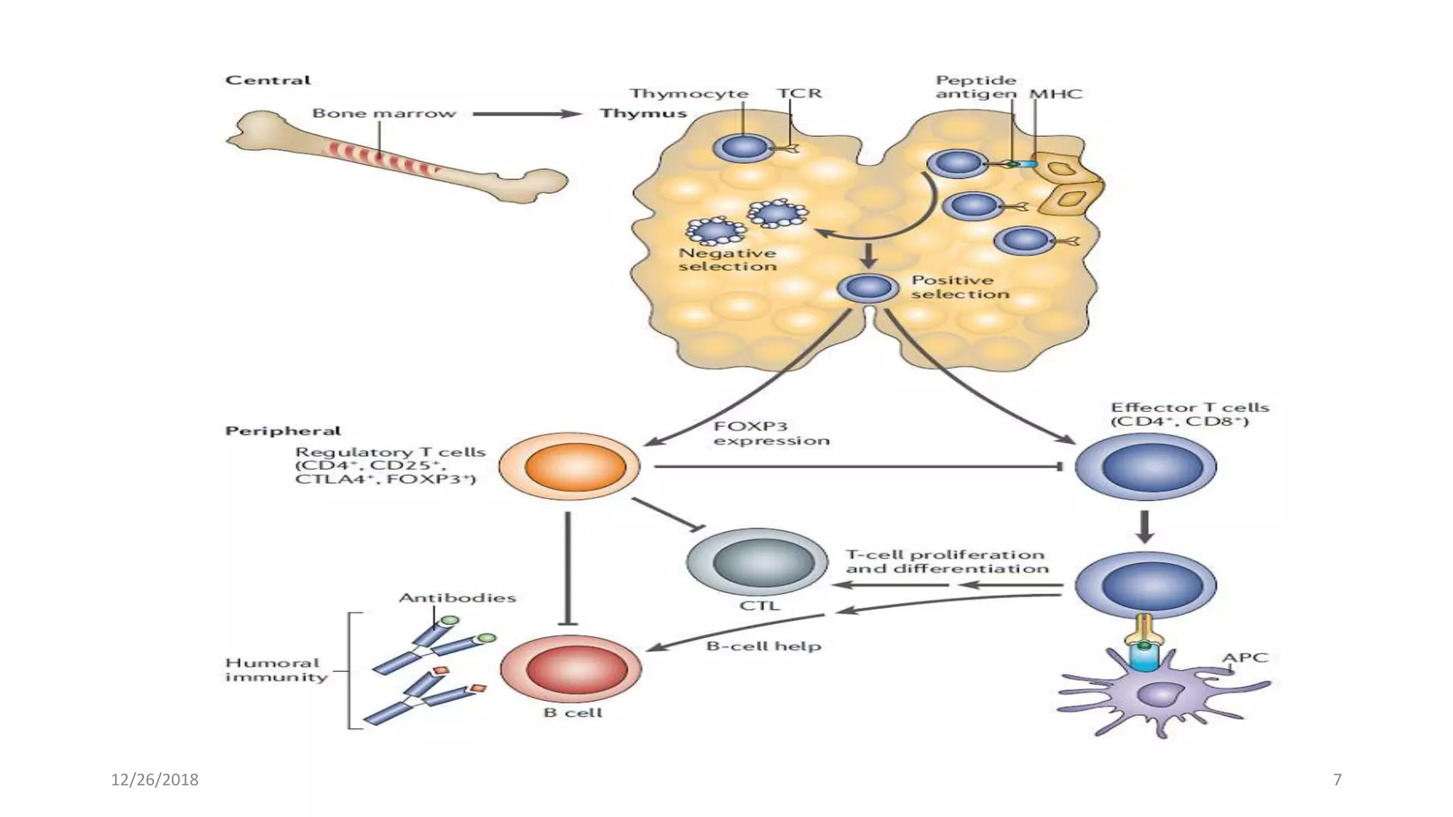
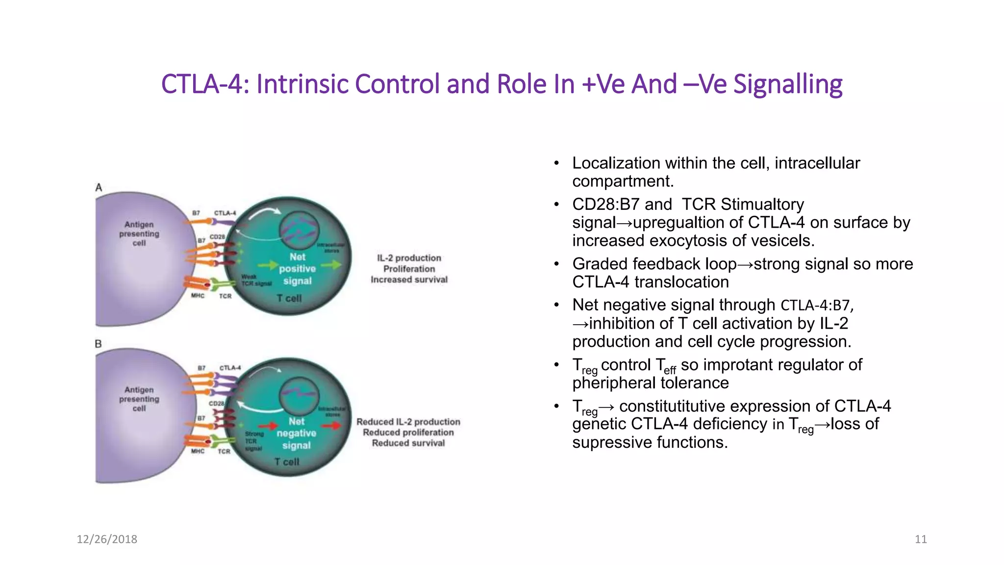
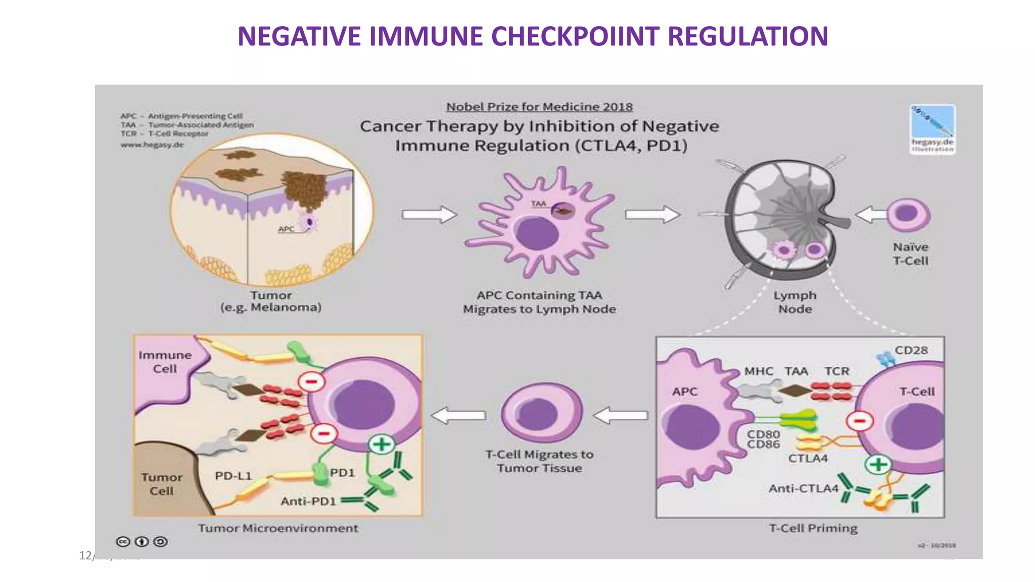
CTLA-4: IMMUNE CHECKPOINT BLOCKADE THERAPY,,,, THE 2018 NOBEL PRIZE WINNING STUDY IN THE FIELD OF PHYSIOLOGY/ MEDICINE,,, JOINTLY AWARDED TO JAMES P. ALLISON OF USA and TASAKU HONJU OF JAPAN .
1. CURRENT TRENDS IN IMMUNOCHEMISTRY
PRESENTATION
Anti-CTLA-4: IMMUNE CHECK POINT BLOCKADE THERAPY
Presented by: Saima Barki
DEPARTMENT OF BIOCHEMISTRY
QUAID-E-AZAM UNIVERISTY, ISLAMABAD
3. Cytotoxic T-lymphocyte antigen 4(CTLA-4)
HISTORY:
• 1st identified by Pierre Golstein and colleagues In 1987.
• November 1995, the labs of Tak Wah Mak and Arlene H. Sharpe independent discovery→ function of CTLA-
4 as a negative regulator of T-cell activation, by knocking out gene in mice.
• In the early 1990s, Jim Allison→CTLA-4 acts as an inhibitory molecule to restrict T cell responses.
• In 1996, Allison was the first to show that antibody blockade of a T-cell inhibitory molecule (known as CTLA-
4) could lead to enhanced anti-tumor immune responses and tumor rejection.
GENOMIC LOCATION IN HUMAN: 2q33.2
STRUCTURE:
• 3 domained structure, a) extracellualar, b). TM, 3) cytoplasmic tail (homology to CD28)→No intrinsic
catalytic activity and contian 2 motifs, one YVKM motif able to bind PI3K, PP2A and SHP-2 and 2nd one
proline-rich motif .
• The first role of CTLA-4 in inhibiting T cell responses seem to be directly via SHP-2 and PP2A
dephosphorylation of TCR-proximal signalling proteins as CD3 and LAT.
• Membrane boundform dimeric, monomers bound toghether by disulphide bond
• Free form as monomer
12/26/2018 3
4. BASICS OF IMMUNE SYSTEM
• To distinguish self from non self.
• Complex system.
• Main compenent:
• Recognition and binding of TCR to antigen displayed in connection with MHC of APC.
• Binding leads to→ Activation or Anergy
• T cell development:
• Progenitor cell→ T cell in bone marrow.
• Commitment to lineage in thymus, by exposing to Ag on the surface of APCs.
• In start T cell double negative(DN) i.e, CD4-CD8-TCR-.
• Then gene rearrangment unique TCR.
• Then selection or check for tolerance.
12/26/2018 4
5. T CELL TOLERANCE
Two Types Of Tolerance Mechanism:
• central tolerance
• peripheral tolerance
• CENTRAL TOLERANCE/NEGATIVE SELECTION:
• Location→ primary lymphoid organ.
• Process of eliminating any developing T or B lymphocyte reactive to self Ag.
• Ensure immune system not reactive to self molecules.
• Not a perfect system.
• SIGNALS/AFFINITY FOR SELECTION:
• Strong signal→ apoptotic death
• Intermediate signal→ T regulatory cells
• Weak signal→ T effector cells
• All cells either Treg or Teff depending on affinity towards self peptide.
12/26/2018 5
6. • PERIPHERAL TOLERANCE:
• Secondary mechanism
• Weak affinity bearing or Teff cells released as naive T cells to circulate in
blood, spleen, lymphatic organs.
• Its main purpose is to ensure that self-reactive T and B cells which escaped
central tolerance do not cause autoimmune disease.
• Then exposure to proffesional APCs bearing self mutated/altered antigen or
foreign antigen.
• Some self reactive so autoimmunity, prevented by immune checkpoint, being
regulated at multiple steps.
• IMMUNE CHECKPOINT PATHWAY MOLECULES: CTLA-4 AND PD-1
• Cytotoxic T lymphocyte like antigen-4(CTLA-4)→ leader→stop autoreactive
T cell at priming stage of the naive cell activation
• Programmed death protein 1 (PD-1)→later working→ in pheripheral tissues
• Key similarites and differences between both immune checkpoint pathways
• WHY A HOT TOPIC IN CANCER RESEARCH?
Tumors cell developed pathway to evade host immune system taking
advantage of peripheral tolerance.
12/26/2018 6
8. Normal T Cell Positve Costimulatory Signal
• T cell activation→complex→>1 stimualtory signal.
• TCR+MHC+Ag→T cell activation specificity but not complete signal.
• APC bearing B7-1(CD80) or B7-2(CD86) and T cell CD28→ +ve
costimualtory signal.
• Sufficient level of CD28:B7-1/2 →+ve signal.
• Proliferation,
• Increased T cell survival,
• Differentiation through growth cytokines(IL-2),
• Increased energy metabolism,
• Upregulation of cell survival genes, etc.,
12/26/2018 8
9. IMMUNE CHECKPOINT BLOCKADE
• Removes inhibitory signals of T cell activation.
• Enables tumor reactive cell to overcome regulatory mechanism and mounting
of anti tumor response.
• Inhibition result in increased activation of immune system.
• Induce durable response across multiple types of cancers.
• Discovery of immune checkpoint blockade and durable response, a
transformative event.
• Different inhibitors approval from FDA since 2011.
• Inhibitors approved being anti-CTLA-4 i.e. Ipilimumab and Tremilimumab for
metastatic melanoma.
• Combination therapy of ipilimumab and novolumab(anti PD-1)→synergistic
effect in advance melanoma patients.
12/26/2018 9
10. CTLA-4 PATHWAY: NEGATIVE COSTIMUALTORY SIGNAL
• CTLA-4 : CD28 HOMOLOG also known as CD125
• Regulate at T Cell priming in secnodary lymphoid organs,
• Higher avidity and affinity for B7 as compared to CD28,
• Rapid binding kinetics withB7-1,
• Competative binding,
• Relative amount of CD28 :B7 binding verses CTLA-4:B7 →T cell Activation or
Anergy respectively
• Attenuates positive signalling by CD28 and limits downstream signalling
mediated by PI3K and AKT,
• CTLA-4:B7 binding→inhibitory signals
• INHIBITORY MECHANISM:
• Direct inhibiton at TCR immune synapse
• Inhibition of CD28 or its signalling pathway
12/26/2018 10
11. CTLA-4: Intrinsic Control and Role In +Ve And –Ve Signalling
• Localization within the cell, intracellular
compartment.
• CD28:B7 and TCR Stimualtory
signal→upregualtion of CTLA-4 on surface by
increased exocytosis of vesicels.
• Graded feedback loop→strong signal so more
CTLA-4 translocation
• Net negative signal through CTLA-4:B7,
→inhibition of T cell activation by IL-2
production and cell cycle progression.
• Treg control Teff so improtant regulator of
pheripheral tolerance
• Treg→ constitutitutive expression of CTLA-4
genetic CTLA-4 deficiency in Treg→loss of
supressive functions.
12/26/2018 11
15. Mechanism of Tregcontrolling Teff
by:
• Downregulation of B7 ligands
on APCs→reduced CD28
costimualtion
MECHANISM OF NEGATIVE
COSTIMULATION VERSES MECHANISM
OF CHECKPOINT BLOCKADE:
• Cancer highly analogous to
infectious disease so principles of
virus system applicabe here where
chronic antigen stimulation results
in Tcell exhaustion
12/26/2018
15
16. MECHANISM OF ACTION OF CTLA-4 BLOCKADE INDUCED
TUMOR REJECTION
CTLA-4 blockade→tumor rejection
Exact Mechanisms not clear yet,, possibly these 3 mechanisms:
1. Primarily through direct blockade of CTLA-4 competition for B7-1/2 costimulatory
signal→unrestrained CD28 +ve signalling sp. activation.
Tumor cell→ No B7 ligands expression , so occur in tumor draining lymph nodes and tumor
microenvironment, require tumor cell death→ tumor antigen(neoantigens, TAA) cross
represented by APC to prime tumor reactive T cells.
Tumor presentation possibily in the tertiarry lymphoid structure TLS (presence associated
with improved survival), Context dependent antitumor functionality,Treg presence suppress
antitumor response
• CTLA-4 blockade specific response, expansion of specifically tumor neoantigen-CD28
(TIL) in tumor microenvironment not in lymphoid organ. TIL population include exhauseted
CD8 T cells and PD-1+ICOS+TBET+Th1 like CD4 effector cells.
• ICOS+CD4 Teff cells→pharmacodynamic marker of anti CTLA-4 therapy.
• CD4 Teff cells →Anti Ctla-4 affect differentiation along the activation.
12/26/2018 16
17. 2. DEPLETION of Treg population: mechanism of anti-CTLA-4 therapy
• Treg depletion partial contribution towards efficacy
• Differential efficiancy, as context dependent
• Blockade of both effector and reg t cell compaartment derived ctla-4 is required for effective tumor
rejection
• Ipilimumab: purely human IgG1,blocking antibody not depleting so no treg depletion evidence with
treatment, induce antibody dependent cellular cytotoxicity mediated killiing of Treg
• High affinity Fc receptor→improved response with iplilimumab so efficacy dependend on effector
funciton not depletion
• Tremelimumab:human IgG2→less effective ADCC than ipilumumab
3. Modulation of TCR repertoir:
3. Ipilimumab remodelling of peripheral TCR repertoir i.e. CD8 T cells but associated with irAEs.
4. Loss of CTLA-4, lowering of threshold for T cell activation, effective recognition of tumor
specific antigen→major driver of tumor immunogenicity.
• Survival associated with the maintainess of clones present in high frequency , more the frequency of clones
more the survival chances
• Effective anti CTLA-4 depend on the retention of pre-existing high avidtity cell with relevance to antitumor
response
12/26/2018 17
19. Clinical Efficacy and Characteristics of Response
with Immunecheckpoint Inhibitors
• Ipilimumab,1st anti CTLA-4 , prolong overall survival in melanoma.
• Phase ll/lll→survival curve plateau about 3 years
• Immune checkpoint blockade response efficacy vary with:
a.) Variation in kinetics and efficacy of ind. patient immune system.
b.) Interplay with tumor and metastasis
• Initial increase in tumor volume before generation of effective
tumor response so faster activation lead to inflammation and
influx of immune cells to tumor site.
12/26/2018 19
21. BIOMARKERS and IMMUNOLOGIC TOXICITIES
Retrospective studies reveal biomarkers including:
1. Absoulte lynphocyte count
2. Upregulation of T cell inducable costimulator(ICOS)-activation marker
3. Development of polyfunctional T cell response
IMMUNOLOGIC TOXICITIES:
• Immune checkpoint inhibitors →most promising drugs→toxicities
• Effect→skin,colon, endocrine glands,liver,lungs(common) and neurological
and myocardial disorders(rare)
• Ipilimumab irAEs: anti-ctla-4 3mg/kg in 60-85% paitent of grade 1 and 2 but
27% develop grade 3 to 4 toxicities, 2.1% deaath in 1st phase l trials
• Onset varies, 8 to 12 weeks of treatment initiation,
• Toxicity dose dependent
COMBINATION THERAPY ASSOCIATED TOXICITIES:
95% of patients→adverse effects→55%pf grade 3 to 4
12/26/2018 21
22. Summary Of Biological And Molecular
Functions Of T-cell
MOLECULE LIGANDS RECEPTOR
EXPRESSION
PATTERN
BIOLOGICAL
FUNCTION
MOLECULAR
FUNCTION
CTLA-4 B7-1(CD80),
B7-2(CD86)
Activated T cells,
Treg
Naïve T-cell
costimulation
(primarily at
priming): prevent
tonic signalling
and/or attenuate
high affinity clones
Competative
inhibitors of CD28
costimualtion
(binding of B7-
1(CD80),
B7-2(CD86)
12/26/2018 22
23. CLTA-4 Pathway Inhibitors Approved Or In Phase ll
and/or Phase lll Clinical Trail Stage Of Development
TARGET NAME STATUS COMPANY
CTLA-4 Ipilimumab •Approved for metastatic
melanoma
•Phase lll: lung, kidney
and prostate cancer
•Phase ll: cervical,
colorectal, gastric,
pancreatic, ovarian and
urothelial cancers
•Bristol-myers
•Squibb
CTLA-4 Tremelimumab •Phase lll studies in lung
cancer
•Medimmune/
•AstraaZenecca
12/26/2018 23
24. REFRENCES:
1. .Sharma P, Allison JP. The future of immune checkpoint therapy. Science 2015
2. Topalian SL, Drake CG, Pardoll DM Immune checkpoint blockade: a common denominator approach to cancer therapy. Cancer Cell 201
3. Pardoll DM. The blockade of immune checkpoints in cancer immunotherapy. Nat Rev Cancer 201
4. Dunn GP, Old LJ, Schreiber RD. The three Es of cancer immunoediting. Ann Rev Immunol 2004
5. Tang J, Shalabi A, Hubbard-Lucey VM. Comprehensive analysis of the clinical immuno-oncology landscape. Ann Oncol 2018
6. Sharma P, Hu-Lieskovan S, Wargo JA, Ribas A. Primary, adaptive, and acquired resistance to cancer immunotherapy. Cell 2017
7. Ribas A, Wolchok JD. Cancer immunotherapy using checkpoint blockade. Science 2018
8. Egen JG, Allison JP. Cytotoxic T lymphocyte antigen-4 accumulation in the immunological synapse is regulated by TCR signal strength.
Immunity 2002
9. Pentcheva-Hoang T, Egen JG, Wojnoonski K, Allison JP. B7-1 and B7-2 selectively recruit CTLA-4 and CD28 to the immunological synapse.
Immunity 2004
10. Kane LP, Andres PG, Howland KC, Abbas AK, Weiss A. Akt provides the CD28 costimulatory signal for up-regulation of IL-2 and IFN-gamma
but not TH2 cytokines. Nat Immunol 2001
11. waterhouse P, Penninger JM, Timms E, Wakeham A, Shahinian A, Lee KP, et al. Lymphoproliferative disorders with early lethality in mice
deficient in Ctla-4. Science 1995
12. Tivol EA, Borriello F, Schweitzer AN, Lynch WP, Bluestone JA, Sharpe AH. Loss of CTLA-4 leads to massive lymphoproliferation and fatal
multiorgan tissue destruction, revealing a critical negative regulatory role of CTLA-4. Immunity 1995. Goldrath AW, Bevan MJ. Selecting
and maintaining a diverse T-cell repertoire. Nature. 1999
13. Fife BT, Bluestone JA. Control of peripheral T-cell tolerance and autoimmunity via the CTLA-4 and PD-1 pathways. Immunol Rev. 2008
14. Greenwald RJ, Freeman GJ, Sharpe AH. The B7 family revisited. Annu Rev Immunol. 200512/26/2018 24
25. CONTINUED....
15. Poschke I, Mougiakakos D, Kiessling R. Camouflage and sabotage: tumor escape from the immune system. Cancer Immunol Immunother. 2011
16. Chambers CA, Kuhns MS, Egen JG, et al. CTLA-4-mediated inhibition in regulation of T cell responses: mechanisms and manipulation in tumor immunotherapy.
Annu Rev Immunol. 2001
17. Collins AV, Brodie DW, Gilbert RJ, et al. The interaction properties of costimulatory molecules revisited. Immunity. 2002
18. Egen JG, Kuhns MS, Allison JP. CTLA-4: new insights into its biological function and use in tumor immunotherapy. Nat Immunol. 2002
19. Parry RV, Chemnitz JM, Frauwirth KA, et al. CTLA-4 and PD-1 receptors inhibit T-cell activation by distinct mechanisms. Mol Cell Biol. 2005
20. Fallarino F, Fields PE, Gajewski TF. B7–1 engagement of cytotoxic T lymphocyte antigen 4 inhibits T cell activation in the absence of CD28. J Exp Med. 1998
21. Masteller EL, Chuang E, Mullen AC, et al. Structural analysis of CTLA-4 function in vivo. J Immunol. 2000
22. Schneider H, Downey J, Smith A, et al. Reversal of the TCR stop signal by CTLA-4. Science. 2006
23. Linsley PS, Bradshaw J, Greene J, et al. Intracellular trafficking of CTLA-4 and focal localization towards sites of TCR engagement. Immunity. 1996
24. Krummel MF, Allison JP. CTLA-4 engagement inhibits IL-2 accumulation and cell cycle progression upon activation of resting T cells. J Exp Med. 1996
25. Krummel MF, Allison JP. CTLA-4 engagement inhibits IL-2 accumulation and cell cycle progression upon activation of resting T cells. J Exp Med. 1996
27. Takahashi T, Tagami T, Yamazaki S, et al. Immunologic self-tolerance maintained by CD25(+)CD4(+) regulatory T cells constitutively expressing cytotoxic T
lymphocyte-associated antigen 4. J Exp Med. 2000
28. Wing K, Onishi Y, Prieto-Martin P, et al. CTLA-4 control over Foxp3+ regulatory T cell function. Science. 200829. Qureshi OS, Zheng Y, Nakamura K, et al. Trans-
endocytosis of CD80 and CD86: a molecular basis for the cell-extrinsic function of CTLA-4. Science. 2011
29. Keir ME, Butte MJ, Freeman GJ, et al. PD-1 and its ligands in tolerance and immunity. Annu Rev Immunol. 2008
30. Bennett F, Luxenberg D, Ling V, et al. Program death-1 engagement upon TCR activation has distinct effects on costimulation and cytokine-driven proliferation:
attenuation of ICOS, IL-4, and IL-21, but not CD28, IL-7, and IL-15 responses. J Immunol. 2003
31. Wherry EJ. T cell exhaustion. Nat Immunol. 2011
12/26/2018 25
26. CONTINUED.......
27. Takahashi T, Tagami T, Yamazaki S, et al. Immunologic self-tolerance maintained by CD25(+)CD4(+) regulatory T cells constitutively
expressing cytotoxic T lymphocyte-associated antigen 4. J Exp Med. 2000
28. Wing K, Onishi Y, Prieto-Martin P, et al. CTLA-4 control over Foxp3+ regulatory T cell function. Science. 200829. Qureshi OS, Zheng Y,
Nakamura K, et al. Trans-endocytosis of CD80 and CD86: a molecular basis for the cell-extrinsic function of CTLA-4. Science. 2011
29. Keir ME, Butte MJ, Freeman GJ, et al. PD-1 and its ligands in tolerance and immunity. Annu Rev Immunol. 2008
30. Bennett F, Luxenberg D, Ling V, et al. Program death-1 engagement upon TCR activation has distinct effects on costimulation and cytokine-
driven proliferation: attenuation of ICOS, IL-4, and IL-21, but not CD28, IL-7, and IL-15 responses. J Immunol. 2003
31. Wherry EJ. T cell exhaustion. Nat Immunol. 2011
32. Youngnak P, Kozono Y, Kozono H, et al. Differential binding properties of B7-H1 and B7-DC to death-1. Biochem Biophys Res Commun.
200333. Akbari O, Stock P, Singh
33. Ak, et al. PD-L1 and PD-L2 modulate airway inflammation and iNKT-cell-dependent airway hyperreactivity in opposing directions. Mucosal
Immunol. 2010
34. Huber S, Hoffmann R, Muskens F, et al. Alternatively activated macrophages inhibit T-cell proliferation by Stat6-dependent expression of
PD-L2. Blood. 2010
34. Butte MJ, Keir ME, Phamduy TB, et al. Programmed death-1 ligand 1 interacts specifically with the B7–1 costimulatory molecule to inhibit T
cell responses. Immunity. 2007
35. Wang L, Pino-Lagos K, de Vries VC, et al. Programmed death 1 ligand signaling regulates the generation of adaptive Foxp3+CD4+ regulatory
T cells. Proc Natl Acad Sci. 2008
36. Francisco LM, Salinas VH, Brown KE, et al. PD-L1 regulates the development, maintenance, and function of induced regulatory T cells. J Exp
Med. 2009
12/26/2018 26
CTLA-4-mediated inhibition of T cells. T cells are activated when TCRs bind antigen displayed in the MHC on antigenpresenting cells in concert with CD28:B7-mediated costimulation. A, In the case of a weak TCR stimulus, CD28:B7 binding predominates, resulting in a net positive activating signal and IL-2 production, proliferation, and increased survival. B, In the case of a strong TCR
stimulus, CTLA-4 expression is upregulated by increased transport to the cell surface from intracellular stores and decreased internalization. CTLA-4 competes with CD28 for binding of B7 molecules. Increased CTLA-4:B7 binding can result in a net negative signal,
which limits IL-2 production and proliferation, and limits survival of the T cell. CTLA-4 indicates cytotoxic T-lymphocyte–associated
antigen 4; IL-2, interleukin-2; MHC, major histocompatibility complex; TCR, T-cell receptor
Molecular mechanism of CTLA-4 attenuation of T-cell activation, schematic of the molecular interactions and downstream signalling induced by ligation of CTLA-4 to respecitve ligands. the possibility of additional downstream cell intrinsic signallling mechanisms hioghlighted.
Schematic of the molecular mechanisms of action of CTLA4 and PD-1 blockade. The step-wise progression of T-cell activation, attenuation by normal regulatory mechanisms, and release of such negative regulation by therapeutic intervention using anti-CTLA4 or anti–PD-1 antibodies
is outlined (left). In addition to cell-intrinsic enhancement of effector function, several additional mechanisms are thought to contribute to the effcacy
of anti-CTLA4 and anti–PD-1 therapy (right). These include antibody-mediated depletion of Tregs, enhancement of T-cell positive costimulation within
the tumor microenvironment, blockade of host-derived PD-L1 signals from nontumor cells in the microenvironment (as opposed to tumor cell–derived
PD-L1), and blockade of interactions between PD-L1 and B7-1.
Potential cellular mechanisms that mediate tumor rejection in response to combination anti-CTLA4 and anti–PD-1 checkpoint blockade. Multiple non–mutually exclusive models of the cellular mechanisms underlying combination anti-CTLA4 plus anti–PD-1 therapy of action are proposed. Models
described from left to right: (i) the same T cells may be targeted at the site of priming, leading to enhanced penetrance of effective blockade (i.e., a greater
proportion of target cells receive suffcient signal to increase activation) and/or enhanced costimulatory signals beyond normal limits, (ii) different T-cell
populations are targeted within the site of priming, potentially leading to synergistic effects through cell-extrinsic processes (e.g., providing CD4 help to
CD8 effector T cells), (iii) the same T cells are targeted but with different spatiotemporal kinetics leading to perhaps prolonged costimulatory signaling,
and (iv) different T-cell populations are targeted in different tissues (e.g., PD-1 blockade primarily acting on preexisting tumor-infltrating CD8 T cells
whereas CTLA4 acts on CD4 effector T cells in secondary lymphoid organs). T-cell subsets are denoted as “A” and “B” given that the precise populations
that are directly targeted remain to be fully defned, particularly in the context of kinetics of therapy and different tissue sites. Conceptually, T-cell “A” and
“B” could, for example, represent particular subsets of tumor-specifc CD8 T cells and CD4 effector T cells, respectively. Potential effects are noted below
each scenario; however, there is certain to be additional aggregate effects and differences between these models. Only secondary lymphoid organs (e.g.,
draining lymph node) and tumor are described, but other tissue sites may have functional contributions to this process as well.