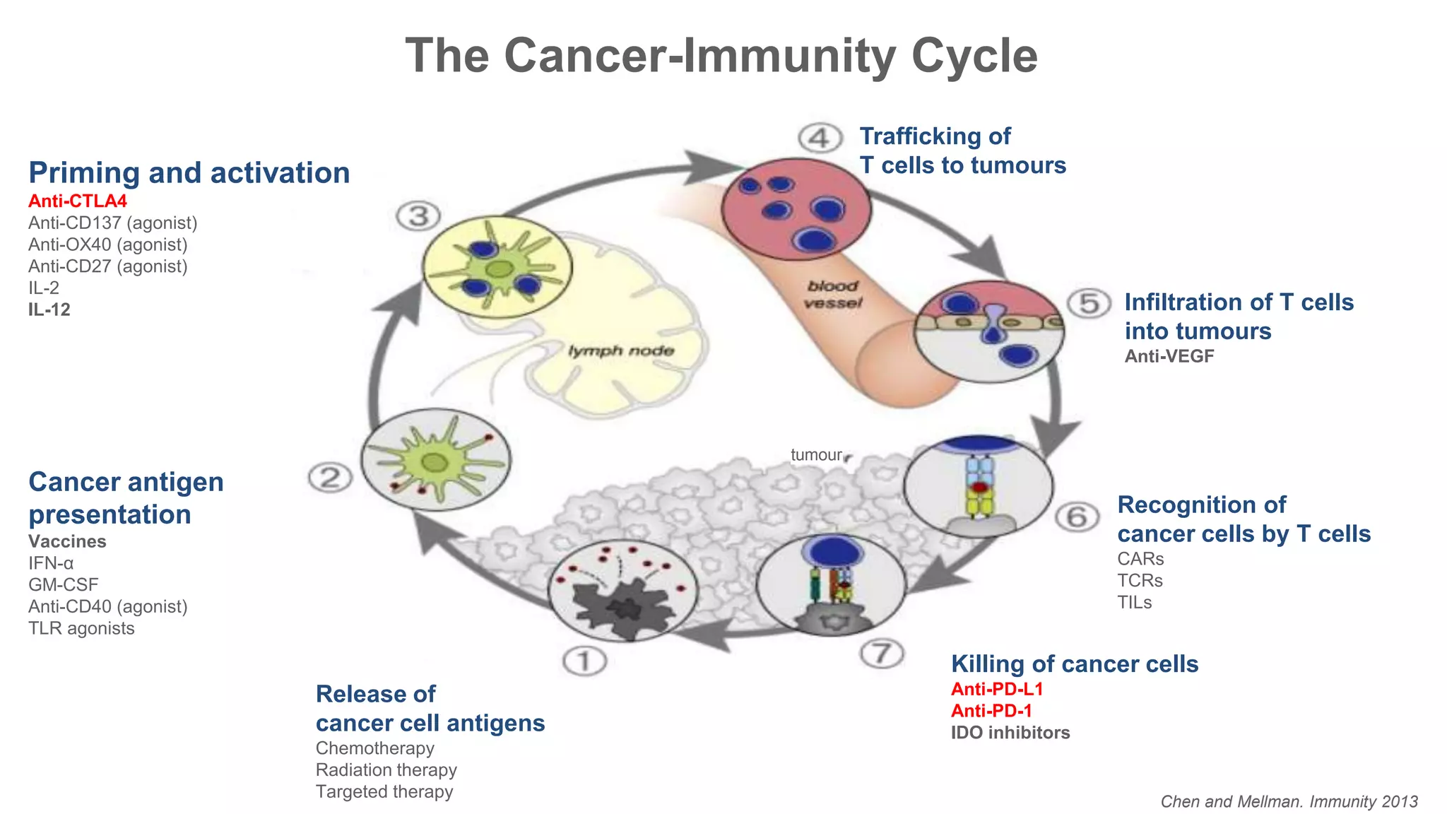
Immunological Checkpoints and Cancer Immunotherapy:
Review of Data and Issues of Interest for Imaging Community
Elad Sharon, MD, MPH
Senior Investigator/Medical Officer
Cancer Therapy Evaluation Program
Division of Cancer Therapy & Diagnosis
National Cancer Institute
August 7, 2017
2
Overview of Immunotherapy and
How Immune Checkpoint
Inhibitors Fit into the Picture
Options for Immune Intervention in Cancer
 Vaccines (induce immune response against presumed cancer antigen)
 Defined antigen and delivery method
 Promote Ag presentation in vivo
 Cytokines to promote T-cell activation, proliferation and function
 Provide T cell co-stimulatory signals
 Block T cell inhibitory signals
 Adoptively transfer antigen-specific T cells
 Bispecific antibodies
 Give antibodies that kill by CDC or ADCC
 Activate NK cell function to kill tumor cells
Atkins, M. B. Clin Cancer Res 2006;12:2353s-
2358s
High Dose IL-2: Durable Responses
• RR: 16% (43/270)
• CR: 6.2%
• Durable responses
– Median 8.9
mos
– CR: not reached
Fyfe G, et al. J Clin Oncol. 1995;13:688-696
Melanoma Renal Cell
Carcinoma
Chen and Mellman. Immunity 2013
Killing of cancer cells
Anti-PD-L1
Anti-PD-1
IDO inhibitors
Priming and activation
Anti-CTLA4
Anti-CD137 (agonist)
Anti-OX40 (agonist)
Anti-CD27 (agonist)
IL-2
IL-12
Cancer antigen
presentation
Vaccines
IFN-α
GM-CSF
Anti-CD40 (agonist)
TLR agonists
Release of
cancer cell antigens
Chemotherapy
Radiation therapy
Targeted therapy
Recognition of
cancer cells by T cells
CARs
TCRs
TILs
Infiltration of T cells
into tumours
Anti-VEGF
Trafficking of
T cells to tumours
tumour
The Cancer-Immunity Cycle
Key Features of Immune Checkpoint Inhibitors
 Associated with immune-related adverse events
 Any organ, but rash, colitis, hepatitis and endocrinopathies are most common
 May require steroids +/- additional immunosuppressive agents
 Unique kinetics of response in some patients
 Responses may improve with time
 Occasional responses after initial increase in total tumor volume
 Response in index plus new lesions at or after the appearance of new lesions
 Continued benefit after therapy of discordant progressing lesions
Biological Function and Details of CTLA-4
 CTLA-4 is (almost) exclusively on T cells
 Primarily regulates the amplitude of the early stages of T-cell activation
 CTLA-4 knockout mice die within 3 weeks from immune destruction of multiple
organs
 CTLA-4 counteracts the activity of the T cell costimulatory receptor CD28
 CD28 does not affect T-cell activation unless the TCR is first engaged by
cognate antigen
CD28, CTLA-4, and the TCR
 CD28 signaling strongly amplifies
the TCR signal to activate T cells
 CD28 and CTLA-4 share
identical ligands:
 CD80 (B7.1) and CD86 (B7.2)
 CTLA-4 has a much higher
overall affinity for both ligands
 Studies suggest that CTLA-4
signaling disrupts kinase signals
induced by TCR and CD28
Alegre et al. Nature Reviews Immunology. 2001
How does CTLA-4 work?
 CTLA-4 is expressed by activated CD8 killer T cells
 Distinct effects on the two major subsets of CD4 T cells
• Down-modulation of helper T-cell activity
• Enhancement of regulatory T-cell suppressive activity
• CTLA-4 blockade results in a broad enhancement of immune
responses dependent on helper T cells
• CTLA-4 is a target gene of the transcription factor Foxp3, the
expression of which determines the Treg lineage
 Tregs express CTLA-4 constitutively
Ipilimumab: The Beginning of an Era
Survival Rate Ipilimumab + gp100 Ipilimumab alone gp100 alone
1-yr 44% 46% 25%
2-yr 22% 24% 14%
Biological Function and Details of PD1
 Limits activity of T cells in the peripheral
tissues at the time of an inflammatory
response to stimulus
 Expression is induced when T cells become
activated
 PD-1 inhibits kinases involved in T-cell
activation via the phosphatase SHP2
 PD-1 is highly expressed on Tregs, where it
may enhance proliferation in the presence of
ligand
Mario Sznol, and Lieping Chen Clin Cancer Res 2013;19:1021-1034
Effects of Expression of PD-1
 PD-1 is more broadly expressed than CTLA-4
 Can be induced on B cells and NK cells
 Two ligands for PD-1 are PD-L1 and PD-L2
 Arose via gene duplication
 Positioned them within 100 kB
 Chronic antigen exposure, (including viral infections) can lead to high levels of
persistent PD-1 expression, which induces a state of exhaustion or anergy
among cognate antigen-specific T cells
 PD-1 can also shift the balance from T-cell activation to tolerance at early
stages in T-cell responses to antigen within secondary lymphoid tissues
Pardoll DM. Seminars in Oncology. 2015
PD-1 Expression in the Tumor Microenvironment
 PD-1 expressed on a large number of TILs in many tumor types
 Tregs can be a large fraction
 Expression on CD8 TILs may reflect an anergic/exhausted state (e.g.
decreased cytokine production)
 Primary PD-1 role is inhibition in the tumor microenvironment
• Inhibition only occurs with cognate ligand
 PD-L2 has also been reported to be upregulated on a number of tumors
 Upregulated on certain B-cell lymphomas
45%
Type 1
17%
Type 2
26%
Type 3
12%
Type 4
Presence of PD-L1 or TILs1
PD-L1/TIL
PPD-L1 /TIL+
D-L1 /TIL+
PD-L1/TIL PD-L1+/TIL+
PD-L1 /TIL+ PD-L1+/TIL
45%
Type 1
17%
Type 2
26%
Type 3
12%
Type 4NSCLC
Melanoma 45% 41% 13% 1%
Schalper &
Rimm, Yale U.
Taube et
al
Spectrum of PD-1/PD-L1 Antagonist Activity
 Melanoma
 Renal cancer (clear cell and non-clear cell)
 NSCLC – adenocarcinoma and squamous cell
 Head and neck cancer
 Hodgkin Lymphoma
 Bladder Cancer
 Merkel Cell
 Mismatch repair deficient tumors
 Small cell lung cancer
 Gastric and GE junction
 Triple negative breast cancer
 Ovarian
 Hepatocellular carcinoma
 Mesothelioma
 Nasopharyngeal Cancer
 Cervical
 Diffuse large B-cell lymphoma
 Follicular lymphoma
Major PD-1/PD-L1 antagonists
• Nivolumab (anti-PD-1)
• Pembrolizumab (anti-PD-1)
• Atezolizumab (MPDL3280A, anti-PD-L1)
• Durvalumab (MEDI-4736, anti-PD-L1)
• Avelumab (anti-PD-L1)
• Other PD-1 efforts ongoing from:
• CureTech/Medivation (CT-011)
• Sanofi/Regeneron (REGN2810)
• Novartis (PDR001)
• Aurigene/Pierre Fabre (AUNP-12)
Minimal to no activity to single agents:
• Prostate cancer
• Microsatellite Stable Colon cancer
• Multiple Myeloma
• Pancreatic Cancer
Active
FDA Approvals of Antibodies Targeting Immune Checkpoints
 2011 Ipilimumab (BMS) – Melanoma
 2014 Pembrolizumab (Merck) – Melanoma
 2014 Nivolumab (BMS) – Melanoma
• 2015 Nivolumab (BMS) – Non-Small Cell Lung Cancer
• 2015 Ipilimumab + Nivolumab (BMS) – Melanoma
• 2015 Pembrolizumab (Merck) – Non-Small Cell Lung Cancer
• 2015 Ipilimumab (BMS) – Adjuvant Melanoma
• 2015 Nivolumab (BMS) – Renal Cell Carcinoma
• 2016 Nivolumab (BMS) – Hodgkin Lymphoma
• 2016 Atezolizumab (Roche) – Bladder Cancer
• 2016 Pembrolizumab (Merck) – Head and Neck Cancer
• 2016 Atezolizumab (Roche) – Non-Small Cell Lung Cancer
• 2017 Avelumab (Pfizer/EMD Serono) – Merkel Cell Cancer
• 2017 Durvalumab (AstraZeneca) – Bladder Cancer
Pembrolizumab in MMR-Deficient Cancer
Le DT et al. N Engl J Med
2015;372:2509-2520.
Gajewski TF et al. Current Opinion in Immunology. 2013
20
Use Case: How PD1/PDL1
Inhibitors Have Performed in
Merkel Cell Cancer
Epidemiology
 About 1600 new cases per year in USA
 Increasing incidence (tripled in past 15 years)
 Uncommon cutaneous malignancy with epithelial and neuroendocrine
features
 Found on UV exposed areas
 Median age 76
 More common in Caucasians and males
 High risk of developing nodal and distant metastases
 Overall mortality of 45%
1. Youlden DR et al. JAMA Dermatol. 2014;150:864-872.
Merkel Cell Polyomavirus
 Identified in 2008 at U of Pittsburgh
 Highly prevalent in normal skin
 One of 13 known human polyomaviruses
 Virus present in approximately 80% of MCC tumor DNA
 Serum antibodies to MCPyV T-antigens more specifically associated
with MCC
1. Feng H et al. Science. 2008;319:1096-1100.
2. Moshiri AS, Nghiem P. J Natl Compr Canc Netw. 2014;12:1255-1262.
3. Grundhoff A, Fischer N. Curr Opin Virol. 2015;14:129-137.
Clinical Presentation
Prognosis of MCC
Key collaborations:
Academic
(8 universities)
Government
(NCI-CTEP-CITN)
Industry
(Merck)
Available on-line at NEJM.org
Tuesday April 19
Trials Avelumab Pembrolizumab
Target PD-L1 PD-1
Median age, y 72.5 68
Patients 88 25
Stage IV IIIB/IV
MCPyV+ 52% (46/88) 65% (17/26)
Prior lines of therapy ≥1 0
Response rate
32% (CR 8)
MCPyV+ (26%) vs – (36%)
PD-L1+ (35%) vs – (19%)
56% (CR 4)
MCPyV+ (62%) vs – (44%)
PD-L1+ (32%) vs – (24%)
6-month PFS 40% (median PFS: 2.7 mo) 67% (median PFS: 9 mo)
6-month OS 69% (median OS: 11.3 mo) Not reported
Median follow-up, mo 10.4 7.6
Comparison of Both Trials
Source: Guilherme Rabinowits. DFCI
Avelumab Results
Waterfall plot: Pembrolizumab Responses in virus-pos &
virus-neg MCCs
-
40
-
80
Maximum Change In Sum of Target Lesion Diameters
PercentChangeinTargetLesions
-100-60-20020406080
(N=24)
Viral Status
Negative
Positive
100120140160
Percentchangeintargetlesions Tumor viral status
negative
positive
n = 24
Fraction responding:
44% of virus-neg
62% of virus-pos
(difference not significant)
Pembrolizumab Swimmer plot: most responses persist
Months from treatment initiationMonths from Treatment Initiation
0 2 4 6 8 10 12 14
➔
➔
Complete Response
Partial Response
Stable Disease
Progressive Disease
Ongoing CR
Ongoing PR
On Treatment
Most responses
are durable
(86%; 12 of 14
confirmed
responses persist)
Even 1-2 doses
of anti-PD-1 can
induce sustained
response
Baseline 3 wks after
pembrolizumab
Pembrolizumab Case example: partial response
After 1 dose:
SQ lesion barely palpable at 3 weeks;
biopsy showed. . .
Primary on R knee...10 mo later 
Bulky tumors in pelvis: bladder compression
Subcutaneous metastases on leg
Baseline 12 weeks
Pembrolizumab Partial response on anti-PD-1
Bulky pelvic disease
Bladder symptoms resolved
Therapy & response ongoing (13 months)
No side effects
Tumors continue to shrink
PD-L1 expression in pre-treatment tumor biopsies does
not predict response to Pembrolizumab
Virus Negative (n=8)
0
20
40
60
80
100
%ofpatientswithineach
viralcategory
p= 0.61
Non-Responders (n=9) Responders (n=14)
0
20
40
60
80
100
%ofpatientswithin
eachresponsecategory
PD-L1 Positive
PD-L1 Negative
*
*
n=23
Responses with Pembrolizumab with
PD-L1 positive / negative tumors
Months from treatment initiation
PercentChangeinTarget
Lesions
Tumor PD-L1 status
negative
positive
n = 23
Goh et al. Oncotarget 2015.
MCPyV positive vs.
negative MCC: at the
extremes of
mutational frequency
compared to TCGA
data for other
cancers
Virus Positive Virus Negative
UVmutations
Goh et al, Oncotarget 2015
Virus-negative MCCs  high mutational load and many
predicted neoantigens
Conclusions
• Anti-PD-1/PDL-1 induces a high response rate in a rare tumor in the
metastatic setting, both frontline and relapsed
• Responses in both virus positive and virus negative MCC
• Encouraging response durability
• Correlative studies are ongoing for anti-MCPyV antibody and T cell
responses
• Expansion of pembrolizumab trial ongoing
• Further efforts are necessary to reduce relapse at earlier settings of
disease to improve overall survival
40
How to Approach Combinations
and Resistance in the Clinic
Day, Monjazeb, Sharon, Ivy, Rubin, Rosner, Butler. CCR Focus, [Accepted]
Options for combinations
 Immune Checkpoints
 CTLA-4, PD-1, PD-L1, LAG3, TIM3, BTLA, TIGIT, VISTA, KIR
 Immune Stimulators
 OX40 (CD134), GITR, CD137, CD40, ICOS, 4-1BB, CD27
 Immunosuppressive Soluble Factors
 IDO-1, Adenosine
 Oncolytic Viruses
 Adoptive Cell Transfer
 T-cell engaging bispecific agents (blinatumomab)
 Endogenous Adjuvants
 STING, TLR
 Vaccines
 Radiation
 Treg depleting strategies
 Chemotherapy, CCR4, anti-CD25
 Other Targeted Therapies (BRAF, MEK)
 IMiDs
Pardoll DM. Nature Reviews Cancer. 2012
How to Establish Efficacy
 Use of poor endpoints can be just as problematic in immuno-oncology
drug development as in all other parts of drug development
 Single arm PFS or OS trials can be subject to significant bias as a result of
patient selection
 Trials in general involve a “sample” of the total cancer population – samples
can be misleading…
 Recommend discrete, measurable and definitive endpoints
 Randomized trials: OS, PFS, Response Rate, pCR
 Single arm trials: Some proxy for biologic activity – Response Rate, pCR
 TCR Analysis and Changes in Repertoire? TILs?
45
Implications for Imaging
Community
Issues for the Immunotherapy Community
 iRECIST and related immunologically-modified Imaging Response
Criteria
 Pseudoprogression
 Novel Imaging Problems
 Hyperprogression
 Novel Imaging Products
 Imaging T cells by PET
 Imaging radiolabeled PDL1
 Radiomics
iRECIST, irRECIST, iChesson, irRC
 Alphabet soup of new imaging criteria for immunotherapy
 What is the point?
 Will this help patients, or patient management?
 Does this involve lowering the bar for efficacy?
 Does iPFS better predict OS than current standards?
Hyperprogression
 Does it exist?
 Is there a tendency to overcall hyperprogression?
 How to prove it?
www.cancer.gov www.cancer.gov/espanol
Contact Information:
Elad Sharon, MD, MPH
sharone@mail.nih.gov
240-276-6565
Twitter: @EladSharonMD

Immunological Checkpoints and Cancer Immunotherapy

  • 1.
    Immunological Checkpoints andCancer Immunotherapy: Review of Data and Issues of Interest for Imaging Community Elad Sharon, MD, MPH Senior Investigator/Medical Officer Cancer Therapy Evaluation Program Division of Cancer Therapy & Diagnosis National Cancer Institute August 7, 2017
  • 2.
    2 Overview of Immunotherapyand How Immune Checkpoint Inhibitors Fit into the Picture
  • 3.
    Options for ImmuneIntervention in Cancer  Vaccines (induce immune response against presumed cancer antigen)  Defined antigen and delivery method  Promote Ag presentation in vivo  Cytokines to promote T-cell activation, proliferation and function  Provide T cell co-stimulatory signals  Block T cell inhibitory signals  Adoptively transfer antigen-specific T cells  Bispecific antibodies  Give antibodies that kill by CDC or ADCC  Activate NK cell function to kill tumor cells
  • 4.
    Atkins, M. B.Clin Cancer Res 2006;12:2353s- 2358s High Dose IL-2: Durable Responses • RR: 16% (43/270) • CR: 6.2% • Durable responses – Median 8.9 mos – CR: not reached Fyfe G, et al. J Clin Oncol. 1995;13:688-696 Melanoma Renal Cell Carcinoma
  • 5.
    Chen and Mellman.Immunity 2013 Killing of cancer cells Anti-PD-L1 Anti-PD-1 IDO inhibitors Priming and activation Anti-CTLA4 Anti-CD137 (agonist) Anti-OX40 (agonist) Anti-CD27 (agonist) IL-2 IL-12 Cancer antigen presentation Vaccines IFN-α GM-CSF Anti-CD40 (agonist) TLR agonists Release of cancer cell antigens Chemotherapy Radiation therapy Targeted therapy Recognition of cancer cells by T cells CARs TCRs TILs Infiltration of T cells into tumours Anti-VEGF Trafficking of T cells to tumours tumour The Cancer-Immunity Cycle
  • 6.
    Key Features ofImmune Checkpoint Inhibitors  Associated with immune-related adverse events  Any organ, but rash, colitis, hepatitis and endocrinopathies are most common  May require steroids +/- additional immunosuppressive agents  Unique kinetics of response in some patients  Responses may improve with time  Occasional responses after initial increase in total tumor volume  Response in index plus new lesions at or after the appearance of new lesions  Continued benefit after therapy of discordant progressing lesions
  • 7.
    Biological Function andDetails of CTLA-4  CTLA-4 is (almost) exclusively on T cells  Primarily regulates the amplitude of the early stages of T-cell activation  CTLA-4 knockout mice die within 3 weeks from immune destruction of multiple organs  CTLA-4 counteracts the activity of the T cell costimulatory receptor CD28  CD28 does not affect T-cell activation unless the TCR is first engaged by cognate antigen
  • 8.
    CD28, CTLA-4, andthe TCR  CD28 signaling strongly amplifies the TCR signal to activate T cells  CD28 and CTLA-4 share identical ligands:  CD80 (B7.1) and CD86 (B7.2)  CTLA-4 has a much higher overall affinity for both ligands  Studies suggest that CTLA-4 signaling disrupts kinase signals induced by TCR and CD28 Alegre et al. Nature Reviews Immunology. 2001
  • 9.
    How does CTLA-4work?  CTLA-4 is expressed by activated CD8 killer T cells  Distinct effects on the two major subsets of CD4 T cells • Down-modulation of helper T-cell activity • Enhancement of regulatory T-cell suppressive activity • CTLA-4 blockade results in a broad enhancement of immune responses dependent on helper T cells • CTLA-4 is a target gene of the transcription factor Foxp3, the expression of which determines the Treg lineage  Tregs express CTLA-4 constitutively
  • 10.
    Ipilimumab: The Beginningof an Era Survival Rate Ipilimumab + gp100 Ipilimumab alone gp100 alone 1-yr 44% 46% 25% 2-yr 22% 24% 14%
  • 11.
    Biological Function andDetails of PD1  Limits activity of T cells in the peripheral tissues at the time of an inflammatory response to stimulus  Expression is induced when T cells become activated  PD-1 inhibits kinases involved in T-cell activation via the phosphatase SHP2  PD-1 is highly expressed on Tregs, where it may enhance proliferation in the presence of ligand Mario Sznol, and Lieping Chen Clin Cancer Res 2013;19:1021-1034
  • 12.
    Effects of Expressionof PD-1  PD-1 is more broadly expressed than CTLA-4  Can be induced on B cells and NK cells  Two ligands for PD-1 are PD-L1 and PD-L2  Arose via gene duplication  Positioned them within 100 kB  Chronic antigen exposure, (including viral infections) can lead to high levels of persistent PD-1 expression, which induces a state of exhaustion or anergy among cognate antigen-specific T cells  PD-1 can also shift the balance from T-cell activation to tolerance at early stages in T-cell responses to antigen within secondary lymphoid tissues Pardoll DM. Seminars in Oncology. 2015
  • 13.
    PD-1 Expression inthe Tumor Microenvironment  PD-1 expressed on a large number of TILs in many tumor types  Tregs can be a large fraction  Expression on CD8 TILs may reflect an anergic/exhausted state (e.g. decreased cytokine production)  Primary PD-1 role is inhibition in the tumor microenvironment • Inhibition only occurs with cognate ligand  PD-L2 has also been reported to be upregulated on a number of tumors  Upregulated on certain B-cell lymphomas
  • 14.
    45% Type 1 17% Type 2 26% Type3 12% Type 4 Presence of PD-L1 or TILs1 PD-L1/TIL PPD-L1 /TIL+ D-L1 /TIL+ PD-L1/TIL PD-L1+/TIL+ PD-L1 /TIL+ PD-L1+/TIL 45% Type 1 17% Type 2 26% Type 3 12% Type 4NSCLC Melanoma 45% 41% 13% 1% Schalper & Rimm, Yale U. Taube et al
  • 15.
    Spectrum of PD-1/PD-L1Antagonist Activity  Melanoma  Renal cancer (clear cell and non-clear cell)  NSCLC – adenocarcinoma and squamous cell  Head and neck cancer  Hodgkin Lymphoma  Bladder Cancer  Merkel Cell  Mismatch repair deficient tumors  Small cell lung cancer  Gastric and GE junction  Triple negative breast cancer  Ovarian  Hepatocellular carcinoma  Mesothelioma  Nasopharyngeal Cancer  Cervical  Diffuse large B-cell lymphoma  Follicular lymphoma Major PD-1/PD-L1 antagonists • Nivolumab (anti-PD-1) • Pembrolizumab (anti-PD-1) • Atezolizumab (MPDL3280A, anti-PD-L1) • Durvalumab (MEDI-4736, anti-PD-L1) • Avelumab (anti-PD-L1) • Other PD-1 efforts ongoing from: • CureTech/Medivation (CT-011) • Sanofi/Regeneron (REGN2810) • Novartis (PDR001) • Aurigene/Pierre Fabre (AUNP-12) Minimal to no activity to single agents: • Prostate cancer • Microsatellite Stable Colon cancer • Multiple Myeloma • Pancreatic Cancer Active
  • 16.
    FDA Approvals ofAntibodies Targeting Immune Checkpoints  2011 Ipilimumab (BMS) – Melanoma  2014 Pembrolizumab (Merck) – Melanoma  2014 Nivolumab (BMS) – Melanoma • 2015 Nivolumab (BMS) – Non-Small Cell Lung Cancer • 2015 Ipilimumab + Nivolumab (BMS) – Melanoma • 2015 Pembrolizumab (Merck) – Non-Small Cell Lung Cancer • 2015 Ipilimumab (BMS) – Adjuvant Melanoma • 2015 Nivolumab (BMS) – Renal Cell Carcinoma • 2016 Nivolumab (BMS) – Hodgkin Lymphoma • 2016 Atezolizumab (Roche) – Bladder Cancer • 2016 Pembrolizumab (Merck) – Head and Neck Cancer • 2016 Atezolizumab (Roche) – Non-Small Cell Lung Cancer • 2017 Avelumab (Pfizer/EMD Serono) – Merkel Cell Cancer • 2017 Durvalumab (AstraZeneca) – Bladder Cancer
  • 18.
    Pembrolizumab in MMR-DeficientCancer Le DT et al. N Engl J Med 2015;372:2509-2520.
  • 19.
    Gajewski TF etal. Current Opinion in Immunology. 2013
  • 20.
    20 Use Case: HowPD1/PDL1 Inhibitors Have Performed in Merkel Cell Cancer
  • 21.
    Epidemiology  About 1600new cases per year in USA  Increasing incidence (tripled in past 15 years)  Uncommon cutaneous malignancy with epithelial and neuroendocrine features  Found on UV exposed areas  Median age 76  More common in Caucasians and males  High risk of developing nodal and distant metastases  Overall mortality of 45% 1. Youlden DR et al. JAMA Dermatol. 2014;150:864-872.
  • 22.
    Merkel Cell Polyomavirus Identified in 2008 at U of Pittsburgh  Highly prevalent in normal skin  One of 13 known human polyomaviruses  Virus present in approximately 80% of MCC tumor DNA  Serum antibodies to MCPyV T-antigens more specifically associated with MCC 1. Feng H et al. Science. 2008;319:1096-1100. 2. Moshiri AS, Nghiem P. J Natl Compr Canc Netw. 2014;12:1255-1262. 3. Grundhoff A, Fischer N. Curr Opin Virol. 2015;14:129-137.
  • 23.
  • 24.
  • 27.
  • 29.
    Trials Avelumab Pembrolizumab TargetPD-L1 PD-1 Median age, y 72.5 68 Patients 88 25 Stage IV IIIB/IV MCPyV+ 52% (46/88) 65% (17/26) Prior lines of therapy ≥1 0 Response rate 32% (CR 8) MCPyV+ (26%) vs – (36%) PD-L1+ (35%) vs – (19%) 56% (CR 4) MCPyV+ (62%) vs – (44%) PD-L1+ (32%) vs – (24%) 6-month PFS 40% (median PFS: 2.7 mo) 67% (median PFS: 9 mo) 6-month OS 69% (median OS: 11.3 mo) Not reported Median follow-up, mo 10.4 7.6 Comparison of Both Trials Source: Guilherme Rabinowits. DFCI
  • 30.
  • 31.
    Waterfall plot: PembrolizumabResponses in virus-pos & virus-neg MCCs - 40 - 80 Maximum Change In Sum of Target Lesion Diameters PercentChangeinTargetLesions -100-60-20020406080 (N=24) Viral Status Negative Positive 100120140160 Percentchangeintargetlesions Tumor viral status negative positive n = 24 Fraction responding: 44% of virus-neg 62% of virus-pos (difference not significant)
  • 32.
    Pembrolizumab Swimmer plot:most responses persist Months from treatment initiationMonths from Treatment Initiation 0 2 4 6 8 10 12 14 ➔ ➔ Complete Response Partial Response Stable Disease Progressive Disease Ongoing CR Ongoing PR On Treatment Most responses are durable (86%; 12 of 14 confirmed responses persist) Even 1-2 doses of anti-PD-1 can induce sustained response
  • 33.
    Baseline 3 wksafter pembrolizumab Pembrolizumab Case example: partial response After 1 dose: SQ lesion barely palpable at 3 weeks; biopsy showed. . . Primary on R knee...10 mo later  Bulky tumors in pelvis: bladder compression Subcutaneous metastases on leg
  • 34.
    Baseline 12 weeks PembrolizumabPartial response on anti-PD-1 Bulky pelvic disease Bladder symptoms resolved Therapy & response ongoing (13 months) No side effects Tumors continue to shrink
  • 35.
    PD-L1 expression inpre-treatment tumor biopsies does not predict response to Pembrolizumab Virus Negative (n=8) 0 20 40 60 80 100 %ofpatientswithineach viralcategory p= 0.61 Non-Responders (n=9) Responders (n=14) 0 20 40 60 80 100 %ofpatientswithin eachresponsecategory PD-L1 Positive PD-L1 Negative * * n=23
  • 36.
    Responses with Pembrolizumabwith PD-L1 positive / negative tumors Months from treatment initiation PercentChangeinTarget Lesions Tumor PD-L1 status negative positive n = 23
  • 37.
    Goh et al.Oncotarget 2015. MCPyV positive vs. negative MCC: at the extremes of mutational frequency compared to TCGA data for other cancers Virus Positive Virus Negative UVmutations
  • 38.
    Goh et al,Oncotarget 2015 Virus-negative MCCs  high mutational load and many predicted neoantigens
  • 39.
    Conclusions • Anti-PD-1/PDL-1 inducesa high response rate in a rare tumor in the metastatic setting, both frontline and relapsed • Responses in both virus positive and virus negative MCC • Encouraging response durability • Correlative studies are ongoing for anti-MCPyV antibody and T cell responses • Expansion of pembrolizumab trial ongoing • Further efforts are necessary to reduce relapse at earlier settings of disease to improve overall survival
  • 40.
    40 How to ApproachCombinations and Resistance in the Clinic
  • 42.
    Day, Monjazeb, Sharon,Ivy, Rubin, Rosner, Butler. CCR Focus, [Accepted]
  • 43.
    Options for combinations Immune Checkpoints  CTLA-4, PD-1, PD-L1, LAG3, TIM3, BTLA, TIGIT, VISTA, KIR  Immune Stimulators  OX40 (CD134), GITR, CD137, CD40, ICOS, 4-1BB, CD27  Immunosuppressive Soluble Factors  IDO-1, Adenosine  Oncolytic Viruses  Adoptive Cell Transfer  T-cell engaging bispecific agents (blinatumomab)  Endogenous Adjuvants  STING, TLR  Vaccines  Radiation  Treg depleting strategies  Chemotherapy, CCR4, anti-CD25  Other Targeted Therapies (BRAF, MEK)  IMiDs Pardoll DM. Nature Reviews Cancer. 2012
  • 44.
    How to EstablishEfficacy  Use of poor endpoints can be just as problematic in immuno-oncology drug development as in all other parts of drug development  Single arm PFS or OS trials can be subject to significant bias as a result of patient selection  Trials in general involve a “sample” of the total cancer population – samples can be misleading…  Recommend discrete, measurable and definitive endpoints  Randomized trials: OS, PFS, Response Rate, pCR  Single arm trials: Some proxy for biologic activity – Response Rate, pCR  TCR Analysis and Changes in Repertoire? TILs?
  • 45.
  • 46.
    Issues for theImmunotherapy Community  iRECIST and related immunologically-modified Imaging Response Criteria  Pseudoprogression  Novel Imaging Problems  Hyperprogression  Novel Imaging Products  Imaging T cells by PET  Imaging radiolabeled PDL1  Radiomics
  • 47.
    iRECIST, irRECIST, iChesson,irRC  Alphabet soup of new imaging criteria for immunotherapy  What is the point?  Will this help patients, or patient management?  Does this involve lowering the bar for efficacy?  Does iPFS better predict OS than current standards?
  • 49.
    Hyperprogression  Does itexist?  Is there a tendency to overcall hyperprogression?  How to prove it?
  • 52.
    www.cancer.gov www.cancer.gov/espanol Contact Information: EladSharon, MD, MPH sharone@mail.nih.gov 240-276-6565 Twitter: @EladSharonMD