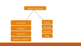
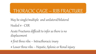
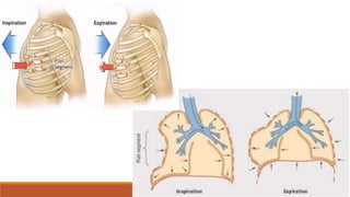
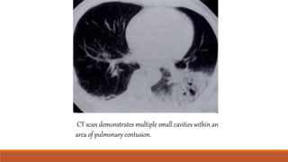
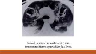
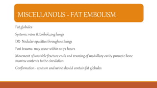
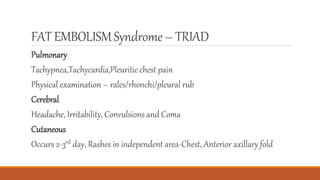
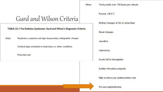
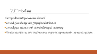
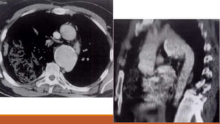
CTBLUNTCHESTTRAUMA
DR.SAHANA.S
MODERATOR - DR.UDHAYAKUMAR.K
DIRECT TRAUMA
FALLS
BLOWS
BLASTS
RTA
SHOOTING
STABBING
SHRAPNEL
THORACIC SURGERY
INJURY
SKELETAL TRAUMA
DIAPHRAGM
PLEURA
LUNG
TRACHEA AND BRONCHI
MEDIASTINUM
VASCULAR
THORACIC CAGE – RIB FRACTURE
May be single/multiple and unilateral/bilateral
Healed # - CXR
Acute Fractures difficult to infer as there is no
displacement
# first three ribs – Intracthoracic inury
# Lower three ribs – Hepatic, Splenic or Renal injury
• Flail segment
• Pneumothorax
• Hemothorax
• Subcutaneous Emphysema
COMPLICATIONS
THORACIC CAGE – RIB FRACTURE
FLAILSEGMENT
Apparent clinically
Affected part
Sucked inspiration compromising underlying lung
Several adjacent ribs or Bilateral Rib #
Pneumothorax
Hemothorax
Hemopneumothorax
Intrapulmonary hemorrhage
Fractured ends
Penetrate
Pleura
AIR enters Chest Wall – Subcutaneous Emphysema
CT axial section contrast study shows displaced right rib fracture with extensive subcutaneous emphysema. There is also a
sternal fracture with associated retrosternal hematoma and lung window settings shows a large right pneumothorax.
CT scan demonstrates an anterior mediastinal hematoma with a preserved fat plane between the
hematoma and the aorta. Arrow indicates a sternal fracture.
MISCELLANOUS
Stress #- First and Second ribs
Cough #- 6th – 9th in the posterior axillary line{not seen
until callus formation}
Sternum # - CXR Lateral view (occasionally)
Thoracic Spine with Para spinal shadow – HEMATOMA
Clavicle # maybe associated with Subclavian/ brachial injury
Axial CT scan shows mild irregularity of the
anterior aspect of T12 Sagittal reconstructed
image demonstrates an acute anterior wedge
fracture of T12.
Axial CT scan shows multiple fragments of
the fractured T5 vertebral body. The spinal
canal is narrowed at this level. Sagittal
reconstructed image clearly demonstrates
the loss of vertebral body height and bone
fragments posteriorly displaced into the
spinal canal. The posterior elements are also
fractured.
SubclavianArtery
Right and left have different courses
Right is one of the terminal branch of brachiocephalic artery
Left arises as the third branch of the aortic arch after the left common carotid artery
Course:
Exits the thorax via the superior thoracic aperature between the anterior scalene and
posterior scalene before passing between the first rib and clavicle.
Then at the lateral border it continues as axillary artery
Parts
First part – From its origin to the medial border of anterior scalene
Second part-Posterior to anterior scalene
Third part-from lateral border of anterior scalene to lateral border of first rib
MISCELLANOUS
Posterior dislocation of Sternoclavicular joint
Injury to Trachea/Oesophagus/Great Vessels/Superior Mediastinum
Herniation of Lung Tissue
CT of the chest reveals contusion of
the right lung and left lobe.
Also effect is noted on the left side
in the rib with herniation of lung
tissue parenchyma through an
intercostal space.
INJURY
SKELETAL TRAUMA
DIAPHRAGM
PLEURA
LUNG
TRACHEA AND BRONCHI
MEDIASTINUM
VASCULAR
DIAPHRAGM
• Laceration
•Most common on the left side
•CXR – Obscuration of the affected diaphragm with increasing shadowing off
ipsilateral hemothorax
•Ultrasound – May show laceration / #both pleura
•Barium studies
Diaphragmatic tear bowel herniation. CT scan shows intrathoracic herniation of bowel and
fat. Note the torn end of the midiaphragm (arrow).
Diaphragmatic tear herniation of the kidney. CT scan demonstrates abrupt discontinuity of
the right hemidiaphragm. Note the poor enhancement of the right kidney.
Spiral CT scan shows an elevated liver with abnormal orientation and shows an abrupt discontuniity of the
right hemidiaphragm (arrow).
Diaphragmatic tear (collar sign)-scan demonstrates a focal indentation of the greater curvature of the
stomach (arrow) and shows a sharp termination of the left hemidiaphragm inferiorly (arrow).
Diaphragmatic tear (absent
diaphragm sign) in a patient who
had been involved in a motor
vehicle accident 5 years earlier.
INJURY
SKELETAL TRAUMA
DIAPHRAGM
PLEURA
LUNG
TRACHEA AND BRONCHI
MEDIASTINUM
VASCULAR
PLEURA
•Rib# - Pneumothorax
•If no Rib# - Secondary to Pneumomediastinum
Pulmonary laceration/Penetrating chest trauma
Pressure
Tension Pneumothorax
PLEURA
Hemothorax <With or without RIB #>
Mostly due to laceration of intercostal and pleural vessels
INJURY
SKELETAL TRAUMA
DIAPHRAGM
PLEURA
LUNG
TRACHEA AND BRONCHI
MEDIASTINUM
VASCULAR
LUNG
Pulmonary Contusion
Pulmonary Laceration
Torsion of Lung
Atelectasis
PULMONARY CONTUSION
Hemorrhagic exudation into alveoli and interstitial space
Appears Patchy/Non segmented consolidation
CXR
•First few hours - Shadowing
•2-3 days - Improves
•3-4 days – Clears
•Contusion bullet clears
PULMONARYCONTUSION
CONTUSION – focal parenchymal
injury consisting of edema and
interstitial and alveolar hemorrhage
PULMONARY LACERATION
Non penetrating Round walled thin cystic space
Pulmonary laceration refers to a traumatic disruption of alveolar spaces with
formation of a cavity filled with blood or air.
Can be unilateral or bilateral
Laceration filled with blood (homogenous round opacity)
Gradually decreased in size
Few months to resolve
CT scan demonstrates multiple small cavities within an
area of pulmonary contusion.
PNEUMATOCOELE
Pneumatocoeles are intrapulmonary air filled cystic spaces whith varying sizes
and appearances
RESOLUTION: one to six weeks
Smooth inner margins
Contains little fluid if present
Wall is thin and regular
DD:
Pulm.abscess – Difficult to distinguish
Thick irregular walls
Cavitating lung mass
Post pneumonia d/t S.aureus, S.pneumoniae, H.influenza
Bilateral traumatic pneumatoceles. CT scan
demonstrates bilateral cysts with air-fluid levels.
TORSION OF LUNG
Rare
Iatrogenic mostly post transplant surgeries
Lung twists at hilum @180 degrees
Unrelieved Gangrenous Opaque
TORSION OF LUNG
Rapidly progressing signs and symptoms with resultant infarction
Shock
Sepsis
Deterioration of the patient
Unrecognized or Surgical fixation/ Excision of the involved lobe- If delayed may
lead to death.
TORSION OF LUNG
The degree of rotation in pulmonary torsion is generally 180°, although on
occasion, 90°or 360° torsion has been recorded.
Torsion of hilar structures and pulmonary veins leads to impaired circulation
with reflux into the venous pathways, resulting in interstitial edema and
alveolar exudation
Mostly happens when the anchorage is improper
shows a torsed pulmonary artery and venous congestion
ATELECTASIS/ COMPENSATORY HYPERINFLATION
Secondary due to decrease in respiration
&
Due to aspiration of blood /mucus into bronchi
MISCELLANOUS
•Major trauma- Pulm.edema after ARDS
MISCELLANOUS - FAT EMBOLISM
Fat globules
Systemic veins & Embolizing lungs
DX- Nodular opacities throughout lungs
Post trauma may occur within 12-72 hours
Movement of unstable fracture ends and reaming of medullary cavity promote bone
marrow contents to the circulation
Confirmation - sputum and urine should contain fat globules
FAT EMBOLISMSyndrome– TRIAD
Pulmonary
Tachypnea,Tachycardia,Pleuritic chest pain
Physical examination – rales/rhonchi/pleural rub
Cerebral
Headache, Irritability, Convulsions and Coma
Cutaneous
Occurs 2-3rd day, Rashes in independent area-Chest, Anterior axillary fold
Gurd and Wilson Criteria
FAT Embolism
Three predominatepatterns are observed
Ground-glass change with geographic distribution
Ground glass opacities with interlobular septal thickening
Nodular opacities: no zone predominance or gravity dependence in the nodular pattern
CT at left shows
multiplenodular
densities and patchy
airspace disease
scatteredthroughout
both lungs
HRCT scan obtained at the lower lung
zones reveals a predominantly peripheral
distribution of ground-glass opacities
associated with smooth and nodular septal
thickening.
HRCT obtained at a lower level shows
relative sparing of some secondary lobules
INJURY
SKELETAL TRAUMA
DIAPHRAGM
PLEURA
LUNG
TRACHEA AND BRONCHI
MEDIASTINUM
VASCULAR
TRACHEA & BRONCHI
Severe chest trauma
#Fracture first three ribs Mediastinal emphysema and Pneumothorax
Most common sites
•Trachea just above the carina
•Main bronchus just distal to carina
Bronchoscopy
CT scan shows normal distal trachea.
CT scan obtained at the level of the carina shows focal
narrowing of the left main bronchus that resulted
from the bronchial tear.
CT scan obtained 1 cm lower than b shows normal
diameter of the left lower lobe bronchus.
INJURY
THORACIC CAGE
DIAPHRAGM
PLEURA
LUNG
TRACHEA AND BRONCHI
MEDIASTINUM
VASCULAR
PNEUMOMEDIATINUM
Pneumomediastinum is presence of extraluminal gas within the mediastinum
Continuous diaphragm sign
Ring around artery sign
Tubular artery sign
Double bronchial sign
Ring aroundarterysign – airsurroundingthe pulmonaryartery
Tubulararterysign – airoutliningthe ascendingaorta,aorticarchand
majorbranches of aorta
Double bronchialsign – airoutlining thetracheaandproximalbronchi
Pneumopericardium– air anteriorto pericardium
INJURY
THORACIC CAGE
DIAPHRAGM
PLEURA
LUNG
TRACHEA AND BRONCHI
MEDIASTINUM
VASCULAR
THORACIC AORTA
Its most critical and about 25% of patients will die if untreated
Most common site is aortic isthumus
Occasionally can occur at level of diaphragm so descending aorta should also be evaluated
CXR Findings-Mediastinal hematoma
Aortogram and MDCT
1. Subtle tears
2. Precise point of psuedoaneurysm
OTHER CAUSES- MEDIASTINALWIDENING
Hematoma d/t sternal #
Tortuous vessels
Pleural fluid
IF NO ANSWER TO MEDIASTINAL WIDENING THEN AORTOGRAPHY MAY
BE HELPFUL
IMAGING TECHNIQUES
Trauma – High Kv techniques used
Ultrasound – Very useful in examining the pleura/diaphragm and sub
phrenic space
Vascular injuries – Aortography/CT
Post operative- Highest kv/ma technique
CT PROTOCOL FOR VASCULARINJURY
If suscpected CT should be done using smart prep
ROI should be kept in ascending aorta with attenuation ~90HU
An immediate scan should be done to rule out any dissection/extravastion
Flow rate: 5ml/sec
Preferably right hand anterior medial cubital vein (to prevent cross over
artefact)
Volume 1.5 times body weight
THANK YOU
Right upper lobe torsion in a 55-year-old woman who had undergone right middle lobectomy for bronchioloalveolar
carcinoma. (a) Chest radiograph obtained immediately after surgery shows chest tubes in the right hemithorax,
subcutaneous emphysema in the right chest wall, and an epidural catheter in the sternal area. No significant
abnormality is seen in the lungs. (b) Chest radiograph obtained 1 day later shows rapidly developing consolidation
with volume expansion in the right upper lobe. A neofissure is seen bulging downward (arrow), indicating a volume
increase in the right upper lobe. A small amount of right pleural effusion is also noted. One chest tube was
subsequently removed. (c) Intravenous contrast-enhanced CT scan (7-mm collimation, mediastinal windowing)
obtained at the carinal level at the same time as b shows airspace consolidation in the right upper lobe without
enhancement of the parenchyma or pulmonary vessels. Note also the posterior bulging of the neofissure (arrow). (d)
CT scan obtained at the level of the bronchus intermedius shows obstruction of the right upper lobar pulmonary artery
(arrow) with surrounding soft-tissue attenuation. Note also the thickening of the posterior wall of the bronchus
intermedius (arrowhead). (e) Thin-section CT scan (1-mm collimation) obtained at the level of the aortic arch shows
ground-glass attenuation and consolidation and interlobular septal thickening with volume expansion in the right
upper lobe. A chest tube has been inserted (arrow). (f) Photograph of the gross specimen shows hemorrhagic infarction
with congestion (arrows). (g) Photomicrograph (original magnification, 100; H-E stain) shows a torsed pulmonary
artery (arrows) and venous congestion (arrowheads). (h) Photomicrograph (original magnification, 100; H-E stain)
shows intra-alveolar hemorrhage (arrowheads) and diffuse edematous thickening of the interlobular septa (arrow).

Ct blunt chest trauma

  • 1.
  • 2.
  • 3.
  • 4.
    THORACIC CAGE –RIB FRACTURE May be single/multiple and unilateral/bilateral Healed # - CXR Acute Fractures difficult to infer as there is no displacement # first three ribs – Intracthoracic inury # Lower three ribs – Hepatic, Splenic or Renal injury
  • 5.
    • Flail segment •Pneumothorax • Hemothorax • Subcutaneous Emphysema COMPLICATIONS THORACIC CAGE – RIB FRACTURE
  • 6.
    FLAILSEGMENT Apparent clinically Affected part Suckedinspiration compromising underlying lung Several adjacent ribs or Bilateral Rib #
  • 7.
  • 10.
    CT axial sectioncontrast study shows displaced right rib fracture with extensive subcutaneous emphysema. There is also a sternal fracture with associated retrosternal hematoma and lung window settings shows a large right pneumothorax.
  • 11.
    CT scan demonstratesan anterior mediastinal hematoma with a preserved fat plane between the hematoma and the aorta. Arrow indicates a sternal fracture.
  • 12.
    MISCELLANOUS Stress #- Firstand Second ribs Cough #- 6th – 9th in the posterior axillary line{not seen until callus formation} Sternum # - CXR Lateral view (occasionally) Thoracic Spine with Para spinal shadow – HEMATOMA Clavicle # maybe associated with Subclavian/ brachial injury
  • 13.
    Axial CT scanshows mild irregularity of the anterior aspect of T12 Sagittal reconstructed image demonstrates an acute anterior wedge fracture of T12. Axial CT scan shows multiple fragments of the fractured T5 vertebral body. The spinal canal is narrowed at this level. Sagittal reconstructed image clearly demonstrates the loss of vertebral body height and bone fragments posteriorly displaced into the spinal canal. The posterior elements are also fractured.
  • 14.
    SubclavianArtery Right and lefthave different courses Right is one of the terminal branch of brachiocephalic artery Left arises as the third branch of the aortic arch after the left common carotid artery Course: Exits the thorax via the superior thoracic aperature between the anterior scalene and posterior scalene before passing between the first rib and clavicle. Then at the lateral border it continues as axillary artery
  • 15.
    Parts First part –From its origin to the medial border of anterior scalene Second part-Posterior to anterior scalene Third part-from lateral border of anterior scalene to lateral border of first rib
  • 18.
    MISCELLANOUS Posterior dislocation ofSternoclavicular joint Injury to Trachea/Oesophagus/Great Vessels/Superior Mediastinum
  • 20.
    Herniation of LungTissue CT of the chest reveals contusion of the right lung and left lobe. Also effect is noted on the left side in the rib with herniation of lung tissue parenchyma through an intercostal space.
  • 21.
  • 22.
    DIAPHRAGM • Laceration •Most commonon the left side •CXR – Obscuration of the affected diaphragm with increasing shadowing off ipsilateral hemothorax •Ultrasound – May show laceration / #both pleura •Barium studies
  • 23.
    Diaphragmatic tear bowelherniation. CT scan shows intrathoracic herniation of bowel and fat. Note the torn end of the midiaphragm (arrow). Diaphragmatic tear herniation of the kidney. CT scan demonstrates abrupt discontinuity of the right hemidiaphragm. Note the poor enhancement of the right kidney.
  • 24.
    Spiral CT scanshows an elevated liver with abnormal orientation and shows an abrupt discontuniity of the right hemidiaphragm (arrow).
  • 25.
    Diaphragmatic tear (collarsign)-scan demonstrates a focal indentation of the greater curvature of the stomach (arrow) and shows a sharp termination of the left hemidiaphragm inferiorly (arrow).
  • 26.
    Diaphragmatic tear (absent diaphragmsign) in a patient who had been involved in a motor vehicle accident 5 years earlier.
  • 28.
  • 29.
    PLEURA •Rib# - Pneumothorax •Ifno Rib# - Secondary to Pneumomediastinum Pulmonary laceration/Penetrating chest trauma Pressure Tension Pneumothorax
  • 30.
    PLEURA Hemothorax <With orwithout RIB #> Mostly due to laceration of intercostal and pleural vessels
  • 32.
  • 33.
  • 34.
    PULMONARY CONTUSION Hemorrhagic exudationinto alveoli and interstitial space Appears Patchy/Non segmented consolidation CXR •First few hours - Shadowing •2-3 days - Improves •3-4 days – Clears •Contusion bullet clears
  • 35.
    PULMONARYCONTUSION CONTUSION – focalparenchymal injury consisting of edema and interstitial and alveolar hemorrhage
  • 36.
    PULMONARY LACERATION Non penetratingRound walled thin cystic space Pulmonary laceration refers to a traumatic disruption of alveolar spaces with formation of a cavity filled with blood or air. Can be unilateral or bilateral Laceration filled with blood (homogenous round opacity) Gradually decreased in size Few months to resolve
  • 37.
    CT scan demonstratesmultiple small cavities within an area of pulmonary contusion.
  • 38.
    PNEUMATOCOELE Pneumatocoeles are intrapulmonaryair filled cystic spaces whith varying sizes and appearances RESOLUTION: one to six weeks Smooth inner margins Contains little fluid if present Wall is thin and regular
  • 39.
    DD: Pulm.abscess – Difficultto distinguish Thick irregular walls Cavitating lung mass Post pneumonia d/t S.aureus, S.pneumoniae, H.influenza
  • 40.
    Bilateral traumatic pneumatoceles.CT scan demonstrates bilateral cysts with air-fluid levels.
  • 41.
    TORSION OF LUNG Rare Iatrogenicmostly post transplant surgeries Lung twists at hilum @180 degrees Unrelieved Gangrenous Opaque
  • 42.
    TORSION OF LUNG Rapidlyprogressing signs and symptoms with resultant infarction Shock Sepsis Deterioration of the patient Unrecognized or Surgical fixation/ Excision of the involved lobe- If delayed may lead to death.
  • 43.
    TORSION OF LUNG Thedegree of rotation in pulmonary torsion is generally 180°, although on occasion, 90°or 360° torsion has been recorded. Torsion of hilar structures and pulmonary veins leads to impaired circulation with reflux into the venous pathways, resulting in interstitial edema and alveolar exudation Mostly happens when the anchorage is improper
  • 46.
    shows a torsedpulmonary artery and venous congestion
  • 47.
    ATELECTASIS/ COMPENSATORY HYPERINFLATION Secondarydue to decrease in respiration & Due to aspiration of blood /mucus into bronchi MISCELLANOUS •Major trauma- Pulm.edema after ARDS
  • 49.
    MISCELLANOUS - FATEMBOLISM Fat globules Systemic veins & Embolizing lungs DX- Nodular opacities throughout lungs Post trauma may occur within 12-72 hours Movement of unstable fracture ends and reaming of medullary cavity promote bone marrow contents to the circulation Confirmation - sputum and urine should contain fat globules
  • 50.
    FAT EMBOLISMSyndrome– TRIAD Pulmonary Tachypnea,Tachycardia,Pleuriticchest pain Physical examination – rales/rhonchi/pleural rub Cerebral Headache, Irritability, Convulsions and Coma Cutaneous Occurs 2-3rd day, Rashes in independent area-Chest, Anterior axillary fold
  • 51.
  • 52.
    FAT Embolism Three predominatepatternsare observed Ground-glass change with geographic distribution Ground glass opacities with interlobular septal thickening Nodular opacities: no zone predominance or gravity dependence in the nodular pattern
  • 53.
    CT at leftshows multiplenodular densities and patchy airspace disease scatteredthroughout both lungs
  • 54.
    HRCT scan obtainedat the lower lung zones reveals a predominantly peripheral distribution of ground-glass opacities associated with smooth and nodular septal thickening. HRCT obtained at a lower level shows relative sparing of some secondary lobules
  • 55.
  • 56.
    TRACHEA & BRONCHI Severechest trauma #Fracture first three ribs Mediastinal emphysema and Pneumothorax Most common sites •Trachea just above the carina •Main bronchus just distal to carina Bronchoscopy
  • 57.
    CT scan showsnormal distal trachea. CT scan obtained at the level of the carina shows focal narrowing of the left main bronchus that resulted from the bronchial tear. CT scan obtained 1 cm lower than b shows normal diameter of the left lower lobe bronchus.
  • 58.
  • 59.
    PNEUMOMEDIATINUM Pneumomediastinum is presenceof extraluminal gas within the mediastinum Continuous diaphragm sign Ring around artery sign Tubular artery sign Double bronchial sign
  • 61.
    Ring aroundarterysign –airsurroundingthe pulmonaryartery
  • 62.
    Tubulararterysign – airoutliningtheascendingaorta,aorticarchand majorbranches of aorta
  • 63.
    Double bronchialsign –airoutlining thetracheaandproximalbronchi
  • 64.
  • 65.
  • 66.
    THORACIC AORTA Its mostcritical and about 25% of patients will die if untreated Most common site is aortic isthumus Occasionally can occur at level of diaphragm so descending aorta should also be evaluated CXR Findings-Mediastinal hematoma Aortogram and MDCT 1. Subtle tears 2. Precise point of psuedoaneurysm
  • 70.
    OTHER CAUSES- MEDIASTINALWIDENING Hematomad/t sternal # Tortuous vessels Pleural fluid IF NO ANSWER TO MEDIASTINAL WIDENING THEN AORTOGRAPHY MAY BE HELPFUL
  • 71.
    IMAGING TECHNIQUES Trauma –High Kv techniques used Ultrasound – Very useful in examining the pleura/diaphragm and sub phrenic space Vascular injuries – Aortography/CT Post operative- Highest kv/ma technique
  • 72.
    CT PROTOCOL FORVASCULARINJURY If suscpected CT should be done using smart prep ROI should be kept in ascending aorta with attenuation ~90HU An immediate scan should be done to rule out any dissection/extravastion Flow rate: 5ml/sec Preferably right hand anterior medial cubital vein (to prevent cross over artefact) Volume 1.5 times body weight
  • 73.
  • 74.
    Right upper lobetorsion in a 55-year-old woman who had undergone right middle lobectomy for bronchioloalveolar carcinoma. (a) Chest radiograph obtained immediately after surgery shows chest tubes in the right hemithorax, subcutaneous emphysema in the right chest wall, and an epidural catheter in the sternal area. No significant abnormality is seen in the lungs. (b) Chest radiograph obtained 1 day later shows rapidly developing consolidation with volume expansion in the right upper lobe. A neofissure is seen bulging downward (arrow), indicating a volume increase in the right upper lobe. A small amount of right pleural effusion is also noted. One chest tube was subsequently removed. (c) Intravenous contrast-enhanced CT scan (7-mm collimation, mediastinal windowing) obtained at the carinal level at the same time as b shows airspace consolidation in the right upper lobe without enhancement of the parenchyma or pulmonary vessels. Note also the posterior bulging of the neofissure (arrow). (d) CT scan obtained at the level of the bronchus intermedius shows obstruction of the right upper lobar pulmonary artery (arrow) with surrounding soft-tissue attenuation. Note also the thickening of the posterior wall of the bronchus intermedius (arrowhead). (e) Thin-section CT scan (1-mm collimation) obtained at the level of the aortic arch shows ground-glass attenuation and consolidation and interlobular septal thickening with volume expansion in the right upper lobe. A chest tube has been inserted (arrow). (f) Photograph of the gross specimen shows hemorrhagic infarction with congestion (arrows). (g) Photomicrograph (original magnification, 100; H-E stain) shows a torsed pulmonary artery (arrows) and venous congestion (arrowheads). (h) Photomicrograph (original magnification, 100; H-E stain) shows intra-alveolar hemorrhage (arrowheads) and diffuse edematous thickening of the interlobular septa (arrow).

Editor's Notes

  • #5 Acute fractures can be found if there is surrounding hematoma --- causes extapleural opacity
  • #23 Laceration may be due to penetrating or non penetrating trauma
  • #35 A longitudinal hematoma bullet track is visible
  • #36 CT axial section a poorly defined local area of consolidation in the periphery of the left lower lobe and small anterior pneumothorax
  • #37 Often multiple
  • #42 Severe thoracic trauma
  • #50 Fat from bone marrow Confirmation – SPUTUM & URINE FAT GLOBULES
  • #68 h/o MVC ct axial sections shows an intraluminal flap in the proximal descending aorta and a mediastinal hematoma which iis compressing itself against the ng tube Subcutaneous emphysema and rib fracture ad hemothorax ? Descendig aorta an obvious tear with peri aortic hematoma.
  • #69 CECT axial section of thorax shows a large hemothorax and disruption of the proximal descending aorta CECT axial section shows active extravasation of contrast into the left pleural space’ TUBES?
  • #70  CECT thorax axial section shows the aortic injury 32 years earlier in which the psuedoaneurysm is contained in the proximal descending aorta There is a ruptured right hemidiaphragm with herniation of bowel loops into the thoracic cavity SAG aneurysm just distal to the left subclavaian artery
  • #72 CR – standard portable generator -> image captured on a phosphor plate detector -> scanned DR More advantage---- Wide detector Aortagraphy- Smart prep usually 4cc/sec Delay done in s/p endograft repair of AAA to see for endoleak High kv/ma- minimize motion blurring