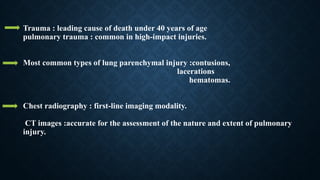
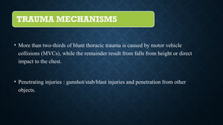
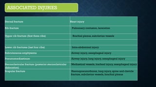
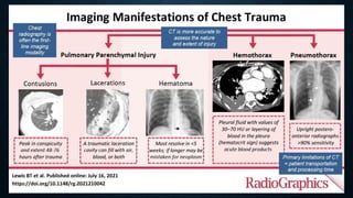
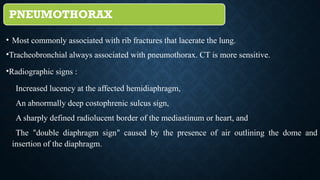
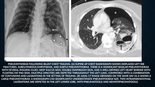
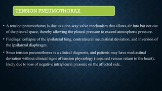
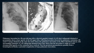
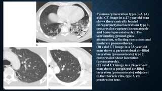
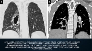
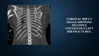
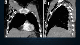
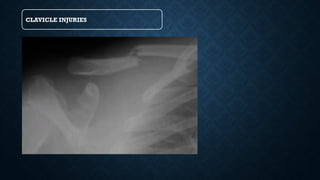
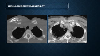
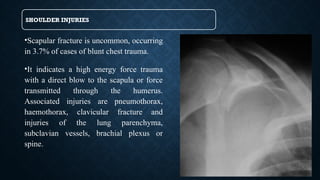
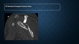
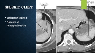
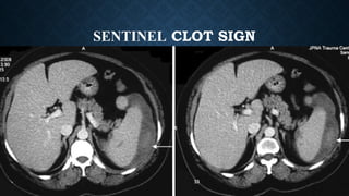
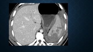
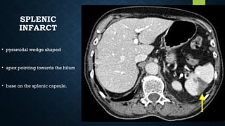
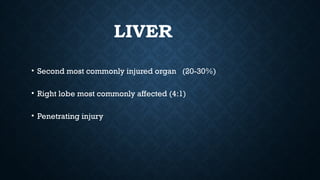
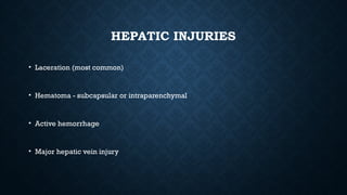
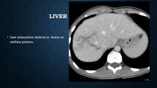
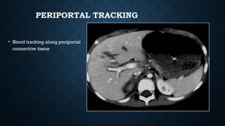
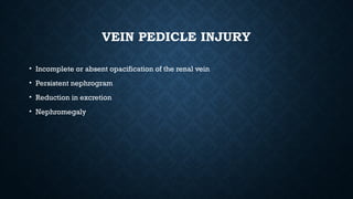
CT SCAN IN EVALATUATION OF THORACIC
AND ABDOMINA TRAUMA
• MODERATORS: DR. L. NATH
(PROFESSOR & HOD) DR. P. BORAH
DR. D. J. BORPATROGOHAIN
DR. P N TAYE
DR. S. S. BISWAS
• PRESENTER : DR.VENKATESH
Trauma : leading cause of death under 40 years of age
pulmonary trauma : common in high-impact injuries.
Most common types of lung parenchymal injury :contusions,
lacerations
hematomas.
Chest radiography : first-line imaging modality.
CT images :accurate for the assessment of the nature and extent of pulmonary
injury.
TRAUMA MECHANISMS
• More than two-thirds of blunt thoracic trauma is caused by motor vehicle
collisions (MVCs), while the remainder result from falls from height or direct
impact to the chest.
• Penetrating injuries : gunshot/stab/blast injuries and penetration from other
objects.
ASSOCIATED INJURIES
Sternal fracture Heart injury
Rib fracture Pulmonary contusion, laceration
Upper rib fracture (first three ribs) Brachial plexus, subclavian vessels
Lower rib fractures (last four ribs) Intra-abdominal injury
Subcutaneous emphysema Airway injury, oesophageal injury
Pneumomediastinum Airway injury, lung injury, oesophageal injury
Sternoclavicular fracture (posterior sternoclavicular
dislocation)
Mediastinal vessels, tracheal injury, oesophageal injury
Scapular fracture Haemopneumothorax, lung injury, spine and clavicle
fracture, subclavian vessels, brachial plexus
IMAGING TECHNIQUES FOR PULMONARY
TRAUMA
 The chest radiograph is the primary initial screening examination performed in thoracic
trauma, although some centres also perform extended focused assessment in sonography
for trauma (eFAST).
 Chest radiography :1]Identify rib fractures
2] Foreign bodies/ballistic fragments
3] Contusions,
4] Pneumothorax
5] Hemothorax &mediastinal injuries
 Contrast-enhanced CT :standard imaging tool in the evaluation of trauma patients.
(Greater sensitivity and specificity)
PNEUMOTHORAX
• Most commonly associated with rib fractures that lacerate the lung.
•Tracheobronchial always associated with pneumothorax. CT is more sensitive.
•Radiographic signs :
(1)Increased lucency at the affected hemidiaphragm,
(2)An abnormally deep costophrenic sulcus sign,
(3)A sharply defined radiolucent border of the mediastinum or heart, and
(4)The “double diaphragm sign” caused by the presence of air outlining the dome and
insertion of the diaphragm.
PNEUMOTHORAX FOLLOWING BLUNT CHEST TRAUMA. (A) SUPINE AP CHEST RADIOGRAPH SHOWS DISPLACED LEFT RIB
FRACTURES, SUBCUTANEOUS EMPHYSEMA, AND SUBTLE PNEUMOTHORAX. THERE IS A SIGNIFICANT BASILAR PNEUMOTHORAX
WITH SEVERAL IMAGING CLUES: DEEP SULCUS SIGN, DOUBLE DIAPHRAGM SIGN, AND A WELL-DEFINED LEFT HEART BORDER WITH
FLOATING FAT PAD SIGN. MULTIPLE OPACITIES ARE DEPICTED THROUGHOUT THE LEFT LUNG, COMPATIBLE WITH A COMBINATION
OF CONTUSIONS AND LACERATIONS IN THE SETTING OF TRAUMA. (B) AXIAL CT IMAGE OBTAINED ON THE SAME DAY AS A SHOWS A
LARGE PNEUMOTHORAX; A RADIOGRAPH CAN SIGNIFICANTLY UNDERESTIMATE PNEUMOTHORAX SIZE. TWO INTRAPARENCHYMAL
LACERATIONS ARE DEPICTED IN THE LEFT LOWER LOBE, WITH PNEUMATOCELE AND HEMATOPNEUMATOCELE.
TENSION PNEUMOTHORAX
• A tension pneumothorax is due to a one-way valve mechanism that allows air into but not out
of the pleural space, thereby allowing the pleural pressure to exceed atmospheric pressure.
• Findings: collapse of the ipsilateral lung, contralateral mediastinal deviation, and inversion of
the ipsilateral diaphragm.
• Since tension pneumothorax is a clinical diagnosis, and patients may have mediastinal
deviation without clinical signs of tension physiology (impaired venous return to the heart),
likely due to loss of negative intrapleural pressure on the affected side.
TENSION PNEUMOTHORAX
• Erect AP/PA view is best
• Shift of mediastinum /heart/trachea away from pneumothorax side
• Depressed hemidiaphragm
• Degree of lung collapse is variable
Posteroanterior chest radiograph in a 29-
year-old man with shortness of breath shows
findings of tension pneumothorax, including
collapse of the ipsilateral lung, contralateral
mediastinal deviation, and inversion of the
ipsilateral diaphragm.
Tension haemopneumothorax. Axial contrast-
enhanced CT at mediastinal window shows a
right tension haemopneumothorax with
heterogeneous increased density due to
presence of blood clots and a significant shift
of the mediastinum contralaterally.
Tension pneumothorax. Sagittal reformatted CT image
at lung window showing tension pneumothorax with
significantly collapsed lung at the posterior part of the
hemithorax associated with ipsilateral pleural effusion.
HEMOTHORAX
• Haemothorax: In 50% of chest trauma cases. Blood pooling into the pleural space from
variable sources: the lung parenchyma, the chest wall, the great vessels, the heart or even
the liver and spleen through diaphragmatic rupture.
• Arterial bleeding more common.
• CT: very sensitive in detecting even a small haemothorax.
TRACHEA/BRONCHI INJURY
• Tracheobronchial injuries are rare, occurring in 0.2–8% of all cases of chest trauma.
• Bronchial injuries: more commonly than tracheal, usually on the right side and within 2.5 cm
from the carina.
• 85% of tracheal lacerations occur 2 cm above the carina.
• A direct CT finding of tracheobronchial injuries is the cutoff of the tracheal and bronchial wall
with extraluminal air surrounding the airway. Indirect findings are the “fallen lung” sign,
corresponding to the collapsed lung resting away from the hilum towards the dependent portion
of the hemithorax.
Bronchial transection. A 22-year-old
man involved in a car accident.
Volume-rendered image of the
tracheobronchial tree showing
complete transection of the right
intermediate bronchus.
TRACHEAL INJURY IN A 32-YEAR-OLD MAN AFTER A THORACIC GUNSHOT WOUND. (A) AP RADIOGRAPH
SHOWS ENTRY AND EXIT SITES. THE AP BULLET TRAJECTORY WOULD BE EXPECTED TO INVOLVE MIDLINE
STRUCTURES SUCH AS THE TRACHEA. PARAMEDIASTINAL HAZINESS IS DEPICTED, REFLECTING PULMONARY
CONTUSIONS. (B) AXIAL CONTRAST-ENHANCED CHEST CT IMAGE SHOWS EXTENSIVE DIFFUSE
PNEUMOMEDIASTINUM. NOTE THE SMALL ANTERIOR TRACHEAL WALL DEFECT. TRACHEAL INJURY FROM A
PENETRATING WOUND WOULD LIKELY BE ASSOCIATED WITH INJURY TO OTHER ADJACENT MEDIASTINAL
STRUCTURES AS WELL. (C) PARASAGITTAL CONTRAST-ENHANCED REFORMATTED CT IMAGE SHOWS
EXTENSIVE ANTERIOR AND POSTERIOR PNEUMOMEDIASTINUM FROM THE VISUALIZED NECK TO THE ROOT
PULMONARY CONTUSION AND
LACERATION
• Contusion: Blood in intact lung parenchyma
• Laceration: Blood in torn lung parenchyma
• Chest film cannot differentiate them.
Contusions peak in 2-3 days, begin to resolve in a week; lacerations
take much longer to resolve and may leave scars.
AAST
Organ
Injury
Scale for
Lung
Injury
Grade Injury Type Description of Injury
I Contusion Unilateral contusion, segmental or subsegmental involvement
II Contusion Unilateral contusion, lobar involvement
Laceration Peripheral laceration, with or without simple pneumothorax
III Contusion Unilateral contusion with more than single lobe involvement or bilateral
contusions
Laceration Persistent laceration (>72 hours); injury to distal airways, with or without air
leak
Hematoma Intraparenchymal hematoma (nonexpanding)
IV Laceration Major laceration (segmental or lobar) with imaging findings suggestive of air
leak
Hematoma Expanding intraparenchymal hematoma (expanding over consecutive imaging
examinations or with active contrast material extravasation)
Vascular injury Primary branch intrapulmonary vessel disruption
V Vascular injury Hilar vascular injury (contained, without active contrast material extravasation)
VI Vascular injury Transection of pulmonary hilum or hilar vessel injury with uncontained bleeding
PULMONARY
CONTUSION
• Lung contusion is a focal parenchymal injury caused by disruption of the capillaries of the
alveolar walls and septa, and leakage of blood into the alveolar spaces and interstitium.
• It is the most common type of lung injury in blunt chest trauma.
• Main mechanism: compression and tearing of the lung parenchyma at the site of impact
against osseous structures, rib fractures or pre-existing pleural adhesions.
• Lung contusion occurs at the time of injury, but it may be undetectable on chest
radiography for the first 6 h after trauma.
• The pooling of haemorrhage and oedema will occur at 24 h, rendering the contusion
radiographically more evident, although CT may readily reveal it from the initial imaging.
• The appearance of consolidation on chest radiography after the first 24 h should
raise suspicion of other pathological conditions such as aspiration, pneumonia and
fat embolism.
• Contusions appear as geographic, non-segmental areas of ground-glass or nodular
opacities or consolidation on CT that do not respect the lobar boundaries and may
manifest air bronchograms if the bronchioles are not filled with blood.
• Subpleural sparing of 1–2 mm may be seen, especially in children. Clearance of an
uncomplicated contusion begins at 24 to 48 h with complete resolution after 3 to 14
days.
• Lack of resolution within the expected time frame should raise the suspicion of
complications such as pneumonia, abscess or ARDS.
Lung contusion. Axial (a, b) and coronal
(c) CT images at lung window show
nodular opacities of ground-glass
opacity that do not respect the lung
boundaries of the right upper lobe. (A),
diffuse areas of ground-glass opacity in
the upper lobes bilaterally with
subpleural sparing (b) and multiple
areas of consolidation with air
bronchograms and small lacerations in
both lungs consistent with lung
contusions.
PULMONARY LACERATIONS
• Pulmonary laceration occurs in major chest trauma when disruption and tearing of the lung parenchyma follows
shearing forces, caused by direct impact, compression or inertial deceleration.
• Classified into the following four types according to the mechanism of injury:
• type I - compression rupture: most common type of laceration that usually occurs as a 2-8 cm lesion in the
central lung.
• type II - compression shear: occurs after sudden compression of the lower chest when the lung suffers from a
shear injury to the spine; the lung is compressed by lateral compression, against the spine leading to a
paravertebral tubular lesion in lower part of the lung.
• type III - direct puncture / rib penetration: occur with a penetrating fractured rib; these lesions are commonly
multiple.
• type IV - adhesion tears: occurs in sudden injuries of the chest wall where prior pleuropulmonary adhesions
have been created.
Pulmonary laceration in a 38-year-old man after a thoracic gunshot wound. (A) AP chest radiograph obtained at
presentation shows a hazy opacity in the left upper lobe, reflecting laceration obscured by pulmonary contusions. (B)
axial CT image obtained on the same day better shows the pulmonary laceration cavity filled with blood and air, with
surrounding contusion. (C) AP radiograph obtained 8 days later shows that the laceration is visible as a well-
circumscribed opacity, as the contusions have resolved. Note the persistent pneumothorax, possibly from a
bronchopleural fistula, a potential complication of large pulmonary lacerations.
Pulmonary laceration types 1–3. (A)
axial CT image in a 27-year-old man
shows three centrally located
intraparenchymal lacerations type 1,
compression rupture (pneumatocele
and hematopneumatocele). The
surrounding ground-glass
attenuation, reflecting contusions and
moderate pneumothorax.
(B) axial CT image in a 33-year-old
man shows a paravertebral air-filled
laceration (pneumatocele) type 2,
compression shear laceration
(pneumatocele).
(C) axial CT image in a 24-year-old
man shows a peripheral air-filled
laceration (pneumatocele) subjacent
to the thoracic ribs, type 3, rib
penetration tear.
LUNG LACERATION, TYPE II. CORONAL REFORMATTED CT IMAGE AT LUNG WINDOW (A) SHOWS
A LOBULATED PARASPINAL PNEUMATOCELE SURROUNDED BY GROUND-GLASS OPACITY
(CONTUSION) IN THE RIGHT LUNG CONSISTENT WITH LUNG LACERATION (TYPE II?). ON
MEDIASTINAL WINDOW LUNG LACERATION IS SEEN TO HAVE BEEN COMPLICATED BY ACUTE
PULMONARY EMBOLISM.
LUNG LACERATION, TYPE IV.
AXIAL CT IMAGE OF THE LEFT
LUNG AT LUNG WINDOW
SHOWS A SMALL PERIPHERAL
LACERATION BENEATH A RIB
FRACTURE SURROUNDED BY
GROUND-GLASS OPACITY
(LUNG CONTUSION) AND
ASSOCIATED WITH A SMALL
IPSILATERAL
PNEUMOTHORAX.
PNEUMOMEDIASTINUM
• Usually from ruptured alveoli
• Can also be from trachea, bronchi,
esophagus, bowel and neck injuries
SIGNS
• Linear paratracheal lucencies
• Air along heart border
• “V” sign at aortic-diaphragm
junction
• Continuous diaphragm sign
CHEST XRAY FOLLOWING TRAUMA SHOWING AIR OUTLINING
MEDIASTINAL STRUCTURES AND SUBCUTANEOUS EMPHYSEMA
V SIGN OF
NECLERIO
PNEUMOMEDIASTINUM: CT
PNEUMOMEDIASTINUMWITH SUBCUTANEOUS EMPHYSEMA
Pneumopericardium
HEMOPERICARDIUM
• Hemopericardium refers to the presence of blood within the pericardial cavity. Can occur
from blunt/penetrating/deceleration trauma.
• Plain radiograph
• enlargement of the cardiac silhouette may be present but chest x-rays are insensitive and
non-specific.
• the "straight left heart border" is an infrequent sign with low sensitivity (~40%) for
hemopericardium in penetrating trauma patients.
• ​
the Oreo cookie sign on lateral CXR.
HEMOPERICARDIUM: CT
RIBS FRACTURE
• Rib fractures are the most common injury in blunt chest trauma. A single rib fracture is
usually not clinically significant, whereas multiple rib fractures indicate severe injury.
• Fractures of the first three ribs imply high-energy trauma that may be associated with
injury of the brachial plexus or subclavian vessels.
• Fractures of the fourth up to the eighth ribs are the most common, while fractures of the
last four ribs are usually associated with intra-abdominal injury.
• Flail chest: An injury that occurs typically
following a blunt trauma to the chest.
When three or more ribs in a row have
multiple fractures [atleast two] within each
rib, it can cause a part of your chest wall to
become separated and out of sync from the
rest of your chest wall.
• The diagnosis is clinical based on the
paradoxical motion during respiration,
which may result in ventilatory
compromise. More than 50% of cases
require surgical treatment and prolonged
mechanical ventilation.
CORONAL MIP CT
IMAGE SHOWING
MULTIPLE
CONTIGUOUS LEFT
RIB FRACTURES.
CORONAL (A) AND SAGITTAL (B) RECONSTRUCTED CT IMAGES SHOW
FRACTURES OF THREE CONTIGUOUS RIGHT RIBS (ARROWS) THAT WERE
ASSOCIATED WITH PARADOX MOTION OF THE CHEST DURING RESPIRATION.
SPINE INJURY
• Thoracic spine fractures account for up to 30% of all spine fractures.
• 62% of spine fractures will result in neurological deficits. The most vulnerable site is
between the ninth and twelfth vertebra.
• The main mechanism is hyperflexion and axial loading.
• Sagittal and coronal MDCT reformats readily reveal even small spinal fractures.
• MDCT of the spine is highly indicated for spinal survey for possible fractures and
determine the type of fracture. However, in the case of suspected compressive myelopathy,
MRI is the method of choice.
Thoracic spine fracture.
Coronal (a) and sagittal (b) CT
reconstructed images of two different
patients show fractures of the upper
thoracic vertebrae.
STERNUM FRACTURE
• Sternal fractures: 3–8% in blunt chest trauma.
• Main mechanism: deceleration injury/direct blow to the anterior chest wall.
• Difficult to detect on lateral chest radiographs .
• Almost always accompanied by anterior mediastinal haemorrhage, which has a preserved fat
plane with the aorta, as opposed to an anterior mediastinal haemorrhage secondary to aortic
injury, which will present with a lost fat plane with the aorta.
STERNAL FRACTURE. SAGITTAL
RECONSTRUCTED CT IMAGE SHOWS
MULTIPLE FRACTURES OF THE
MANUBRIUM AND THE BODY OF THE
STERNUM ACCOMPANIED BY
EXTENSIVE RETROSTERNAL
HAEMATOMA.
STERNAL FRACTURE. AXIAL CT IMAGE AT MEDIASTINAL WINDOW SHOWS STERNAL
FRACTURE ASSOCIATED WITH RETROSTERNAL HAEMATOMA. NOTE THE PRESERVED
FAT PLANE WITH THE AORTA, EXCLUDING THE PRESENCE OF AORTIC INJURY.
CLAVICLE INJURIES
STERNO-CLAVICLE DISLOCATION: CT
SHOULDER INJURIES
•Scapular fracture is uncommon, occurring
in 3.7% of cases of blunt chest trauma.
•It indicates a high energy force trauma
with a direct blow to the scapula or force
transmitted through the humerus.
Associated injuries are pneumothorax,
haemothorax, clavicular fracture and
injuries of the lung parenchyma,
subclavian vessels, brachial plexus or
spine.
CT Needed if Scapula Fracture Seen
DIAPHRAGM INJURIES
• 5% of major blunt trauma.
• Left clinically injured more than right 60/40.
• CT is a sensitive modality.
• Signs of diaphragmatic injury:
• The collar sign (or hourglass sign) : a waist-like constriction of the herniating hollow viscus
from the abdomen into the chest at the site of the diaphragmatic tear, which is classical for
diaphragmatic rupture.
• The dependent viscera sign: when a patient with a ruptured diaphragm lies supine at ct
examination, the herniated viscera (bowel or solid organs) are no longer supported
posteriorly by the injured diaphragm and fall to a dependent position against the posterior
ribs.
• Segmental non-recognition of the diaphragm.
• Focal diaphragmatic thickening.
POSITION OF NG TUBE
VASCULAR INJURY
• Thoracic aortic injury can result from either blunt or penetrating trauma:
1.blunt trauma (more common)
1.rapid deceleration (eg. motor vehicle accident, fall from great height)
2.crush injury
2.penetrating trauma
1.stab wound
2.gunshot wound
THORACIC AORTIC INJURY
• Grading
• Thoracic aortic injury can be graded according to the severity of
injury. One grading system is
• grade 1: intimal tear
• grade 2: intramural hematoma
• grade 3: pseudoaneurysm formation
• grade 4: free rupture
VASCULAR INJURY: SIGNS
• Signs of mediastinal hematoma
Widened mediastinum
Indistinct or abnormal aortic contour
Deviation of trachea or NGT to the right
Depression of left main bronchus
Widened paraspinal stripe
X RAY: VASCULAR
INJURY
CT
Indirect signs of aortic injury:
Mediastinal hematoma
Periaortic fat stranding
CT Angiography:
100% sensitivity and specificity
Signs of mediastinal hematoma
Abnormal soft tissue density around mediastinal structures
Location – periaortic hematoma than isolated mediastinal hematoma remote from the
aorta.
SIGNS OF AORTIC
INJURY:
Intraluminal filling defect (intimal
flap or clot)
Abnormal aortic contour (mural
hematoma)
Pseudoaneurysm & extravasation
of contrast
TRAUMATIC ESOPHAGEAL RUPTURE
• Can occur secondary to both blunt or
penetrating trauma.
• CT is the preferred modality.
• Signs on CT:
• extraluminal gas locules in the
mediastinum or abdominal cavity,
adjacent to the esophagus: highly
suggestive.
• pleural or mediastinal fluid.
• pneumomediastinum or pneumothorax.
• pericardial or pleural effusions can be
seen.
TRAUMATIC LUNG HERNIATION
• Herniation of the lung beyond the confines of the thoracic cage.
• Usually associated with rib fracture.
• Results in subcutaneous emphysema.
SUBCUTANEOUS EMPHYSEMA
• Causes:
• Usually from ruptured
alveoli
• Can also be from
trachea, bronchi,
esophagus, bowel and
neck injuries
IMAGING OF ABDOMINAL
TRAUMA
ABDOMINAL TRAUMA
• Trauma causes I0% of deaths worldwide
• Blunt trauma is more common
ROLE OF CT SCAN
• More accurate for solid visceral assessment
• Quantification of hemorrhage
• Assessment of retroperitoneal injuries
• Most sensitive and imaging modality o choice for evaluation of
abdominal trauma
SPLEEN
• Most common injured organ in blunt trauma (40% of all solid organ injuries)
• CECT is the investigation of choice
Integrity of organs
Extent of injury / Localization of parenchymal contusions, hematoma
SPLENIC HEMATOMA
• Parenchymal
• Subcapsular
SUBCAPSULAR
HEMATOMA
• Indents splenic
parenchyma
DELAYED SPLENIC RUPTURE
• Bleeding due to splenic injury occurring more than 48 h after blunt
trauma
• Due to ruptures of subcapsular splenic hematomas.
SPLENIC LACERATION
• Linear low attenuation
defects between high
attenuation vascular
spleen.
SPLENIC CLEFT
• Superiorly located
• Absence of
hemoperitoneum
SENTINEL CLOT SIGN
SPLENIC
INFARCT
• pyramidal wedge shaped
• apex pointing towards the hilum
• base on the splenic capsule.
IMAGING CONSIDERATIONS
• Images should be take 50-60 seconds after contrast for uniform
enhancement of the spleen
CONTRAST BLUSH
• The differential diagnosis is:
• Active arterial extravasation
• Post-traumatic pseudo-aneurysm
• Post-traumatic AV fistula
AMERICAN ASSOCIATION FOR THE
SURGERY OF TRAUMA (AAST) GRADE
I
• Subcapsular hematoma <10%
• capsular laceration <1 cm depth
GRADE II
• Subcapsular hematoma 10-50%
• Parenchymal hematoma <5 cm
• Laceration 1-3 cm depth
GRADE III
• Subcapsular hematoma >50%
• Intraparenchymal hematoma >5 cm
• Laceration >3 cm
GRADE IV
• laceration hilar vessels
• major devascularisation
(>25% of spleen)
GRADE V
• Shattered spleen
• Hilar vascular injury with splenic
devascularisation
• Axial CECT scan shows
nonenhancing spleen
LIVER
• Second most commonly injured organ (20-30%)
• Right lobe most commonly affected (4:1)
• Penetrating injury
HEPATIC INJURIES
• Laceration (most common)
• Hematoma - subcapsular or intraparenchymal
• Active hemorrhage
• Major hepatic vein injury
LIVER LACERATION
• Low attenuation defects in linear or
stellate pattern.
PERIPORTAL TRACKING
• Blood tracking along periportal
connective tissue
Grade I
•Subcapsular hematoma : <10%
•Laceration: capsular tear <1 cm
•Subcapsular hematoma: 10-50%
•Intraparenchymal hematoma <10 cm
•Laceration: 1-3 cm in parenchymal depth
Grade II
•Subcapsular hematoma: >50%
•Intraparenchymal hematoma >10 cm
•Laceration: capsular tear >3 cm
parenchymal depth
Grade III
GRADE IV
•Parenchymal disruption
involving 25-75% hepatic lobe
•Active bleeding
GRADE V
•Laceration: parenchymal
disruption involving >75% of
hepatic lobe or involves >3
Couinaud’s segments (one lobe)
PANCREAS
• Least commonly injured solid organ (3-12 %).
• Seatbelt injuries : Compression against the vertebral column.
• Rarely an isolated injury
CLINICAL AND BIOCHEMICAL FINDINGS
• Epigastric pain
• Elevated serum amylase in 60 %
• ( 95% Negative Predictive Value)
CT FINDINGS
• 40 % of pancreatic injuries may not
be visible on CT in first 12 hrs
• laceration/transection involving neck/proximal body of pancreas
• Grade I- Minor contusion or laceration
without duct injury
• Grade II- Major contusion or
laceration without duct injury
GRADE III
Distal pancreatic transection
with duct injury
• Grade IV :Proximal transection
• Grade V : Massive disruption of pancreatic head
FINDINGS THAT MAY SUGGEST
PANCREATIC TRAUMA
• Localized edema
• Retroperitoneal hematoma / fat infiltration.
• Fluid in lesser sac
• Thickening of anterior pararenal fascia
BOWEL AND MESENTERY INJURY
• Bowel and mesenteric injuries in 5% of blunt trauma
• MDCT is more sensitive than Clinical exam, USG, DPL
DIRECT FINDINGS –PNEUMOPERITONEUM OR
PNEUMO-RETROPERITONEUM
DIRECT FINDINGS
• Contrast extravastation
• Interloop fluid
• Bowel wall discontinuity
LESS COMMON
SIGNS
• Focal bowel wall
thickening greater than 3
mm
• Mesenteric infiltration,
thickening
• Abnormal bowel wall
enhancement
SHOCK BOWEL
• Hypoperfusion state
MESENTERIC
INJURY
Signs
• Contrast extravasation
• Beaded mesenteric
vessels
• Hematoma
• Infiltration
VASCULAR INJURY – CT FINDINGS
• IVC injuries lumen is irregular
and compressed by hematoma
• Active contrast extravasation.
AORTIC INJURIES
• Contrast extravasation
• Large psoas or mesenteric hemorrhage, pseudo-aneurysm.
Pelvic vasculature injuries are associated with pelvic fractures.
IMAGING OF RENAL TRAUMA
• Computed tomography (CT) is the modality of choice
• Injury to the kidney is seen in approximately 8%– 10% of patients with blunt or
penetrating abdominal injuries
SUBCAPSULAR HEMATOMA (CATEGORY I)
Crescent shaped hyperdensity, located
in the periphery of the kidney
LACERATION
• Hypodense, irregularly linear areas, typically distributed along the vessels and filled
with blood.
• They are best analyzed at arterial phase
• Superficial (<1 cm from the renal cortex)
• Deep (>1 cm from the renal cortex)
SIMPLE RENAL LACERATION (CATEGORY I)
MAJOR RENAL LACERATION WITHOUT INVOLVEMENTOF
THE COLLECTING SYSTEM (CATEGORY II <1 CM)
MAJOR RENAL LACERATION INVOLVING
THE COLLECTING SYSTEM (CATEGORY II)
MULTIPLE RENAL LACERATIONS
(CATEGORY III >1 CM)
SHATTERED KIDNEY (CATEGORY III)
SEGMENTAL RENAL INFARCTION
(CATEGORY II)
TRAUMATIC
OCCLUSION OF THE
MAIN RENAL ARTERY
(CATEGORY III)
ACTIVE ARTERIAL EXTRAVASATION
(CATEGORY III)
VEIN PEDICLE INJURY
• Incomplete or absent opacification of the renal vein
• Persistent nephrogram
• Reduction in excretion
• Nephromegaly
LACERATION OF THE RENAL VEIN (CATEGORY III)
URINOMA/UROHEMATOMA
• Presence of a more or less significant breach of the collecting tube
system, with urine escape reflected by extravasation of contrast medium
on delayed imaging, in an extrarenal location
AVULSION OF THE URETEROPELVIC
JUNCTION (CATEGORY IV)
thoracic and abdominal trauma  modified.pptx

thoracic and abdominal trauma modified.pptx

  • 1.
    CT SCAN INEVALATUATION OF THORACIC AND ABDOMINA TRAUMA • MODERATORS: DR. L. NATH (PROFESSOR & HOD) DR. P. BORAH DR. D. J. BORPATROGOHAIN DR. P N TAYE DR. S. S. BISWAS • PRESENTER : DR.VENKATESH
  • 2.
    Trauma : leadingcause of death under 40 years of age pulmonary trauma : common in high-impact injuries. Most common types of lung parenchymal injury :contusions, lacerations hematomas. Chest radiography : first-line imaging modality. CT images :accurate for the assessment of the nature and extent of pulmonary injury.
  • 3.
    TRAUMA MECHANISMS • Morethan two-thirds of blunt thoracic trauma is caused by motor vehicle collisions (MVCs), while the remainder result from falls from height or direct impact to the chest. • Penetrating injuries : gunshot/stab/blast injuries and penetration from other objects.
  • 4.
    ASSOCIATED INJURIES Sternal fractureHeart injury Rib fracture Pulmonary contusion, laceration Upper rib fracture (first three ribs) Brachial plexus, subclavian vessels Lower rib fractures (last four ribs) Intra-abdominal injury Subcutaneous emphysema Airway injury, oesophageal injury Pneumomediastinum Airway injury, lung injury, oesophageal injury Sternoclavicular fracture (posterior sternoclavicular dislocation) Mediastinal vessels, tracheal injury, oesophageal injury Scapular fracture Haemopneumothorax, lung injury, spine and clavicle fracture, subclavian vessels, brachial plexus
  • 5.
    IMAGING TECHNIQUES FORPULMONARY TRAUMA  The chest radiograph is the primary initial screening examination performed in thoracic trauma, although some centres also perform extended focused assessment in sonography for trauma (eFAST).  Chest radiography :1]Identify rib fractures 2] Foreign bodies/ballistic fragments 3] Contusions, 4] Pneumothorax 5] Hemothorax &mediastinal injuries  Contrast-enhanced CT :standard imaging tool in the evaluation of trauma patients. (Greater sensitivity and specificity)
  • 7.
    PNEUMOTHORAX • Most commonlyassociated with rib fractures that lacerate the lung. •Tracheobronchial always associated with pneumothorax. CT is more sensitive. •Radiographic signs : (1)Increased lucency at the affected hemidiaphragm, (2)An abnormally deep costophrenic sulcus sign, (3)A sharply defined radiolucent border of the mediastinum or heart, and (4)The “double diaphragm sign” caused by the presence of air outlining the dome and insertion of the diaphragm.
  • 8.
    PNEUMOTHORAX FOLLOWING BLUNTCHEST TRAUMA. (A) SUPINE AP CHEST RADIOGRAPH SHOWS DISPLACED LEFT RIB FRACTURES, SUBCUTANEOUS EMPHYSEMA, AND SUBTLE PNEUMOTHORAX. THERE IS A SIGNIFICANT BASILAR PNEUMOTHORAX WITH SEVERAL IMAGING CLUES: DEEP SULCUS SIGN, DOUBLE DIAPHRAGM SIGN, AND A WELL-DEFINED LEFT HEART BORDER WITH FLOATING FAT PAD SIGN. MULTIPLE OPACITIES ARE DEPICTED THROUGHOUT THE LEFT LUNG, COMPATIBLE WITH A COMBINATION OF CONTUSIONS AND LACERATIONS IN THE SETTING OF TRAUMA. (B) AXIAL CT IMAGE OBTAINED ON THE SAME DAY AS A SHOWS A LARGE PNEUMOTHORAX; A RADIOGRAPH CAN SIGNIFICANTLY UNDERESTIMATE PNEUMOTHORAX SIZE. TWO INTRAPARENCHYMAL LACERATIONS ARE DEPICTED IN THE LEFT LOWER LOBE, WITH PNEUMATOCELE AND HEMATOPNEUMATOCELE.
  • 9.
    TENSION PNEUMOTHORAX • Atension pneumothorax is due to a one-way valve mechanism that allows air into but not out of the pleural space, thereby allowing the pleural pressure to exceed atmospheric pressure. • Findings: collapse of the ipsilateral lung, contralateral mediastinal deviation, and inversion of the ipsilateral diaphragm. • Since tension pneumothorax is a clinical diagnosis, and patients may have mediastinal deviation without clinical signs of tension physiology (impaired venous return to the heart), likely due to loss of negative intrapleural pressure on the affected side.
  • 10.
    TENSION PNEUMOTHORAX • ErectAP/PA view is best • Shift of mediastinum /heart/trachea away from pneumothorax side • Depressed hemidiaphragm • Degree of lung collapse is variable
  • 11.
    Posteroanterior chest radiographin a 29- year-old man with shortness of breath shows findings of tension pneumothorax, including collapse of the ipsilateral lung, contralateral mediastinal deviation, and inversion of the ipsilateral diaphragm.
  • 12.
    Tension haemopneumothorax. Axialcontrast- enhanced CT at mediastinal window shows a right tension haemopneumothorax with heterogeneous increased density due to presence of blood clots and a significant shift of the mediastinum contralaterally.
  • 14.
    Tension pneumothorax. Sagittalreformatted CT image at lung window showing tension pneumothorax with significantly collapsed lung at the posterior part of the hemithorax associated with ipsilateral pleural effusion.
  • 15.
    HEMOTHORAX • Haemothorax: In50% of chest trauma cases. Blood pooling into the pleural space from variable sources: the lung parenchyma, the chest wall, the great vessels, the heart or even the liver and spleen through diaphragmatic rupture. • Arterial bleeding more common. • CT: very sensitive in detecting even a small haemothorax.
  • 17.
    TRACHEA/BRONCHI INJURY • Tracheobronchialinjuries are rare, occurring in 0.2–8% of all cases of chest trauma. • Bronchial injuries: more commonly than tracheal, usually on the right side and within 2.5 cm from the carina. • 85% of tracheal lacerations occur 2 cm above the carina. • A direct CT finding of tracheobronchial injuries is the cutoff of the tracheal and bronchial wall with extraluminal air surrounding the airway. Indirect findings are the “fallen lung” sign, corresponding to the collapsed lung resting away from the hilum towards the dependent portion of the hemithorax.
  • 18.
    Bronchial transection. A22-year-old man involved in a car accident. Volume-rendered image of the tracheobronchial tree showing complete transection of the right intermediate bronchus.
  • 19.
    TRACHEAL INJURY INA 32-YEAR-OLD MAN AFTER A THORACIC GUNSHOT WOUND. (A) AP RADIOGRAPH SHOWS ENTRY AND EXIT SITES. THE AP BULLET TRAJECTORY WOULD BE EXPECTED TO INVOLVE MIDLINE STRUCTURES SUCH AS THE TRACHEA. PARAMEDIASTINAL HAZINESS IS DEPICTED, REFLECTING PULMONARY CONTUSIONS. (B) AXIAL CONTRAST-ENHANCED CHEST CT IMAGE SHOWS EXTENSIVE DIFFUSE PNEUMOMEDIASTINUM. NOTE THE SMALL ANTERIOR TRACHEAL WALL DEFECT. TRACHEAL INJURY FROM A PENETRATING WOUND WOULD LIKELY BE ASSOCIATED WITH INJURY TO OTHER ADJACENT MEDIASTINAL STRUCTURES AS WELL. (C) PARASAGITTAL CONTRAST-ENHANCED REFORMATTED CT IMAGE SHOWS EXTENSIVE ANTERIOR AND POSTERIOR PNEUMOMEDIASTINUM FROM THE VISUALIZED NECK TO THE ROOT
  • 21.
    PULMONARY CONTUSION AND LACERATION •Contusion: Blood in intact lung parenchyma • Laceration: Blood in torn lung parenchyma • Chest film cannot differentiate them. Contusions peak in 2-3 days, begin to resolve in a week; lacerations take much longer to resolve and may leave scars.
  • 22.
    AAST Organ Injury Scale for Lung Injury Grade InjuryType Description of Injury I Contusion Unilateral contusion, segmental or subsegmental involvement II Contusion Unilateral contusion, lobar involvement Laceration Peripheral laceration, with or without simple pneumothorax III Contusion Unilateral contusion with more than single lobe involvement or bilateral contusions Laceration Persistent laceration (>72 hours); injury to distal airways, with or without air leak Hematoma Intraparenchymal hematoma (nonexpanding) IV Laceration Major laceration (segmental or lobar) with imaging findings suggestive of air leak Hematoma Expanding intraparenchymal hematoma (expanding over consecutive imaging examinations or with active contrast material extravasation) Vascular injury Primary branch intrapulmonary vessel disruption V Vascular injury Hilar vascular injury (contained, without active contrast material extravasation) VI Vascular injury Transection of pulmonary hilum or hilar vessel injury with uncontained bleeding
  • 23.
    PULMONARY CONTUSION • Lung contusionis a focal parenchymal injury caused by disruption of the capillaries of the alveolar walls and septa, and leakage of blood into the alveolar spaces and interstitium. • It is the most common type of lung injury in blunt chest trauma. • Main mechanism: compression and tearing of the lung parenchyma at the site of impact against osseous structures, rib fractures or pre-existing pleural adhesions. • Lung contusion occurs at the time of injury, but it may be undetectable on chest radiography for the first 6 h after trauma. • The pooling of haemorrhage and oedema will occur at 24 h, rendering the contusion radiographically more evident, although CT may readily reveal it from the initial imaging.
  • 24.
    • The appearanceof consolidation on chest radiography after the first 24 h should raise suspicion of other pathological conditions such as aspiration, pneumonia and fat embolism. • Contusions appear as geographic, non-segmental areas of ground-glass or nodular opacities or consolidation on CT that do not respect the lobar boundaries and may manifest air bronchograms if the bronchioles are not filled with blood. • Subpleural sparing of 1–2 mm may be seen, especially in children. Clearance of an uncomplicated contusion begins at 24 to 48 h with complete resolution after 3 to 14 days. • Lack of resolution within the expected time frame should raise the suspicion of complications such as pneumonia, abscess or ARDS.
  • 25.
    Lung contusion. Axial(a, b) and coronal (c) CT images at lung window show nodular opacities of ground-glass opacity that do not respect the lung boundaries of the right upper lobe. (A), diffuse areas of ground-glass opacity in the upper lobes bilaterally with subpleural sparing (b) and multiple areas of consolidation with air bronchograms and small lacerations in both lungs consistent with lung contusions.
  • 26.
    PULMONARY LACERATIONS • Pulmonarylaceration occurs in major chest trauma when disruption and tearing of the lung parenchyma follows shearing forces, caused by direct impact, compression or inertial deceleration. • Classified into the following four types according to the mechanism of injury: • type I - compression rupture: most common type of laceration that usually occurs as a 2-8 cm lesion in the central lung. • type II - compression shear: occurs after sudden compression of the lower chest when the lung suffers from a shear injury to the spine; the lung is compressed by lateral compression, against the spine leading to a paravertebral tubular lesion in lower part of the lung. • type III - direct puncture / rib penetration: occur with a penetrating fractured rib; these lesions are commonly multiple. • type IV - adhesion tears: occurs in sudden injuries of the chest wall where prior pleuropulmonary adhesions have been created.
  • 27.
    Pulmonary laceration ina 38-year-old man after a thoracic gunshot wound. (A) AP chest radiograph obtained at presentation shows a hazy opacity in the left upper lobe, reflecting laceration obscured by pulmonary contusions. (B) axial CT image obtained on the same day better shows the pulmonary laceration cavity filled with blood and air, with surrounding contusion. (C) AP radiograph obtained 8 days later shows that the laceration is visible as a well- circumscribed opacity, as the contusions have resolved. Note the persistent pneumothorax, possibly from a bronchopleural fistula, a potential complication of large pulmonary lacerations.
  • 28.
    Pulmonary laceration types1–3. (A) axial CT image in a 27-year-old man shows three centrally located intraparenchymal lacerations type 1, compression rupture (pneumatocele and hematopneumatocele). The surrounding ground-glass attenuation, reflecting contusions and moderate pneumothorax. (B) axial CT image in a 33-year-old man shows a paravertebral air-filled laceration (pneumatocele) type 2, compression shear laceration (pneumatocele). (C) axial CT image in a 24-year-old man shows a peripheral air-filled laceration (pneumatocele) subjacent to the thoracic ribs, type 3, rib penetration tear.
  • 29.
    LUNG LACERATION, TYPEII. CORONAL REFORMATTED CT IMAGE AT LUNG WINDOW (A) SHOWS A LOBULATED PARASPINAL PNEUMATOCELE SURROUNDED BY GROUND-GLASS OPACITY (CONTUSION) IN THE RIGHT LUNG CONSISTENT WITH LUNG LACERATION (TYPE II?). ON MEDIASTINAL WINDOW LUNG LACERATION IS SEEN TO HAVE BEEN COMPLICATED BY ACUTE PULMONARY EMBOLISM.
  • 30.
    LUNG LACERATION, TYPEIV. AXIAL CT IMAGE OF THE LEFT LUNG AT LUNG WINDOW SHOWS A SMALL PERIPHERAL LACERATION BENEATH A RIB FRACTURE SURROUNDED BY GROUND-GLASS OPACITY (LUNG CONTUSION) AND ASSOCIATED WITH A SMALL IPSILATERAL PNEUMOTHORAX.
  • 31.
    PNEUMOMEDIASTINUM • Usually fromruptured alveoli • Can also be from trachea, bronchi, esophagus, bowel and neck injuries
  • 32.
    SIGNS • Linear paratracheallucencies • Air along heart border • “V” sign at aortic-diaphragm junction • Continuous diaphragm sign
  • 33.
    CHEST XRAY FOLLOWINGTRAUMA SHOWING AIR OUTLINING MEDIASTINAL STRUCTURES AND SUBCUTANEOUS EMPHYSEMA
  • 35.
  • 36.
  • 37.
  • 38.
  • 39.
    HEMOPERICARDIUM • Hemopericardium refersto the presence of blood within the pericardial cavity. Can occur from blunt/penetrating/deceleration trauma. • Plain radiograph • enlargement of the cardiac silhouette may be present but chest x-rays are insensitive and non-specific. • the "straight left heart border" is an infrequent sign with low sensitivity (~40%) for hemopericardium in penetrating trauma patients. • ​ the Oreo cookie sign on lateral CXR.
  • 41.
  • 42.
    RIBS FRACTURE • Ribfractures are the most common injury in blunt chest trauma. A single rib fracture is usually not clinically significant, whereas multiple rib fractures indicate severe injury. • Fractures of the first three ribs imply high-energy trauma that may be associated with injury of the brachial plexus or subclavian vessels. • Fractures of the fourth up to the eighth ribs are the most common, while fractures of the last four ribs are usually associated with intra-abdominal injury.
  • 43.
    • Flail chest:An injury that occurs typically following a blunt trauma to the chest. When three or more ribs in a row have multiple fractures [atleast two] within each rib, it can cause a part of your chest wall to become separated and out of sync from the rest of your chest wall. • The diagnosis is clinical based on the paradoxical motion during respiration, which may result in ventilatory compromise. More than 50% of cases require surgical treatment and prolonged mechanical ventilation.
  • 44.
    CORONAL MIP CT IMAGESHOWING MULTIPLE CONTIGUOUS LEFT RIB FRACTURES.
  • 45.
    CORONAL (A) ANDSAGITTAL (B) RECONSTRUCTED CT IMAGES SHOW FRACTURES OF THREE CONTIGUOUS RIGHT RIBS (ARROWS) THAT WERE ASSOCIATED WITH PARADOX MOTION OF THE CHEST DURING RESPIRATION.
  • 46.
    SPINE INJURY • Thoracicspine fractures account for up to 30% of all spine fractures. • 62% of spine fractures will result in neurological deficits. The most vulnerable site is between the ninth and twelfth vertebra. • The main mechanism is hyperflexion and axial loading. • Sagittal and coronal MDCT reformats readily reveal even small spinal fractures. • MDCT of the spine is highly indicated for spinal survey for possible fractures and determine the type of fracture. However, in the case of suspected compressive myelopathy, MRI is the method of choice.
  • 47.
    Thoracic spine fracture. Coronal(a) and sagittal (b) CT reconstructed images of two different patients show fractures of the upper thoracic vertebrae.
  • 48.
    STERNUM FRACTURE • Sternalfractures: 3–8% in blunt chest trauma. • Main mechanism: deceleration injury/direct blow to the anterior chest wall. • Difficult to detect on lateral chest radiographs . • Almost always accompanied by anterior mediastinal haemorrhage, which has a preserved fat plane with the aorta, as opposed to an anterior mediastinal haemorrhage secondary to aortic injury, which will present with a lost fat plane with the aorta.
  • 49.
    STERNAL FRACTURE. SAGITTAL RECONSTRUCTEDCT IMAGE SHOWS MULTIPLE FRACTURES OF THE MANUBRIUM AND THE BODY OF THE STERNUM ACCOMPANIED BY EXTENSIVE RETROSTERNAL HAEMATOMA.
  • 50.
    STERNAL FRACTURE. AXIALCT IMAGE AT MEDIASTINAL WINDOW SHOWS STERNAL FRACTURE ASSOCIATED WITH RETROSTERNAL HAEMATOMA. NOTE THE PRESERVED FAT PLANE WITH THE AORTA, EXCLUDING THE PRESENCE OF AORTIC INJURY.
  • 51.
  • 52.
  • 53.
    SHOULDER INJURIES •Scapular fractureis uncommon, occurring in 3.7% of cases of blunt chest trauma. •It indicates a high energy force trauma with a direct blow to the scapula or force transmitted through the humerus. Associated injuries are pneumothorax, haemothorax, clavicular fracture and injuries of the lung parenchyma, subclavian vessels, brachial plexus or spine.
  • 54.
    CT Needed ifScapula Fracture Seen
  • 55.
    DIAPHRAGM INJURIES • 5%of major blunt trauma. • Left clinically injured more than right 60/40. • CT is a sensitive modality.
  • 56.
    • Signs ofdiaphragmatic injury: • The collar sign (or hourglass sign) : a waist-like constriction of the herniating hollow viscus from the abdomen into the chest at the site of the diaphragmatic tear, which is classical for diaphragmatic rupture. • The dependent viscera sign: when a patient with a ruptured diaphragm lies supine at ct examination, the herniated viscera (bowel or solid organs) are no longer supported posteriorly by the injured diaphragm and fall to a dependent position against the posterior ribs. • Segmental non-recognition of the diaphragm. • Focal diaphragmatic thickening.
  • 59.
  • 61.
    VASCULAR INJURY • Thoracicaortic injury can result from either blunt or penetrating trauma: 1.blunt trauma (more common) 1.rapid deceleration (eg. motor vehicle accident, fall from great height) 2.crush injury 2.penetrating trauma 1.stab wound 2.gunshot wound
  • 62.
    THORACIC AORTIC INJURY •Grading • Thoracic aortic injury can be graded according to the severity of injury. One grading system is • grade 1: intimal tear • grade 2: intramural hematoma • grade 3: pseudoaneurysm formation • grade 4: free rupture
  • 63.
    VASCULAR INJURY: SIGNS •Signs of mediastinal hematoma Widened mediastinum Indistinct or abnormal aortic contour Deviation of trachea or NGT to the right Depression of left main bronchus Widened paraspinal stripe
  • 64.
  • 65.
    CT Indirect signs ofaortic injury: Mediastinal hematoma Periaortic fat stranding CT Angiography: 100% sensitivity and specificity Signs of mediastinal hematoma Abnormal soft tissue density around mediastinal structures Location – periaortic hematoma than isolated mediastinal hematoma remote from the aorta.
  • 66.
    SIGNS OF AORTIC INJURY: Intraluminalfilling defect (intimal flap or clot) Abnormal aortic contour (mural hematoma) Pseudoaneurysm & extravasation of contrast
  • 70.
    TRAUMATIC ESOPHAGEAL RUPTURE •Can occur secondary to both blunt or penetrating trauma. • CT is the preferred modality. • Signs on CT: • extraluminal gas locules in the mediastinum or abdominal cavity, adjacent to the esophagus: highly suggestive. • pleural or mediastinal fluid. • pneumomediastinum or pneumothorax. • pericardial or pleural effusions can be seen.
  • 72.
    TRAUMATIC LUNG HERNIATION •Herniation of the lung beyond the confines of the thoracic cage. • Usually associated with rib fracture. • Results in subcutaneous emphysema.
  • 74.
    SUBCUTANEOUS EMPHYSEMA • Causes: •Usually from ruptured alveoli • Can also be from trachea, bronchi, esophagus, bowel and neck injuries
  • 76.
  • 77.
    ABDOMINAL TRAUMA • Traumacauses I0% of deaths worldwide • Blunt trauma is more common
  • 78.
    ROLE OF CTSCAN • More accurate for solid visceral assessment • Quantification of hemorrhage • Assessment of retroperitoneal injuries • Most sensitive and imaging modality o choice for evaluation of abdominal trauma
  • 79.
    SPLEEN • Most commoninjured organ in blunt trauma (40% of all solid organ injuries) • CECT is the investigation of choice Integrity of organs Extent of injury / Localization of parenchymal contusions, hematoma
  • 80.
  • 81.
  • 82.
    DELAYED SPLENIC RUPTURE •Bleeding due to splenic injury occurring more than 48 h after blunt trauma • Due to ruptures of subcapsular splenic hematomas.
  • 83.
    SPLENIC LACERATION • Linearlow attenuation defects between high attenuation vascular spleen.
  • 84.
    SPLENIC CLEFT • Superiorlylocated • Absence of hemoperitoneum
  • 85.
  • 87.
    SPLENIC INFARCT • pyramidal wedgeshaped • apex pointing towards the hilum • base on the splenic capsule.
  • 88.
    IMAGING CONSIDERATIONS • Imagesshould be take 50-60 seconds after contrast for uniform enhancement of the spleen
  • 89.
    CONTRAST BLUSH • Thedifferential diagnosis is: • Active arterial extravasation • Post-traumatic pseudo-aneurysm • Post-traumatic AV fistula
  • 90.
    AMERICAN ASSOCIATION FORTHE SURGERY OF TRAUMA (AAST) GRADE I • Subcapsular hematoma <10% • capsular laceration <1 cm depth
  • 91.
    GRADE II • Subcapsularhematoma 10-50% • Parenchymal hematoma <5 cm • Laceration 1-3 cm depth
  • 92.
    GRADE III • Subcapsularhematoma >50% • Intraparenchymal hematoma >5 cm • Laceration >3 cm
  • 93.
    GRADE IV • lacerationhilar vessels • major devascularisation (>25% of spleen)
  • 94.
    GRADE V • Shatteredspleen • Hilar vascular injury with splenic devascularisation
  • 95.
    • Axial CECTscan shows nonenhancing spleen
  • 96.
    LIVER • Second mostcommonly injured organ (20-30%) • Right lobe most commonly affected (4:1) • Penetrating injury
  • 97.
    HEPATIC INJURIES • Laceration(most common) • Hematoma - subcapsular or intraparenchymal • Active hemorrhage • Major hepatic vein injury
  • 98.
    LIVER LACERATION • Lowattenuation defects in linear or stellate pattern.
  • 99.
    PERIPORTAL TRACKING • Bloodtracking along periportal connective tissue
  • 100.
    Grade I •Subcapsular hematoma: <10% •Laceration: capsular tear <1 cm
  • 101.
    •Subcapsular hematoma: 10-50% •Intraparenchymalhematoma <10 cm •Laceration: 1-3 cm in parenchymal depth Grade II
  • 102.
    •Subcapsular hematoma: >50% •Intraparenchymalhematoma >10 cm •Laceration: capsular tear >3 cm parenchymal depth Grade III
  • 103.
    GRADE IV •Parenchymal disruption involving25-75% hepatic lobe •Active bleeding
  • 104.
    GRADE V •Laceration: parenchymal disruptioninvolving >75% of hepatic lobe or involves >3 Couinaud’s segments (one lobe)
  • 105.
    PANCREAS • Least commonlyinjured solid organ (3-12 %). • Seatbelt injuries : Compression against the vertebral column. • Rarely an isolated injury
  • 106.
    CLINICAL AND BIOCHEMICALFINDINGS • Epigastric pain • Elevated serum amylase in 60 % • ( 95% Negative Predictive Value)
  • 107.
    CT FINDINGS • 40% of pancreatic injuries may not be visible on CT in first 12 hrs
  • 108.
    • laceration/transection involvingneck/proximal body of pancreas
  • 109.
    • Grade I-Minor contusion or laceration without duct injury • Grade II- Major contusion or laceration without duct injury
  • 110.
    GRADE III Distal pancreatictransection with duct injury
  • 111.
    • Grade IV:Proximal transection • Grade V : Massive disruption of pancreatic head
  • 112.
    FINDINGS THAT MAYSUGGEST PANCREATIC TRAUMA • Localized edema • Retroperitoneal hematoma / fat infiltration. • Fluid in lesser sac • Thickening of anterior pararenal fascia
  • 113.
    BOWEL AND MESENTERYINJURY • Bowel and mesenteric injuries in 5% of blunt trauma • MDCT is more sensitive than Clinical exam, USG, DPL
  • 114.
    DIRECT FINDINGS –PNEUMOPERITONEUMOR PNEUMO-RETROPERITONEUM
  • 115.
    DIRECT FINDINGS • Contrastextravastation • Interloop fluid • Bowel wall discontinuity
  • 116.
    LESS COMMON SIGNS • Focalbowel wall thickening greater than 3 mm • Mesenteric infiltration, thickening • Abnormal bowel wall enhancement
  • 117.
  • 118.
    MESENTERIC INJURY Signs • Contrast extravasation •Beaded mesenteric vessels • Hematoma • Infiltration
  • 119.
    VASCULAR INJURY –CT FINDINGS • IVC injuries lumen is irregular and compressed by hematoma • Active contrast extravasation.
  • 120.
    AORTIC INJURIES • Contrastextravasation • Large psoas or mesenteric hemorrhage, pseudo-aneurysm. Pelvic vasculature injuries are associated with pelvic fractures.
  • 121.
    IMAGING OF RENALTRAUMA • Computed tomography (CT) is the modality of choice • Injury to the kidney is seen in approximately 8%– 10% of patients with blunt or penetrating abdominal injuries
  • 122.
    SUBCAPSULAR HEMATOMA (CATEGORYI) Crescent shaped hyperdensity, located in the periphery of the kidney
  • 123.
    LACERATION • Hypodense, irregularlylinear areas, typically distributed along the vessels and filled with blood. • They are best analyzed at arterial phase • Superficial (<1 cm from the renal cortex) • Deep (>1 cm from the renal cortex)
  • 124.
  • 125.
    MAJOR RENAL LACERATIONWITHOUT INVOLVEMENTOF THE COLLECTING SYSTEM (CATEGORY II <1 CM)
  • 126.
    MAJOR RENAL LACERATIONINVOLVING THE COLLECTING SYSTEM (CATEGORY II)
  • 127.
  • 128.
  • 129.
  • 130.
    TRAUMATIC OCCLUSION OF THE MAINRENAL ARTERY (CATEGORY III)
  • 131.
  • 132.
    VEIN PEDICLE INJURY •Incomplete or absent opacification of the renal vein • Persistent nephrogram • Reduction in excretion • Nephromegaly
  • 133.
    LACERATION OF THERENAL VEIN (CATEGORY III)
  • 134.
    URINOMA/UROHEMATOMA • Presence ofa more or less significant breach of the collecting tube system, with urine escape reflected by extravasation of contrast medium on delayed imaging, in an extrarenal location
  • 135.
    AVULSION OF THEURETEROPELVIC JUNCTION (CATEGORY IV)

Editor's Notes

  • #77 50 % of trauma related deaths are due to RTAs
  • #79 rich vascular supply, fractured ribs splenomegaly//viable parenchyma will enhance.//Identifies great vessels// Factors,.
  • #80 Parenchymal hematomas appear as focal, poorly marginated hypodense areas.
  • #81 Lenticular configuration and flattening of the adjacent splenic parenchyma….
  • #82  following an apparently normal CT examination
  • #83 Superficial, linear hypodensity, usually less than 3 cm in length Fracture - involves two visceral surfaces, or if its length is more than 3 cm
  • #84 Preserved fat planes…On delayed film laceration margins appear to fill in and become less visible, and in splenic
  • #85 Clotted blood adjacent to the site of injury is of higher attenuation (45-70 HU) than unclotted blood (30- 45 HU) which flows Away.. location of highest attenuating blood clot
  • #86 Linear hypodense areas consistent with lacerations. Round and oval hypodense areas consistent with intrasplenic hematoma. Hemoperitoneum.
  • #87 More triangular and more peripheral
  • #88 Portal venous phase as heterogenous enhancement of spleen can simulate injury… Imaging early arterial for active extravastation/ traumatic pseudoaneurysm
  • #89 Active contrast HU 85-350 HU ..High attenuating, poorly marginated, pseudoaneurysm has a well defined margin, enhancement close to 10 hu of artery even on delayed, angiography//hematoma that forms as the result of a leaking hole in an artery
  • #90 Moderate volume of free intraperitoneal fluid, particularly around the spleen which has a small hypodense and superficial cleft posteriorly, suspicious for grade I laceration.
  • #91 not involving trabecular vessels hypodense laceration seen involving the posterior aspect of the spleen with intrasplenic hematoma.
  • #92 expanding or involving trabecular vessels intraparenchymal haematoma/laceration measuring > 5 cm which extends to the splenic hilum
  • #93 Extensive splenic lacerations extending to the hilum with areas of devascularisation. No evidence of active bleeding or pseudo-aneurysm. There is extensive haemoperitoneum throughout the abdominopelvic cavity. Cholecystectomy clips are noted along with extrahepatic and first order intrahepatic duct dilatation.
  • #94 The spleen is shattered. When assessing the need for surgery hemodynamic status is more important than CT appearance
  • #95 (devascularized).
  • #96 As with spleen CT is the diagnostic modality of choice
  • #98 Stellate / radiating lacerations -Bear claw pattern
  • #99 Blood / obstructed lymphatics at hila. D/t Overtransfusion after traum
  • #100 Liver (segment 7) subcapsular haematoma. Subcapsulatr hematomas indent the liver margin and are frequently identified in the right lobe of liver( anterolateral part of right lobe of liver) less chance for delayed rupture as spleen
  • #101 Acute hematomas show higher attenuation than normal liver parenchyma on unenhanced CT Superficial<3 cmDeep >3 cm
  • #102 or expanding Multiple deep linear branching hypodense lacerations are seen involving the posterior segment of right hepatic lobe with associated large hypodense subcapsular parenchymal hematoma. No evidence of contrast blush or active bleeding. Intact vascular pedicle.
  • #103 Large laceration in the right lobe of the liver with a perihepatic haematoma.  Active contrast extravastation from the liver parenchyma into the perihepatic spac
  • #104 vascular: juxtahepatic venous injuries
  • #106 Elevated serum amylase is nonspecific and does not correlate with severity of injury…
  • #107 Axial CECT scans show a contusion involving the head of pancreas
  • #108 Axial CECT scan through pancreas ……… with a large collection seen anterior to pancreas
  • #110 Ct is insensitive to detect duct injuries MRCP may be done
  • #112 Specific sign is tracking of fluid between body of pancreas and splenic vein.
  • #113 Deceleration injuries shearing forces
  • #114 sagittal reformat (B) images of CECT shows a focal defect in wall of ileum (arrows) with air extending outside bowel lumen. An axial section at a different level with changed window settings shows the presence of pneumoperitoneum
  • #115 Presence of these signs mandate urgent laparotomy
  • #116 (intramural hematoma) entire descending colon distended with hyperdense contents representing hematoma with thinned out abnormally enhancing walls and air within the wall suggesting colonic injury
  • #117 Does not necessarily imply bowel rupture. Axial CECT scan through mid abdomen (A) shows diffuse thickening and hyperenhancement of jejunal loops. Flat ivc
  • #118 Shows infiltration and hematoma (arrows) in mesentric root representing mesentric injury
  • #119 Blunt injuries to IVC and aorta are rare..hepatic lacerations may extend to IVC , have a high mortality rate extreme hypovoluemia. Active extravastation in IVC lavceration
  • #120 Infrarenal better prognosis than perihepatic due to tamponading effect of retroperitoneum.