SlideShare a Scribd company logo
1 of 145
Download to read offline
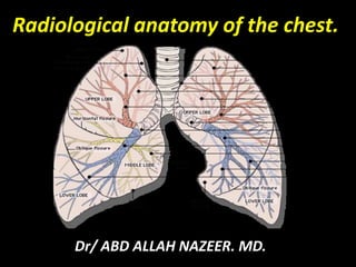
Radiological anatomy of the chest.
Dr/ ABD ALLAH NAZEER. MD.
Imaging techniques used to investigate
pulmonary radiological anatomy include:
Plain film.
Computed Tomography.
Magnetic Resonance Imaging.
Angiography.
Pulmonary Imaging.
Lobar Anatomy
Both lungs are divided into lobes, and these into
segments. The right lung has three lobes: the right
upper lobe (RUL); the right middle lobe (RML); and, the
right lower lobe (RLL).
The left lung has only two lobes: the left upper lobe
(LUL); and the left lower lobe (LLL). There is a segment
of the left upper lobe which is the equivalent of the right
middle lobe called the lingula. Like the middle lobe, the
lingula is composed of two smaller segments. Unlike the
middle lobe, however, there is no fissure separating the
lingula from the remainder of the left upper lobe. In
disease processes, the two regions behave similarly.
Fissures
Fissures (also known as interlobar septa) separate the lobes from
one another. Fissures are two adjacent layers of visceral pleura
with a tiny amount of surfactant in an otherwise potential space.
There are two ways fissures come to be discernible on chest x-rays:
by being aligned parallel to the x-ray beam; and by abutment of
fluid or other increased density on one side of the fissure. In the
latter, the fissure isn't actually seen- it's the sharp margin of
opacity in one lobe adjacent to an air-filled lobe on the other.
On a radiograph the fissures look very much like opaque scratches
on the film. The lateral view often shows both major fissures as
well as the minor fissure.
Why care about the fissures? Because they respond to disease
processes in the chest. For instance, volume loss (atelectasis)
results in structures displacing toward the problem. The minor
fissure is pulled superiorly with RUL atelectasis.
The Right Lung:
A major (oblique) fissure divides the right upper and middle
lobes from the right lower lobe. This fissure is not visible on
a PA view, but is usually visible on the lateral view since
the beam is tangential to it. It tends to be slightly more
vertical than the left major fissure. The best way to
distinguish right from left major fissures is to look for which
hemidiaphragm each fissure ends in.
The minor (horizontal) fissure divides the right upper lobe
from the right middle lobe. This is often visible on both PA
and lateral view of the chest since it is tangential to the
beam in both projections. On the PA view, the lateral aspect
of the fissure is usually found between the 4th and 7th ribs
at the lateral chest wall and converges horizontally toward
the lung hilum.
The Left Lung:
The major fissure divides the upper and lower lobes of the
1.left lung. There is no fissure between the lingula (RML
equivalent) and the remainder of the LUL. Like the right
major fissure, it is visible on the lateral projection only.
The Cardiac Silhouette. Make a determination of size, shape
and location of heart shadow.
Normal cardiac size is less than 1/2 of the internal diameter
of the thorax--measure from the inside of each rib instead
of outside the rib. A cardiothoracic ratio of greater than 1:2
suggests the possibility of cardiomeagly. Remember that
this is not valid on AP thoracic radiographs which naturally
enlarges the appearance of the heart.
Normal cardiac shape. The left side of the heart has three to
four moguls (bumps). The right side has two. The two on
the right are: superior vena cava and right atrium. The
three on the left are: aortic arch, pulmonary artery and left
ventricle. The left atrial appendage, sometimes visible as a
distinct mogul, is located between the pulmonary artery and
the left ventricle.
Normal cardiac position in the chest. Approximately 1/4 to
1/3 of the heart projects to the right of the midline. Pectus
excavatum usually results in a shift of the cardiac shadow
to the left, so that the right heart border is obscured by the
spine. Shift of the heart shadow may also result from
atelectasis.
The Diaphragmatic Contours. Assess the position of the
hemidiaphragm shadows. The dome of the right hemidiaphragm
should be at the level of T10 on full inspiration. The left side is
lower by 1/2 intercostal space. On the lateral view, the right
hemidiaphragm is usually projected higher than the left.
The Costophrenic Angles. Also known as costophrenic sulci.
Check for the presence of blunting from pleural fluid on both the
PA and lateral projections. The two posterior costophrenic
angles (on the lateral view) are superimposed, making it a little
more difficult to detect small amounts of pleural fluid. Also
remember that the posterior costophrenic angles are at the
spinal level of T12.
Trachea. Check that it is within the midline. The distal trachea
(just above the bifurcation) normally deviates slightly to the
right to accommodate the aortic arch. Occasionally the cartilage
rings of the trachea can calcify.
Hilar Shadows and Lung Vascularity- The hili are the "roots" of
each lung, containing large caliber arteries and veins, bronchi and
lymph nodes. This is a rather nebulous region for beginning chest
radiology students largely because the heart itself and the aorta
obscure these structures. Most radiologists would agree that the
hilar shadows are among the most challenging areas of the chest
radiograph to evaluate. Follow the superior aspect of each
pulmonary artery proximally. This gives the approximate location
of the top of the hilus. Check the relative location of the lung hili.
The right hilus should be slightly lower than the left, occasionally
at the same level as the left, but never higher than the left.
The branches of the blood vessels naturally get smaller and
smaller toward the periphery of each lung, so there should be
very few vascular shadows in the lateral-most third of the lung.
The upper lung zones have smaller blood vessels larger because
of gravity. Divide each lung into three zones from medial to the
lateral chest wall.
The Subdiaphragmatic Area. Check for the normal arrangement
of the abdominal visceral shadows, including the stomach bubble, liver
and spleen. Check for calcifications, organomegaly, abnormal
collections of air, etc.
The Osseous Components of the Thorax. Check the spine, ribs, clavicles,
scapulae, and proximal humeri if visible. The Soft Tissues of the Chest
Wall. Check for the presence of both breast shadows in females. In
cases of radical mastectomy, the muscle planes will be altered
consistent with the removal of portions of the pectoral muscle. This is
most notable in the axillary area. A simple mastectomy shows an
absent breast shadow only. Small, asymmetrical breasts can simulate
mastectomy. Check the chest wall in males and females for other
deformities, irregularities. Includes the "companion shadow" for the
clavicles which represents the skin overlying the clavicle visible due to
the fossa created behind the clavicle due to positioning the patient so
that the scapulae are abducted.
The Retrocardiac and Retrosternal Spaces on the Lateral. The volume
and density of these areas should be almost equal.
CT Anatomy
The aorta and its branches, the great veins, the pulmonary
arteries, and the trachea and main bronchi serve as
reliable guides to localizing other important mediastinal
structures. As an aid to understanding regional anatomy,
the mediastinum can be divided into four compartments,
respectively, from superior to inferior: (a) the supraaortic
or superior mediastinum; (b) the region of the aortic arch
and aortopulmonary window (APW); (c) the pulmonary
arteries, subcarinal space, and azygoesophageal recess;
and (d) the heart and paracardiac mediastinum.
Supraaortic (Superior) Mediastinum Just below the
thoracic inlet, the mediastinum is relatively narrow from
anterior to posterior. The trachea is centrally located.
The esophagus lies posterior to the trachea but can be
displaced to the left (more common) or right. It is often
collapsed and appears as a flattened structure of soft-tissue
attenuation. Small amounts of air or fluid may be seen in its
lumen.
The brachiocephalic veins are the most anterior and
lateral vessels visible, lying immediately behind the clavicular
heads. Although they vary in size, their positions are relatively
constant. The right brachiocephalic vein has a nearly vertical
course throughout its length. The left brachiocephalic vein is
longer and courses horizontally as it crosses the mediastinum.
The innominate (brachiocephalic) artery is located in close
proximity to the anterior tracheal wall, near its midline or
slightly to the right of midline in most normals; it is the most
variable of all the great arteries.
The left common carotid artery lies to the left and slightly
posterior to the innominate artery; generally it has the smallest
diameter of the three major arterial branches. The left subclavian
artery is a relatively posterior structure throughout most of its
course, lying to the left of the trachea or slightly posterior to its
midline.
Aortic Arch and Aortopulmonary Window
While the supraaortic region largely contains arterial and venous
branches of the aorta and vena cava, this compartment contains the
undivided mediastinal great vessels, the aorta and superior vena
cava, and also several important mediastinal spaces and lymph
node groups The aortic arch has a characteristic but variable
appearance. The anterior aspect of the arch is located anterior and
to the right of the trachea, with the arch passing to the left and
posteriorly. The posterior arch usually lies anterior and lateral to the
spine. The aortic arch tapers slightly along its length, from anterior
to posterior.
Pulmonary Arteries, Subcarinal Space, and Azygoesophageal
Recess at or near the level of the tracheal carina or main bronchi,
the main pulmonary artery divides into its right and left branches.
The left pulmonary artery is somewhat higher than the right,
usually being seen 1 cm above it, and appears as the continuation
of the main pulmonary artery, directed posterolaterally and to the
left. The right pulmonary artery arises at nearly a right angle to the
main and left pulmonary and crosses the mediastinum from left to
right, anterior to the carina or main bronchi. The right pulmonary
artery limits the most caudal extent of the pre-tracheal space and
the anterior aspect of the subcarinal space. The azygos vein
parallels the esophagus along the right side of the mediastinum
and laterally contacts the medial pleural reflections of the right
lower lobe, defining the posterior border of the azygoesophageal
recess. On the left side, the hemiazygos vein parallels the
descending aorta, lying posterior to it, it is not always visible.
The subcarinal space is the region of the mediastinum immediately
below the carina, marginated laterally by the main bronchi. It contains a
number of lymph nodes and is closely related to the esophagus. Below the
level of the tracheal carina and azygos arch, the medial aspect of the right
lung contacts the mediastinum in close apposition to the azygos vein,
esophagus, and subcarinal space. This region of the mediastinum is called
the azygoesophageal recess. The contour of the azygoesophageal recess is
concave laterally in the large majority of normal subjects; a convexity in
this region should be regarded as suspicious of mass and the scan
examined closely for a pathologic process. However, a convexity in this
region may also be produced by a prominent normal esophagus or azygos
vein and is particularly common in patients with a narrow mediastinum
and in children.
Normal nodes are commonly visible in the subcarinal space, being larger
than normal nodes in other parts of the mediastinum and up to 1.5 cm in
short-axis diameter. The esophagus usually is seen immediately posterior
to the subcarinal space, and distinguishing nodes and esophagus may be
difficult unless the esophagus contains air or contrast material, or its
course is traced on adjacent scans.
Heart and Paracardiac Mediastinum
At the level of the heart, the prevascular mediastinum
becomes thin or is obliterated by the heart contacting the
anterior chest wall. Little soft tissue is visible anterior and
lateral to the cardiac chambers and origins of the main
pulmonary artery and aorta. However, posterior to the
heart, the azygoesophageal recess remains visible to the
level of the diaphragm. As at higher levels, the contour
of the azygoesophageal recess is concave laterally, closely
opposed to the esophagus and azygos vein. Posterior to
the descending aorta, fat, the hemiazygos vein and small
lymph nodes occupy the left paravertebral space. The
right paravertebral space is considerably thinner or
invisible.
1- Clavicle.
2- Spinous process.
3- Trachea.
4- Scapula.
5- Aortic arch.
6- Carina (bifurcation).
7- Pulmonary trunk.
8- Lt. cardiac border (atria).
9- Lt. cardiac border (ventricle).
10- Vertebral body.
11- Descending Aorta.
12- Gastric fundus.
13- Liver.
14- Rt. Hemi-diaphragm.
15- Rt. Main bronchus.
16- Azygous vein.
17- Scapula.
Pleural sac and recesses
Lung Window
Mediastinal Window
CT radiological anatomy.
Pulmonary lobes on CT.
Radiological Anatomy of the Chest.
Radiological Anatomy of the Chest
1
2 3
4
5
6
7 8 9
10
11
12
13
14
15
16
17
11
9
10
10
19
18
14
17
CT OF PULMONARY VASCULTURE
CORONAL SLICES.
Lymph nodes stations of the thorax.
MR CONTRAST ARTERIOGRAM. SUBTRACTION IMAGE.
Intravenous contrast has been injected from a catheter placed from a Lt. subclavian
site with the tip of the catheter in the main pulmonary artery. Rapid imaging while
the opacified blood flows though the pulmonary arterial tree gives this image
rad ana of chest.pdf

More Related Content

What's hot

Basic projections of mammogram
Basic projections of mammogramBasic projections of mammogram
Basic projections of mammogramDonBenny2
Β 
Mammography positioning technique for MLO View
Mammography positioning technique for MLO View Mammography positioning technique for MLO View
Mammography positioning technique for MLO View Selin Prasad
Β 
Spleen Ultrasound anatomy structure scanning techniques and pathologies
Spleen Ultrasound anatomy structure scanning techniques and pathologies Spleen Ultrasound anatomy structure scanning techniques and pathologies
Spleen Ultrasound anatomy structure scanning techniques and pathologies Safi. Khan
Β 
Chest decubitus
Chest decubitusChest decubitus
Chest decubitusSelf
Β 
Imaging of breast pathologies
Imaging of breast pathologiesImaging of breast pathologies
Imaging of breast pathologiesArchana Koshy
Β 
Liver radiology MRI
Liver radiology MRILiver radiology MRI
Liver radiology MRIMahesh Kumar
Β 
Abdominal x ray
Abdominal x rayAbdominal x ray
Abdominal x rayMathew Joseph
Β 
Normal chest x ray- Radiology Basics
Normal chest x  ray- Radiology BasicsNormal chest x  ray- Radiology Basics
Normal chest x ray- Radiology BasicsSandeep Awal
Β 
Mammography Positioning Technique for Additional Views
Mammography Positioning Technique for Additional Views Mammography Positioning Technique for Additional Views
Mammography Positioning Technique for Additional Views Selin Prasad
Β 
The Normal Chest X-ray
The Normal Chest X-rayThe Normal Chest X-ray
The Normal Chest X-raykaphoury
Β 
Developmental Renal Pseudotumours
Developmental Renal PseudotumoursDevelopmental Renal Pseudotumours
Developmental Renal PseudotumoursMuhibullah Bangash
Β 
Imaging breast mammogram
Imaging breast mammogramImaging breast mammogram
Imaging breast mammogramREKHAKHARE
Β 
Presentation1.pptx, radiological anatomy of the chest.
Presentation1.pptx, radiological anatomy of the chest.Presentation1.pptx, radiological anatomy of the chest.
Presentation1.pptx, radiological anatomy of the chest.Abdellah Nazeer
Β 
Presentation1.pptx, radiological anatomy of the abdomen and pelvis.
Presentation1.pptx, radiological anatomy of the abdomen and pelvis.Presentation1.pptx, radiological anatomy of the abdomen and pelvis.
Presentation1.pptx, radiological anatomy of the abdomen and pelvis.Abdellah Nazeer
Β 
Fluoroscopic imaging anatomy and pathology of stomach and duodenum abdul final
Fluoroscopic imaging anatomy and pathology of stomach and duodenum abdul finalFluoroscopic imaging anatomy and pathology of stomach and duodenum abdul final
Fluoroscopic imaging anatomy and pathology of stomach and duodenum abdul finalabduljelil nejmu
Β 
Bi Rads Breast
Bi Rads BreastBi Rads Breast
Bi Rads BreastMadhu Reddy
Β 

What's hot (20)

Basic projections of mammogram
Basic projections of mammogramBasic projections of mammogram
Basic projections of mammogram
Β 
Mammography positioning technique for MLO View
Mammography positioning technique for MLO View Mammography positioning technique for MLO View
Mammography positioning technique for MLO View
Β 
Spleen Ultrasound anatomy structure scanning techniques and pathologies
Spleen Ultrasound anatomy structure scanning techniques and pathologies Spleen Ultrasound anatomy structure scanning techniques and pathologies
Spleen Ultrasound anatomy structure scanning techniques and pathologies
Β 
Chest decubitus
Chest decubitusChest decubitus
Chest decubitus
Β 
Imaging of breast pathologies
Imaging of breast pathologiesImaging of breast pathologies
Imaging of breast pathologies
Β 
Liver radiology MRI
Liver radiology MRILiver radiology MRI
Liver radiology MRI
Β 
Abdominal x ray
Abdominal x rayAbdominal x ray
Abdominal x ray
Β 
Normal chest x ray- Radiology Basics
Normal chest x  ray- Radiology BasicsNormal chest x  ray- Radiology Basics
Normal chest x ray- Radiology Basics
Β 
Mammography Positioning Technique for Additional Views
Mammography Positioning Technique for Additional Views Mammography Positioning Technique for Additional Views
Mammography Positioning Technique for Additional Views
Β 
The Normal Chest X-ray
The Normal Chest X-rayThe Normal Chest X-ray
The Normal Chest X-ray
Β 
Mammography
MammographyMammography
Mammography
Β 
Developmental Renal Pseudotumours
Developmental Renal PseudotumoursDevelopmental Renal Pseudotumours
Developmental Renal Pseudotumours
Β 
Imaging breast mammogram
Imaging breast mammogramImaging breast mammogram
Imaging breast mammogram
Β 
Mammography
MammographyMammography
Mammography
Β 
Presentation1.pptx, radiological anatomy of the chest.
Presentation1.pptx, radiological anatomy of the chest.Presentation1.pptx, radiological anatomy of the chest.
Presentation1.pptx, radiological anatomy of the chest.
Β 
Presentation1.pptx, radiological anatomy of the abdomen and pelvis.
Presentation1.pptx, radiological anatomy of the abdomen and pelvis.Presentation1.pptx, radiological anatomy of the abdomen and pelvis.
Presentation1.pptx, radiological anatomy of the abdomen and pelvis.
Β 
Fluoroscopic imaging anatomy and pathology of stomach and duodenum abdul final
Fluoroscopic imaging anatomy and pathology of stomach and duodenum abdul finalFluoroscopic imaging anatomy and pathology of stomach and duodenum abdul final
Fluoroscopic imaging anatomy and pathology of stomach and duodenum abdul final
Β 
Bi Rads Breast
Bi Rads BreastBi Rads Breast
Bi Rads Breast
Β 
Upper limb
Upper limbUpper limb
Upper limb
Β 
Coronary CT
Coronary CTCoronary CT
Coronary CT
Β 

Similar to rad ana of chest.pdf

Radiographic anatomy of lungs.pptx
Radiographic anatomy of lungs.pptxRadiographic anatomy of lungs.pptx
Radiographic anatomy of lungs.pptxrohanjohnjacob
Β 
Chest X-ray: The Basics
Chest X-ray: The BasicsChest X-ray: The Basics
Chest X-ray: The BasicsAbhineet Dey
Β 
Chest X-ray radiology_Power Point Presentation
Chest X-ray radiology_Power Point PresentationChest X-ray radiology_Power Point Presentation
Chest X-ray radiology_Power Point PresentationAhyaAziz
Β 
Basics of Chest X-Ray
Basics of Chest X-RayBasics of Chest X-Ray
Basics of Chest X-RayKhurram Wazir
Β 
Wayang kulit, no 1 a fundamentals
Wayang kulit, no 1 a fundamentalsWayang kulit, no 1 a fundamentals
Wayang kulit, no 1 a fundamentalsycche19
Β 
BASIC RADIOLOGY
BASIC RADIOLOGYBASIC RADIOLOGY
BASIC RADIOLOGYDJ CrissCross
Β 
Basic Radiology
Basic RadiologyBasic Radiology
Basic RadiologyDJ CrissCross
Β 
Surgery 5th year, 9th, 10th &11th lectures (Dr. Ahmed Al-Azzawi)
Surgery 5th year, 9th, 10th &11th lectures (Dr. Ahmed Al-Azzawi)Surgery 5th year, 9th, 10th &11th lectures (Dr. Ahmed Al-Azzawi)
Surgery 5th year, 9th, 10th &11th lectures (Dr. Ahmed Al-Azzawi)College of Medicine, Sulaymaniyah
Β 
anatomy of heart.pptx
anatomy of heart.pptxanatomy of heart.pptx
anatomy of heart.pptxShivankNaik
Β 
Lungs anatomy, organs in thorax
Lungs anatomy, organs in thoraxLungs anatomy, organs in thorax
Lungs anatomy, organs in thoraxFaarah Yusuf
Β 
CHEST XRAY
CHEST XRAY CHEST XRAY
CHEST XRAY amit jha
Β 
Anatomy of cardiac structures & conducting system in
Anatomy of cardiac structures & conducting system inAnatomy of cardiac structures & conducting system in
Anatomy of cardiac structures & conducting system inRamachandra Barik
Β 
Rad Seminar CHEST IMAGING By Dr Siraj.pptx
Rad Seminar CHEST IMAGING By Dr Siraj.pptxRad Seminar CHEST IMAGING By Dr Siraj.pptx
Rad Seminar CHEST IMAGING By Dr Siraj.pptxImanuIliyas
Β 
Mediastinum - Heart and Lungs
Mediastinum - Heart and LungsMediastinum - Heart and Lungs
Mediastinum - Heart and LungsSado Anatomist
Β 
mediastinal imaging and masses
mediastinal imaging and massesmediastinal imaging and masses
mediastinal imaging and massesArun Singh
Β 
CHEST X-RAY F.pptx
CHEST X-RAY F.pptxCHEST X-RAY F.pptx
CHEST X-RAY F.pptxdevanshi92
Β 
THORAX CLINICAL ANATOMY
THORAX CLINICAL ANATOMYTHORAX CLINICAL ANATOMY
THORAX CLINICAL ANATOMYAkmalSharaf1
Β 
Normal Chest X-Rays & Its Systemic Approach- Anatomy
Normal Chest X-Rays & Its Systemic Approach- AnatomyNormal Chest X-Rays & Its Systemic Approach- Anatomy
Normal Chest X-Rays & Its Systemic Approach- AnatomyDr.Bijay Yadav
Β 

Similar to rad ana of chest.pdf (20)

Radiographic anatomy of lungs.pptx
Radiographic anatomy of lungs.pptxRadiographic anatomy of lungs.pptx
Radiographic anatomy of lungs.pptx
Β 
Chest X-ray: The Basics
Chest X-ray: The BasicsChest X-ray: The Basics
Chest X-ray: The Basics
Β 
Chest X-ray radiology_Power Point Presentation
Chest X-ray radiology_Power Point PresentationChest X-ray radiology_Power Point Presentation
Chest X-ray radiology_Power Point Presentation
Β 
Basics of Chest X-Ray
Basics of Chest X-RayBasics of Chest X-Ray
Basics of Chest X-Ray
Β 
Chest
ChestChest
Chest
Β 
Wayang kulit, no 1 a fundamentals
Wayang kulit, no 1 a fundamentalsWayang kulit, no 1 a fundamentals
Wayang kulit, no 1 a fundamentals
Β 
BASIC RADIOLOGY
BASIC RADIOLOGYBASIC RADIOLOGY
BASIC RADIOLOGY
Β 
Basic Radiology
Basic RadiologyBasic Radiology
Basic Radiology
Β 
Surgery 5th year, 9th, 10th &11th lectures (Dr. Ahmed Al-Azzawi)
Surgery 5th year, 9th, 10th &11th lectures (Dr. Ahmed Al-Azzawi)Surgery 5th year, 9th, 10th &11th lectures (Dr. Ahmed Al-Azzawi)
Surgery 5th year, 9th, 10th &11th lectures (Dr. Ahmed Al-Azzawi)
Β 
anatomy of heart.pptx
anatomy of heart.pptxanatomy of heart.pptx
anatomy of heart.pptx
Β 
Lungs anatomy, organs in thorax
Lungs anatomy, organs in thoraxLungs anatomy, organs in thorax
Lungs anatomy, organs in thorax
Β 
CHEST XRAY
CHEST XRAY CHEST XRAY
CHEST XRAY
Β 
Anatomy of cardiac structures & conducting system in
Anatomy of cardiac structures & conducting system inAnatomy of cardiac structures & conducting system in
Anatomy of cardiac structures & conducting system in
Β 
Rad Seminar CHEST IMAGING By Dr Siraj.pptx
Rad Seminar CHEST IMAGING By Dr Siraj.pptxRad Seminar CHEST IMAGING By Dr Siraj.pptx
Rad Seminar CHEST IMAGING By Dr Siraj.pptx
Β 
Lungs
LungsLungs
Lungs
Β 
Mediastinum - Heart and Lungs
Mediastinum - Heart and LungsMediastinum - Heart and Lungs
Mediastinum - Heart and Lungs
Β 
mediastinal imaging and masses
mediastinal imaging and massesmediastinal imaging and masses
mediastinal imaging and masses
Β 
CHEST X-RAY F.pptx
CHEST X-RAY F.pptxCHEST X-RAY F.pptx
CHEST X-RAY F.pptx
Β 
THORAX CLINICAL ANATOMY
THORAX CLINICAL ANATOMYTHORAX CLINICAL ANATOMY
THORAX CLINICAL ANATOMY
Β 
Normal Chest X-Rays & Its Systemic Approach- Anatomy
Normal Chest X-Rays & Its Systemic Approach- AnatomyNormal Chest X-Rays & Its Systemic Approach- Anatomy
Normal Chest X-Rays & Its Systemic Approach- Anatomy
Β 

More from rohanjohnjacob

hydrocephalus and csf disorders powerpoint
hydrocephalus and csf disorders powerpointhydrocephalus and csf disorders powerpoint
hydrocephalus and csf disorders powerpointrohanjohnjacob
Β 
radiology in dementia powerpoint presentation
radiology in dementia powerpoint presentationradiology in dementia powerpoint presentation
radiology in dementia powerpoint presentationrohanjohnjacob
Β 
barium swallow.pptx
barium swallow.pptxbarium swallow.pptx
barium swallow.pptxrohanjohnjacob
Β 
esophagus 2.pptx
esophagus 2.pptxesophagus 2.pptx
esophagus 2.pptxrohanjohnjacob
Β 
upper limb trauma.pptx
upper limb trauma.pptxupper limb trauma.pptx
upper limb trauma.pptxrohanjohnjacob
Β 
usg artifacts.pptx
usg artifacts.pptxusg artifacts.pptx
usg artifacts.pptxrohanjohnjacob
Β 
radio cassettes and screens.pptx
radio cassettes and screens.pptxradio cassettes and screens.pptx
radio cassettes and screens.pptxrohanjohnjacob
Β 
Normal abd xray rjj.pptx
Normal abd xray rjj.pptxNormal abd xray rjj.pptx
Normal abd xray rjj.pptxrohanjohnjacob
Β 
grids,films.pptx
grids,films.pptxgrids,films.pptx
grids,films.pptxrohanjohnjacob
Β 
ADVERSE EFFECTS OF CONTRAST AGENTS ppt.pptx
ADVERSE EFFECTS OF CONTRAST AGENTS ppt.pptxADVERSE EFFECTS OF CONTRAST AGENTS ppt.pptx
ADVERSE EFFECTS OF CONTRAST AGENTS ppt.pptxrohanjohnjacob
Β 
PET SCANNING.pptx
PET SCANNING.pptxPET SCANNING.pptx
PET SCANNING.pptxrohanjohnjacob
Β 
MAMMOGRAPHY.pptx
MAMMOGRAPHY.pptxMAMMOGRAPHY.pptx
MAMMOGRAPHY.pptxrohanjohnjacob
Β 
upper limb trauma.pptx
upper limb trauma.pptxupper limb trauma.pptx
upper limb trauma.pptxrohanjohnjacob
Β 
linesmediastinalstripes-01-101126132247-phpapp01.pptx
linesmediastinalstripes-01-101126132247-phpapp01.pptxlinesmediastinalstripes-01-101126132247-phpapp01.pptx
linesmediastinalstripes-01-101126132247-phpapp01.pptxrohanjohnjacob
Β 
doppler physics.pptx
doppler physics.pptxdoppler physics.pptx
doppler physics.pptxrohanjohnjacob
Β 
CHEST XRAYS RJJ.pptx
CHEST XRAYS RJJ.pptxCHEST XRAYS RJJ.pptx
CHEST XRAYS RJJ.pptxrohanjohnjacob
Β 
CONTRAST AGENTS PPT.pptx
CONTRAST AGENTS PPT.pptxCONTRAST AGENTS PPT.pptx
CONTRAST AGENTS PPT.pptxrohanjohnjacob
Β 
ADVERSE EFFECTS OF CONTRAST AGENTS ppt.pptx
ADVERSE EFFECTS OF CONTRAST AGENTS ppt.pptxADVERSE EFFECTS OF CONTRAST AGENTS ppt.pptx
ADVERSE EFFECTS OF CONTRAST AGENTS ppt.pptxrohanjohnjacob
Β 

More from rohanjohnjacob (20)

hydrocephalus and csf disorders powerpoint
hydrocephalus and csf disorders powerpointhydrocephalus and csf disorders powerpoint
hydrocephalus and csf disorders powerpoint
Β 
radiology in dementia powerpoint presentation
radiology in dementia powerpoint presentationradiology in dementia powerpoint presentation
radiology in dementia powerpoint presentation
Β 
barium swallow.pptx
barium swallow.pptxbarium swallow.pptx
barium swallow.pptx
Β 
esophagus 2.pptx
esophagus 2.pptxesophagus 2.pptx
esophagus 2.pptx
Β 
upper limb trauma.pptx
upper limb trauma.pptxupper limb trauma.pptx
upper limb trauma.pptx
Β 
usg artifacts.pptx
usg artifacts.pptxusg artifacts.pptx
usg artifacts.pptx
Β 
radio cassettes and screens.pptx
radio cassettes and screens.pptxradio cassettes and screens.pptx
radio cassettes and screens.pptx
Β 
Normal abd xray rjj.pptx
Normal abd xray rjj.pptxNormal abd xray rjj.pptx
Normal abd xray rjj.pptx
Β 
esophagus.pptx
esophagus.pptxesophagus.pptx
esophagus.pptx
Β 
grids,films.pptx
grids,films.pptxgrids,films.pptx
grids,films.pptx
Β 
ADVERSE EFFECTS OF CONTRAST AGENTS ppt.pptx
ADVERSE EFFECTS OF CONTRAST AGENTS ppt.pptxADVERSE EFFECTS OF CONTRAST AGENTS ppt.pptx
ADVERSE EFFECTS OF CONTRAST AGENTS ppt.pptx
Β 
PET SCANNING.pptx
PET SCANNING.pptxPET SCANNING.pptx
PET SCANNING.pptx
Β 
MAMMOGRAPHY.pptx
MAMMOGRAPHY.pptxMAMMOGRAPHY.pptx
MAMMOGRAPHY.pptx
Β 
upper limb trauma.pptx
upper limb trauma.pptxupper limb trauma.pptx
upper limb trauma.pptx
Β 
linesmediastinalstripes-01-101126132247-phpapp01.pptx
linesmediastinalstripes-01-101126132247-phpapp01.pptxlinesmediastinalstripes-01-101126132247-phpapp01.pptx
linesmediastinalstripes-01-101126132247-phpapp01.pptx
Β 
DARK ROOM.pptx
DARK ROOM.pptxDARK ROOM.pptx
DARK ROOM.pptx
Β 
doppler physics.pptx
doppler physics.pptxdoppler physics.pptx
doppler physics.pptx
Β 
CHEST XRAYS RJJ.pptx
CHEST XRAYS RJJ.pptxCHEST XRAYS RJJ.pptx
CHEST XRAYS RJJ.pptx
Β 
CONTRAST AGENTS PPT.pptx
CONTRAST AGENTS PPT.pptxCONTRAST AGENTS PPT.pptx
CONTRAST AGENTS PPT.pptx
Β 
ADVERSE EFFECTS OF CONTRAST AGENTS ppt.pptx
ADVERSE EFFECTS OF CONTRAST AGENTS ppt.pptxADVERSE EFFECTS OF CONTRAST AGENTS ppt.pptx
ADVERSE EFFECTS OF CONTRAST AGENTS ppt.pptx
Β 

Recently uploaded

Sonagachi Call Girls Services 9907093804 @24x7 High Class Babes Here Call Now
Sonagachi Call Girls Services 9907093804 @24x7 High Class Babes Here Call NowSonagachi Call Girls Services 9907093804 @24x7 High Class Babes Here Call Now
Sonagachi Call Girls Services 9907093804 @24x7 High Class Babes Here Call NowRiya Pathan
Β 
Call Girls Service In Shyam Nagar Whatsapp 8445551418 Independent Escort Service
Call Girls Service In Shyam Nagar Whatsapp 8445551418 Independent Escort ServiceCall Girls Service In Shyam Nagar Whatsapp 8445551418 Independent Escort Service
Call Girls Service In Shyam Nagar Whatsapp 8445551418 Independent Escort Serviceparulsinha
Β 
Call Girls Hebbal Just Call 7001305949 Top Class Call Girl Service Available
Call Girls Hebbal Just Call 7001305949 Top Class Call Girl Service AvailableCall Girls Hebbal Just Call 7001305949 Top Class Call Girl Service Available
Call Girls Hebbal Just Call 7001305949 Top Class Call Girl Service Availablenarwatsonia7
Β 
Call Girls In Andheri East Call 9920874524 Book Hot And Sexy Girls
Call Girls In Andheri East Call 9920874524 Book Hot And Sexy GirlsCall Girls In Andheri East Call 9920874524 Book Hot And Sexy Girls
Call Girls In Andheri East Call 9920874524 Book Hot And Sexy Girlsnehamumbai
Β 
Ahmedabad Call Girls CG Road πŸ”9907093804 Short 1500 πŸ’‹ Night 6000
Ahmedabad Call Girls CG Road πŸ”9907093804  Short 1500  πŸ’‹ Night 6000Ahmedabad Call Girls CG Road πŸ”9907093804  Short 1500  πŸ’‹ Night 6000
Ahmedabad Call Girls CG Road πŸ”9907093804 Short 1500 πŸ’‹ Night 6000aliya bhat
Β 
Hemostasis Physiology and Clinical correlations by Dr Faiza.pdf
Hemostasis Physiology and Clinical correlations by Dr Faiza.pdfHemostasis Physiology and Clinical correlations by Dr Faiza.pdf
Hemostasis Physiology and Clinical correlations by Dr Faiza.pdfMedicoseAcademics
Β 
Call Girls Frazer Town Just Call 7001305949 Top Class Call Girl Service Avail...
Call Girls Frazer Town Just Call 7001305949 Top Class Call Girl Service Avail...Call Girls Frazer Town Just Call 7001305949 Top Class Call Girl Service Avail...
Call Girls Frazer Town Just Call 7001305949 Top Class Call Girl Service Avail...narwatsonia7
Β 
High Profile Call Girls Jaipur Vani 8445551418 Independent Escort Service Jaipur
High Profile Call Girls Jaipur Vani 8445551418 Independent Escort Service JaipurHigh Profile Call Girls Jaipur Vani 8445551418 Independent Escort Service Jaipur
High Profile Call Girls Jaipur Vani 8445551418 Independent Escort Service Jaipurparulsinha
Β 
Call Girls Service Chennai Jiya 7001305949 Independent Escort Service Chennai
Call Girls Service Chennai Jiya 7001305949 Independent Escort Service ChennaiCall Girls Service Chennai Jiya 7001305949 Independent Escort Service Chennai
Call Girls Service Chennai Jiya 7001305949 Independent Escort Service ChennaiNehru place Escorts
Β 
Call Girl Bangalore Nandini 7001305949 Independent Escort Service Bangalore
Call Girl Bangalore Nandini 7001305949 Independent Escort Service BangaloreCall Girl Bangalore Nandini 7001305949 Independent Escort Service Bangalore
Call Girl Bangalore Nandini 7001305949 Independent Escort Service Bangalorenarwatsonia7
Β 
Artifacts in Nuclear Medicine with Identifying and resolving artifacts.
Artifacts in Nuclear Medicine with Identifying and resolving artifacts.Artifacts in Nuclear Medicine with Identifying and resolving artifacts.
Artifacts in Nuclear Medicine with Identifying and resolving artifacts.MiadAlsulami
Β 
Book Call Girls in Kasavanahalli - 7001305949 with real photos and phone numbers
Book Call Girls in Kasavanahalli - 7001305949 with real photos and phone numbersBook Call Girls in Kasavanahalli - 7001305949 with real photos and phone numbers
Book Call Girls in Kasavanahalli - 7001305949 with real photos and phone numbersnarwatsonia7
Β 
Mumbai Call Girls Service 9910780858 Real Russian Girls Looking Models
Mumbai Call Girls Service 9910780858 Real Russian Girls Looking ModelsMumbai Call Girls Service 9910780858 Real Russian Girls Looking Models
Mumbai Call Girls Service 9910780858 Real Russian Girls Looking Modelssonalikaur4
Β 
College Call Girls Vyasarpadi Whatsapp 7001305949 Independent Escort Service
College Call Girls Vyasarpadi Whatsapp 7001305949 Independent Escort ServiceCollege Call Girls Vyasarpadi Whatsapp 7001305949 Independent Escort Service
College Call Girls Vyasarpadi Whatsapp 7001305949 Independent Escort ServiceNehru place Escorts
Β 
call girls in green park DELHI πŸ” >ΰΌ’9540349809 πŸ” genuine Escort Service πŸ”βœ”οΈβœ”οΈ
call girls in green park  DELHI πŸ” >ΰΌ’9540349809 πŸ” genuine Escort Service πŸ”βœ”οΈβœ”οΈcall girls in green park  DELHI πŸ” >ΰΌ’9540349809 πŸ” genuine Escort Service πŸ”βœ”οΈβœ”οΈ
call girls in green park DELHI πŸ” >ΰΌ’9540349809 πŸ” genuine Escort Service πŸ”βœ”οΈβœ”οΈsaminamagar
Β 
Kolkata Call Girls Services 9907093804 @24x7 High Class Babes Here Call Now
Kolkata Call Girls Services 9907093804 @24x7 High Class Babes Here Call NowKolkata Call Girls Services 9907093804 @24x7 High Class Babes Here Call Now
Kolkata Call Girls Services 9907093804 @24x7 High Class Babes Here Call NowNehru place Escorts
Β 
Call Girl Koramangala | 7001305949 At Low Cost Cash Payment Booking
Call Girl Koramangala | 7001305949 At Low Cost Cash Payment BookingCall Girl Koramangala | 7001305949 At Low Cost Cash Payment Booking
Call Girl Koramangala | 7001305949 At Low Cost Cash Payment Bookingnarwatsonia7
Β 
VIP Call Girls Mumbai Arpita 9910780858 Independent Escort Service Mumbai
VIP Call Girls Mumbai Arpita 9910780858 Independent Escort Service MumbaiVIP Call Girls Mumbai Arpita 9910780858 Independent Escort Service Mumbai
VIP Call Girls Mumbai Arpita 9910780858 Independent Escort Service Mumbaisonalikaur4
Β 
Low Rate Call Girls Mumbai Suman 9910780858 Independent Escort Service Mumbai
Low Rate Call Girls Mumbai Suman 9910780858 Independent Escort Service MumbaiLow Rate Call Girls Mumbai Suman 9910780858 Independent Escort Service Mumbai
Low Rate Call Girls Mumbai Suman 9910780858 Independent Escort Service Mumbaisonalikaur4
Β 
call girls in Connaught Place DELHI πŸ” >ΰΌ’9540349809 πŸ” genuine Escort Service ...
call girls in Connaught Place  DELHI πŸ” >ΰΌ’9540349809 πŸ” genuine Escort Service ...call girls in Connaught Place  DELHI πŸ” >ΰΌ’9540349809 πŸ” genuine Escort Service ...
call girls in Connaught Place DELHI πŸ” >ΰΌ’9540349809 πŸ” genuine Escort Service ...saminamagar
Β 

Recently uploaded (20)

Sonagachi Call Girls Services 9907093804 @24x7 High Class Babes Here Call Now
Sonagachi Call Girls Services 9907093804 @24x7 High Class Babes Here Call NowSonagachi Call Girls Services 9907093804 @24x7 High Class Babes Here Call Now
Sonagachi Call Girls Services 9907093804 @24x7 High Class Babes Here Call Now
Β 
Call Girls Service In Shyam Nagar Whatsapp 8445551418 Independent Escort Service
Call Girls Service In Shyam Nagar Whatsapp 8445551418 Independent Escort ServiceCall Girls Service In Shyam Nagar Whatsapp 8445551418 Independent Escort Service
Call Girls Service In Shyam Nagar Whatsapp 8445551418 Independent Escort Service
Β 
Call Girls Hebbal Just Call 7001305949 Top Class Call Girl Service Available
Call Girls Hebbal Just Call 7001305949 Top Class Call Girl Service AvailableCall Girls Hebbal Just Call 7001305949 Top Class Call Girl Service Available
Call Girls Hebbal Just Call 7001305949 Top Class Call Girl Service Available
Β 
Call Girls In Andheri East Call 9920874524 Book Hot And Sexy Girls
Call Girls In Andheri East Call 9920874524 Book Hot And Sexy GirlsCall Girls In Andheri East Call 9920874524 Book Hot And Sexy Girls
Call Girls In Andheri East Call 9920874524 Book Hot And Sexy Girls
Β 
Ahmedabad Call Girls CG Road πŸ”9907093804 Short 1500 πŸ’‹ Night 6000
Ahmedabad Call Girls CG Road πŸ”9907093804  Short 1500  πŸ’‹ Night 6000Ahmedabad Call Girls CG Road πŸ”9907093804  Short 1500  πŸ’‹ Night 6000
Ahmedabad Call Girls CG Road πŸ”9907093804 Short 1500 πŸ’‹ Night 6000
Β 
Hemostasis Physiology and Clinical correlations by Dr Faiza.pdf
Hemostasis Physiology and Clinical correlations by Dr Faiza.pdfHemostasis Physiology and Clinical correlations by Dr Faiza.pdf
Hemostasis Physiology and Clinical correlations by Dr Faiza.pdf
Β 
Call Girls Frazer Town Just Call 7001305949 Top Class Call Girl Service Avail...
Call Girls Frazer Town Just Call 7001305949 Top Class Call Girl Service Avail...Call Girls Frazer Town Just Call 7001305949 Top Class Call Girl Service Avail...
Call Girls Frazer Town Just Call 7001305949 Top Class Call Girl Service Avail...
Β 
High Profile Call Girls Jaipur Vani 8445551418 Independent Escort Service Jaipur
High Profile Call Girls Jaipur Vani 8445551418 Independent Escort Service JaipurHigh Profile Call Girls Jaipur Vani 8445551418 Independent Escort Service Jaipur
High Profile Call Girls Jaipur Vani 8445551418 Independent Escort Service Jaipur
Β 
Call Girls Service Chennai Jiya 7001305949 Independent Escort Service Chennai
Call Girls Service Chennai Jiya 7001305949 Independent Escort Service ChennaiCall Girls Service Chennai Jiya 7001305949 Independent Escort Service Chennai
Call Girls Service Chennai Jiya 7001305949 Independent Escort Service Chennai
Β 
Call Girl Bangalore Nandini 7001305949 Independent Escort Service Bangalore
Call Girl Bangalore Nandini 7001305949 Independent Escort Service BangaloreCall Girl Bangalore Nandini 7001305949 Independent Escort Service Bangalore
Call Girl Bangalore Nandini 7001305949 Independent Escort Service Bangalore
Β 
Artifacts in Nuclear Medicine with Identifying and resolving artifacts.
Artifacts in Nuclear Medicine with Identifying and resolving artifacts.Artifacts in Nuclear Medicine with Identifying and resolving artifacts.
Artifacts in Nuclear Medicine with Identifying and resolving artifacts.
Β 
Book Call Girls in Kasavanahalli - 7001305949 with real photos and phone numbers
Book Call Girls in Kasavanahalli - 7001305949 with real photos and phone numbersBook Call Girls in Kasavanahalli - 7001305949 with real photos and phone numbers
Book Call Girls in Kasavanahalli - 7001305949 with real photos and phone numbers
Β 
Mumbai Call Girls Service 9910780858 Real Russian Girls Looking Models
Mumbai Call Girls Service 9910780858 Real Russian Girls Looking ModelsMumbai Call Girls Service 9910780858 Real Russian Girls Looking Models
Mumbai Call Girls Service 9910780858 Real Russian Girls Looking Models
Β 
College Call Girls Vyasarpadi Whatsapp 7001305949 Independent Escort Service
College Call Girls Vyasarpadi Whatsapp 7001305949 Independent Escort ServiceCollege Call Girls Vyasarpadi Whatsapp 7001305949 Independent Escort Service
College Call Girls Vyasarpadi Whatsapp 7001305949 Independent Escort Service
Β 
call girls in green park DELHI πŸ” >ΰΌ’9540349809 πŸ” genuine Escort Service πŸ”βœ”οΈβœ”οΈ
call girls in green park  DELHI πŸ” >ΰΌ’9540349809 πŸ” genuine Escort Service πŸ”βœ”οΈβœ”οΈcall girls in green park  DELHI πŸ” >ΰΌ’9540349809 πŸ” genuine Escort Service πŸ”βœ”οΈβœ”οΈ
call girls in green park DELHI πŸ” >ΰΌ’9540349809 πŸ” genuine Escort Service πŸ”βœ”οΈβœ”οΈ
Β 
Kolkata Call Girls Services 9907093804 @24x7 High Class Babes Here Call Now
Kolkata Call Girls Services 9907093804 @24x7 High Class Babes Here Call NowKolkata Call Girls Services 9907093804 @24x7 High Class Babes Here Call Now
Kolkata Call Girls Services 9907093804 @24x7 High Class Babes Here Call Now
Β 
Call Girl Koramangala | 7001305949 At Low Cost Cash Payment Booking
Call Girl Koramangala | 7001305949 At Low Cost Cash Payment BookingCall Girl Koramangala | 7001305949 At Low Cost Cash Payment Booking
Call Girl Koramangala | 7001305949 At Low Cost Cash Payment Booking
Β 
VIP Call Girls Mumbai Arpita 9910780858 Independent Escort Service Mumbai
VIP Call Girls Mumbai Arpita 9910780858 Independent Escort Service MumbaiVIP Call Girls Mumbai Arpita 9910780858 Independent Escort Service Mumbai
VIP Call Girls Mumbai Arpita 9910780858 Independent Escort Service Mumbai
Β 
Low Rate Call Girls Mumbai Suman 9910780858 Independent Escort Service Mumbai
Low Rate Call Girls Mumbai Suman 9910780858 Independent Escort Service MumbaiLow Rate Call Girls Mumbai Suman 9910780858 Independent Escort Service Mumbai
Low Rate Call Girls Mumbai Suman 9910780858 Independent Escort Service Mumbai
Β 
call girls in Connaught Place DELHI πŸ” >ΰΌ’9540349809 πŸ” genuine Escort Service ...
call girls in Connaught Place  DELHI πŸ” >ΰΌ’9540349809 πŸ” genuine Escort Service ...call girls in Connaught Place  DELHI πŸ” >ΰΌ’9540349809 πŸ” genuine Escort Service ...
call girls in Connaught Place DELHI πŸ” >ΰΌ’9540349809 πŸ” genuine Escort Service ...
Β 

rad ana of chest.pdf

  • 1. Radiological anatomy of the chest. Dr/ ABD ALLAH NAZEER. MD.
  • 2. Imaging techniques used to investigate pulmonary radiological anatomy include: Plain film. Computed Tomography. Magnetic Resonance Imaging. Angiography. Pulmonary Imaging.
  • 3. Lobar Anatomy Both lungs are divided into lobes, and these into segments. The right lung has three lobes: the right upper lobe (RUL); the right middle lobe (RML); and, the right lower lobe (RLL). The left lung has only two lobes: the left upper lobe (LUL); and the left lower lobe (LLL). There is a segment of the left upper lobe which is the equivalent of the right middle lobe called the lingula. Like the middle lobe, the lingula is composed of two smaller segments. Unlike the middle lobe, however, there is no fissure separating the lingula from the remainder of the left upper lobe. In disease processes, the two regions behave similarly.
  • 4. Fissures Fissures (also known as interlobar septa) separate the lobes from one another. Fissures are two adjacent layers of visceral pleura with a tiny amount of surfactant in an otherwise potential space. There are two ways fissures come to be discernible on chest x-rays: by being aligned parallel to the x-ray beam; and by abutment of fluid or other increased density on one side of the fissure. In the latter, the fissure isn't actually seen- it's the sharp margin of opacity in one lobe adjacent to an air-filled lobe on the other. On a radiograph the fissures look very much like opaque scratches on the film. The lateral view often shows both major fissures as well as the minor fissure. Why care about the fissures? Because they respond to disease processes in the chest. For instance, volume loss (atelectasis) results in structures displacing toward the problem. The minor fissure is pulled superiorly with RUL atelectasis.
  • 5. The Right Lung: A major (oblique) fissure divides the right upper and middle lobes from the right lower lobe. This fissure is not visible on a PA view, but is usually visible on the lateral view since the beam is tangential to it. It tends to be slightly more vertical than the left major fissure. The best way to distinguish right from left major fissures is to look for which hemidiaphragm each fissure ends in. The minor (horizontal) fissure divides the right upper lobe from the right middle lobe. This is often visible on both PA and lateral view of the chest since it is tangential to the beam in both projections. On the PA view, the lateral aspect of the fissure is usually found between the 4th and 7th ribs at the lateral chest wall and converges horizontally toward the lung hilum. The Left Lung: The major fissure divides the upper and lower lobes of the 1.left lung. There is no fissure between the lingula (RML equivalent) and the remainder of the LUL. Like the right major fissure, it is visible on the lateral projection only.
  • 6. The Cardiac Silhouette. Make a determination of size, shape and location of heart shadow. Normal cardiac size is less than 1/2 of the internal diameter of the thorax--measure from the inside of each rib instead of outside the rib. A cardiothoracic ratio of greater than 1:2 suggests the possibility of cardiomeagly. Remember that this is not valid on AP thoracic radiographs which naturally enlarges the appearance of the heart. Normal cardiac shape. The left side of the heart has three to four moguls (bumps). The right side has two. The two on the right are: superior vena cava and right atrium. The three on the left are: aortic arch, pulmonary artery and left ventricle. The left atrial appendage, sometimes visible as a distinct mogul, is located between the pulmonary artery and the left ventricle. Normal cardiac position in the chest. Approximately 1/4 to 1/3 of the heart projects to the right of the midline. Pectus excavatum usually results in a shift of the cardiac shadow to the left, so that the right heart border is obscured by the spine. Shift of the heart shadow may also result from atelectasis.
  • 7. The Diaphragmatic Contours. Assess the position of the hemidiaphragm shadows. The dome of the right hemidiaphragm should be at the level of T10 on full inspiration. The left side is lower by 1/2 intercostal space. On the lateral view, the right hemidiaphragm is usually projected higher than the left. The Costophrenic Angles. Also known as costophrenic sulci. Check for the presence of blunting from pleural fluid on both the PA and lateral projections. The two posterior costophrenic angles (on the lateral view) are superimposed, making it a little more difficult to detect small amounts of pleural fluid. Also remember that the posterior costophrenic angles are at the spinal level of T12. Trachea. Check that it is within the midline. The distal trachea (just above the bifurcation) normally deviates slightly to the right to accommodate the aortic arch. Occasionally the cartilage rings of the trachea can calcify.
  • 8. Hilar Shadows and Lung Vascularity- The hili are the "roots" of each lung, containing large caliber arteries and veins, bronchi and lymph nodes. This is a rather nebulous region for beginning chest radiology students largely because the heart itself and the aorta obscure these structures. Most radiologists would agree that the hilar shadows are among the most challenging areas of the chest radiograph to evaluate. Follow the superior aspect of each pulmonary artery proximally. This gives the approximate location of the top of the hilus. Check the relative location of the lung hili. The right hilus should be slightly lower than the left, occasionally at the same level as the left, but never higher than the left. The branches of the blood vessels naturally get smaller and smaller toward the periphery of each lung, so there should be very few vascular shadows in the lateral-most third of the lung. The upper lung zones have smaller blood vessels larger because of gravity. Divide each lung into three zones from medial to the lateral chest wall.
  • 9. The Subdiaphragmatic Area. Check for the normal arrangement of the abdominal visceral shadows, including the stomach bubble, liver and spleen. Check for calcifications, organomegaly, abnormal collections of air, etc. The Osseous Components of the Thorax. Check the spine, ribs, clavicles, scapulae, and proximal humeri if visible. The Soft Tissues of the Chest Wall. Check for the presence of both breast shadows in females. In cases of radical mastectomy, the muscle planes will be altered consistent with the removal of portions of the pectoral muscle. This is most notable in the axillary area. A simple mastectomy shows an absent breast shadow only. Small, asymmetrical breasts can simulate mastectomy. Check the chest wall in males and females for other deformities, irregularities. Includes the "companion shadow" for the clavicles which represents the skin overlying the clavicle visible due to the fossa created behind the clavicle due to positioning the patient so that the scapulae are abducted. The Retrocardiac and Retrosternal Spaces on the Lateral. The volume and density of these areas should be almost equal.
  • 10. CT Anatomy The aorta and its branches, the great veins, the pulmonary arteries, and the trachea and main bronchi serve as reliable guides to localizing other important mediastinal structures. As an aid to understanding regional anatomy, the mediastinum can be divided into four compartments, respectively, from superior to inferior: (a) the supraaortic or superior mediastinum; (b) the region of the aortic arch and aortopulmonary window (APW); (c) the pulmonary arteries, subcarinal space, and azygoesophageal recess; and (d) the heart and paracardiac mediastinum. Supraaortic (Superior) Mediastinum Just below the thoracic inlet, the mediastinum is relatively narrow from anterior to posterior. The trachea is centrally located.
  • 11. The esophagus lies posterior to the trachea but can be displaced to the left (more common) or right. It is often collapsed and appears as a flattened structure of soft-tissue attenuation. Small amounts of air or fluid may be seen in its lumen. The brachiocephalic veins are the most anterior and lateral vessels visible, lying immediately behind the clavicular heads. Although they vary in size, their positions are relatively constant. The right brachiocephalic vein has a nearly vertical course throughout its length. The left brachiocephalic vein is longer and courses horizontally as it crosses the mediastinum. The innominate (brachiocephalic) artery is located in close proximity to the anterior tracheal wall, near its midline or slightly to the right of midline in most normals; it is the most variable of all the great arteries.
  • 12. The left common carotid artery lies to the left and slightly posterior to the innominate artery; generally it has the smallest diameter of the three major arterial branches. The left subclavian artery is a relatively posterior structure throughout most of its course, lying to the left of the trachea or slightly posterior to its midline. Aortic Arch and Aortopulmonary Window While the supraaortic region largely contains arterial and venous branches of the aorta and vena cava, this compartment contains the undivided mediastinal great vessels, the aorta and superior vena cava, and also several important mediastinal spaces and lymph node groups The aortic arch has a characteristic but variable appearance. The anterior aspect of the arch is located anterior and to the right of the trachea, with the arch passing to the left and posteriorly. The posterior arch usually lies anterior and lateral to the spine. The aortic arch tapers slightly along its length, from anterior to posterior.
  • 13. Pulmonary Arteries, Subcarinal Space, and Azygoesophageal Recess at or near the level of the tracheal carina or main bronchi, the main pulmonary artery divides into its right and left branches. The left pulmonary artery is somewhat higher than the right, usually being seen 1 cm above it, and appears as the continuation of the main pulmonary artery, directed posterolaterally and to the left. The right pulmonary artery arises at nearly a right angle to the main and left pulmonary and crosses the mediastinum from left to right, anterior to the carina or main bronchi. The right pulmonary artery limits the most caudal extent of the pre-tracheal space and the anterior aspect of the subcarinal space. The azygos vein parallels the esophagus along the right side of the mediastinum and laterally contacts the medial pleural reflections of the right lower lobe, defining the posterior border of the azygoesophageal recess. On the left side, the hemiazygos vein parallels the descending aorta, lying posterior to it, it is not always visible.
  • 14. The subcarinal space is the region of the mediastinum immediately below the carina, marginated laterally by the main bronchi. It contains a number of lymph nodes and is closely related to the esophagus. Below the level of the tracheal carina and azygos arch, the medial aspect of the right lung contacts the mediastinum in close apposition to the azygos vein, esophagus, and subcarinal space. This region of the mediastinum is called the azygoesophageal recess. The contour of the azygoesophageal recess is concave laterally in the large majority of normal subjects; a convexity in this region should be regarded as suspicious of mass and the scan examined closely for a pathologic process. However, a convexity in this region may also be produced by a prominent normal esophagus or azygos vein and is particularly common in patients with a narrow mediastinum and in children. Normal nodes are commonly visible in the subcarinal space, being larger than normal nodes in other parts of the mediastinum and up to 1.5 cm in short-axis diameter. The esophagus usually is seen immediately posterior to the subcarinal space, and distinguishing nodes and esophagus may be difficult unless the esophagus contains air or contrast material, or its course is traced on adjacent scans.
  • 15. Heart and Paracardiac Mediastinum At the level of the heart, the prevascular mediastinum becomes thin or is obliterated by the heart contacting the anterior chest wall. Little soft tissue is visible anterior and lateral to the cardiac chambers and origins of the main pulmonary artery and aorta. However, posterior to the heart, the azygoesophageal recess remains visible to the level of the diaphragm. As at higher levels, the contour of the azygoesophageal recess is concave laterally, closely opposed to the esophagus and azygos vein. Posterior to the descending aorta, fat, the hemiazygos vein and small lymph nodes occupy the left paravertebral space. The right paravertebral space is considerably thinner or invisible.
  • 16.
  • 17.
  • 18.
  • 19.
  • 20.
  • 21.
  • 22.
  • 23.
  • 24.
  • 25.
  • 26.
  • 27. 1- Clavicle. 2- Spinous process. 3- Trachea. 4- Scapula. 5- Aortic arch. 6- Carina (bifurcation). 7- Pulmonary trunk. 8- Lt. cardiac border (atria). 9- Lt. cardiac border (ventricle). 10- Vertebral body. 11- Descending Aorta. 12- Gastric fundus. 13- Liver. 14- Rt. Hemi-diaphragm. 15- Rt. Main bronchus. 16- Azygous vein. 17- Scapula.
  • 28.
  • 29.
  • 30.
  • 31.
  • 32.
  • 33.
  • 34.
  • 35.
  • 36.
  • 37.
  • 38.
  • 39.
  • 40.
  • 41.
  • 42.
  • 43.
  • 44.
  • 45.
  • 46.
  • 47.
  • 48.
  • 49.
  • 50.
  • 51.
  • 52.
  • 53.
  • 54. Pleural sac and recesses
  • 55. Lung Window Mediastinal Window CT radiological anatomy.
  • 56.
  • 58.
  • 59.
  • 60.
  • 61.
  • 62.
  • 63.
  • 64.
  • 65.
  • 66.
  • 67.
  • 68.
  • 69.
  • 70.
  • 71.
  • 72.
  • 75.
  • 76.
  • 77.
  • 78.
  • 79.
  • 80.
  • 81.
  • 82.
  • 83.
  • 84.
  • 85.
  • 86.
  • 87.
  • 88.
  • 89.
  • 90.
  • 91.
  • 92.
  • 93.
  • 94.
  • 95. 1 2 3 4 5 6 7 8 9 10 11 12 13 14 15 16 17 11 9 10 10 19 18 14 17 CT OF PULMONARY VASCULTURE
  • 97.
  • 98.
  • 99.
  • 100.
  • 101.
  • 102.
  • 103.
  • 104.
  • 105. Lymph nodes stations of the thorax.
  • 106.
  • 107.
  • 108.
  • 109.
  • 110.
  • 111.
  • 112.
  • 113.
  • 114.
  • 115.
  • 116.
  • 117.
  • 118.
  • 119.
  • 120.
  • 121.
  • 122.
  • 123.
  • 124.
  • 125.
  • 126.
  • 127.
  • 128.
  • 129.
  • 130.
  • 131.
  • 132.
  • 133.
  • 134. MR CONTRAST ARTERIOGRAM. SUBTRACTION IMAGE.
  • 135. Intravenous contrast has been injected from a catheter placed from a Lt. subclavian site with the tip of the catheter in the main pulmonary artery. Rapid imaging while the opacified blood flows though the pulmonary arterial tree gives this image