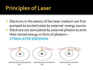
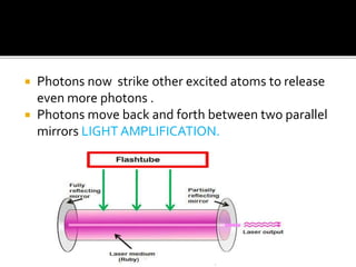
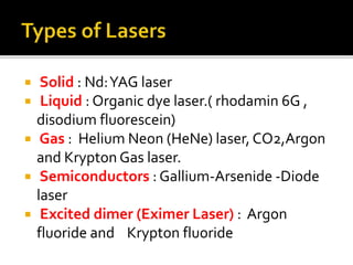
Dr Disha sharma
ENT-HNS
IGMC Shimla
 light amplification by stimulated emission of
radiation
 Albert einstein theodore Maiman
C.Kumar N.Patel
 Electrons in the atoms of the laser medium are first
pumped to excited state by external energy source.
 Electrons are stimulated by external photon to emit
their stored energy in form of photons –
STIMULATED EMISSION
 Photons now strike other excited atoms to release
even more photons .
 Photons move back and forth between two parallel
mirrors LIGHT AMPLIFICATION.
 Monochromatic
.A single pure color emitted by a single wavelength
 Collimated
.A beam in which all photons travel in same parallel
direction
 coherent
.All waves or photons travel in steps, or in phase with
one another.
 Solid : Nd:YAG laser
 Liquid : Organic dye laser.( rhodamin 6G ,
disodium fluorescein)
 Gas : Helium Neon (HeNe) laser, CO2,Argon
and Krypton Gas laser.
 Semiconductors : Gallium-Arsenide -Diode
laser
 Excited dimer (Eximer Laser) : Argon
fluoride and Krypton fluoride
 Continuous : continuously pumped ,emits light
continuously
 • Pulse: laser energy delivered with each peak
over an extremely short period of a few
nanoseconds with rest period (allows time for
tissues to cool down)
 • Q-switched: Allows a high build-up of energy
within the tube which is then released over a
very short duration of a few nanoseconds
 Cavity dumped Lasers- produces slightly
shorter pulse of light
 Mode locked lasers-produces pulses of light
as short as few pico seconds.
 Power
 Spot size
 Exposure time
 Absorption
 Scattering
 Reflection
 transmision
 Photothermal
 Photomechanical
 Photochemical
 Photoablative
 •When laser radiation strikes a tissue, the
temperature begins to rise
 • 10 0 C – 45 0 C : Conformation change of
proteins
 • 50 0 C : Reduction of enzyme activity
 • 60 0 - 99°C : Coagulation begins
 • 100°C and above :Vaporization starts
 • 400 0 -500°C : Char starts to burn
 488 - 514 nm wavelength (Blue green
spectrum) in visible spectrum.
 Oxyhemoglobin is target chromophore
 .
 Small spot size (0 . 1 – 1 mm) , variable in size
and intensity .
 Flexible delivery system
 Mainly used in ophthalmological procedures.
 Selective absorption of light from Laser to
photocoagulate pigmented lesion such as
port wine stains, haemangiomas and
telangiectasias.
 Stapedotomy in otosclerosis
 High intensity beam that is focused on dye
that continously circulates in a second layer
optically coupled with the argon laser.
 Photodynamic therapy
 injection of photosensitizer hematoporphyrin
derivative.
 Therapy for malignant tumors.
 Limitations –
Also absorbed by epidermal and dermal
tissues due to melanin
Continuous mode of operation
Higher fibrosis
prevalence of postoperative pigmentary
alteration
 10,600 nm wavelength
 Built –in coaxial helium neon Laser is necessary.
 Highest power continuous wave laser used for cutting
or ablating tool using water as target chromophore
 Focus to <500 mm and seals blood vessels less than
0.5 mm
 Pulsed to accommodate thermal relaxation time (less
pain and less edema)
 Used in majority of procedures except those requiring
coagulation of larger vessel
 Comparatively a poor hemostat (not being
effective in controlling bleeding from vessels
greater than 0.5 mm in
diameter)
 Not transmissible through the common
optical fibre
 Its use on the cords has the advantage of
producing minimal scarring therefore glottic
competency is rarely jeopardized
 Laser stapedotomy
 Recurrent respiratory papillomatosis
 In paediatric patients surgery for web ,
subglottic stenosis, capillary hemangiomas.
 Laser cordotomy , arytenoidectomy.
 Malignant & benign laryngeal tumours.
 Transoral robotic surgery.
 1064 nm wavelength with Helium-Neon (He-Ne)
beam
 Solid state laser with fiberoptic carrier
 Deeper penetration (up to 4 mm)
 Radiant energy transmitted through clear fluid used in
eye and water filled cavity urinary bladder.
Ideal laser for ablation, coagulation and hemostasis in
vascular malformations

 Ablation palliation of obstructing tracheo
bronchial lesion, oesophageal lesions.
 Removal of malignant tumors in oral cavity.
 Obstruction of tracheobronchial tree –
complication hemorrhage- ND-YAG laser
deep penetration
 Limitations
 Greater scatter than CO2
 Deep thermal injury
 Risk for transmural injury
 532 nm wavelength with Oxyhemoglobin as primary
chromophore
 • Continuous wave (CW) mode to cut tissue
 • Pulsed mode for vascular lesions.

 • Q-Switched mode for red/orange tattoo pigment
 • Delivery
 • Insulated fiber, fiber handpiece, scanner, or
microscope for CW/pulsed mode
 • Articulating arm for Q-Switched mode
 Tosillectomy
 Pigment dermal lesion
 Revision stapedotomy
 Limitation – unintended thermal injury
 Used in larynx absorption peak 577nm
 Targeted chromophore is oxyhemoglobin
 Papilloma ,vascular polyps, varices and vocal
fold polyps
 Unlike CO2 laser ablation effects pulsed dye
laser causes involution of lesion through
disruption of vascular supply .
 Reduced risk of collateral thermal injury
 Articulated arm
 Mirror lens system
 Hollow wave guides
 Micromanipulator
 Fibreoptic fibre
 Fibre tip
 Robotic scanner
 Articulated arm
 use system of hollow
Tubes & mirrors to direct
Laser beam to tissue.
 Micromanipulator
Focussing device connected
To microscope create an
Accurate &reproducible spot
On target tissue
 Fibre optic cable
 Inserted through biopsy channel of a fibreoptic
endoscope.
 End of laser fibre must protrude beyond the end of
endoscope.
 Preliminary check of the length of fibre required to
achieve a satisfactory distal position.
 Hollow wave guide
 fibre tip
 Class 1 lasers pose no safety hazard (e.g., a CD player).
 Class 2 lasers emit only wavelengths in the visible range of
the spectrum and are not hazardous even when shined
directly into the eye(helium-neon laser pointers).
 Class 3a lasers are hazardous to the eye .
 class 3b and 4 laser, looking directly into the beam close to
its emergence from the applicator can injure the eye
regardless of the lens systems used.
 . Medical lasers are in classes 3b and4
 Education- 1 appropriate credential certifying
mechanism required for physician, nurses to
become involved with each.
2 Develop education policies for surgeon
anaesthesiologist and nurses
3 periodic review of all laser related complications
 Lasers absorbed by water (e.g., CO2) damage the
anterior portions of the eye (cornea, lens)
 wavelengths in the visible and NIR range (e.g., argon
and Nd:YAG lasers) pass through the optical media of
the eye and damage the retina.
 Wavelenght specific protected eye glass with side
protectors
 Double layer of saline moistened eye pad
 .
 Saline saturated surgical towel completely drape
 Seperated suction set up in aerodigestive tract
 One for Smoke and steam evacuation from
operative field
 Constant suctioning prevent inhalation by patient
surgeon or personnel.
ET tube ignition & injury to larngotracheal
mucosa
.Tubes are made laser safe in two ways by
using:
 noncombustible or fire-resistant materials
such as a metal spiral tube
compressed foam (Merocel Laser-Guard),
which is made laser resistant by moistening.
Methylene blue colored saline to inflate cuff
1. An endotracheal tube should be kept out of the
operating field if at all possible
2. If this cannot be done, a laser-resistant tube
should be used
3. . If a laser-resistant tube cannot be used, the
surgeon should be able to identify the tube in
the operative field at any time.
4. In this case the part of the tube closest to the
surgical site can be protected by covering it
with wet neurosurgical cotton.
5. Wrapping the tube with aluminum foil can give
a false sense of security and is not advised.
 50ml bulb syringe and basin of saline should
be available
 Stop ventilation immediately
 Withdraw tube and flush saline
 Re establish airway immediately
 Bronchoscopy to assess degree of injury
 I/V steroids
 Remain intubated
 Repeat bronchoscopy
 Keep lasers in standby mode when not in operation
 Bystanders should remain at a safe distance.
 Warning sign & locked doors
 Wear protective glasses (the right kind) in the laser
environment
 Never use the laser as a pointer (coworkers are not a
target)
 Do not aim the beam at other instruments (reflections)
 Do not aim the beam at flammable materials (especially
the endotracheal tube)
 Check your system (be informed)
 Stapes surgery
 chronic hyperplastic mucosal suppuration
 Cholesteatoma
 tympanosclerosis
 malleus fixation
 adhesive processes
 external auditory canal exostoses
 vascular lesions of the middle ear
.
 tympanic membrane -middle ear ventilation problems,
transtympanic endoscopy, and the treatment of perforations

 inner ear- peripheral vestibular disorders , tinnitus and
sensorineural hearing loss .
 Three types of continuous-wave (CW)
thermal laser are currently used in otologic
surgery:
 the argon laser
 the KTP laser
 CO2 laser
 The Er:YAG laser is a pulsed laser that
produces an oligothermal tissue effect.
 Vascular Lesion -hemangiomas and
telangiectasias of the external auditory canal
with argon laser light .
 Polyps and Granulations
 Exostoses: Er:YAG laser
 Stenoses: co2 laser
 Debulking InoperableTumors
laser myringotomy :
 CO2 and Er:YAG lasers
 opening of at least 2 mm should be created
with the CO2 laser
 Secretory Otitis Media
 Acute EustachianTube Dysfunction
 Barotrauma
 Acute Otitis MediaWithVestibulocochlear
Complications
 Transtympanic Endoscopy
 Tympanic Membrane Perforations and Atrophic
Scars
 Graft Fixation forTympanic Membrane Defects
 epidermoid Cysts of theTympanic Membrane
 Medialization of the Malleus
 co2 laser for dividing scar tissue and exposing the
malleus.
 Resecting the distal third of the malleus handle
 Malleus Fixation
 vaporization of the malleus neck or sclerotic foci around
the malleus head with the laser can mobilize the chain
 Tympanosclerosis:
 on the tympanic membrane and on the ossicular chain
and its surroundings causing fixation and obliteration of
the window niches can be removed
 Chronic Otitis Media
 Cholesteatoma
 Vascular Lesions (glomus tumour)
 Using a drill to perforate a thick footplate obliterating the
oval window niche (as in obliterative otosclerosis) can
cause harmful vibrations to be transmitted to the inner
ear.
 goal of laser stapedotomy is to create a precise opening
while protecting the inner ear and avoiding damage to the
remaining middle ear structures
 CO2 laser
 CW mode.
 A power of 1–22W
 pulse duration of 0.03–0.05 s
 power density ranges from 4000W/cm2 to 80,000 W/cm2.
A single laser applicationpoduces precise footplate
opening 0.5–0.7 mm in diameter .
 Obliterative Otosclerosis-The CO2 laser can vaporize a
fenestra in the stapes footplate, regardless of its thickness
or degree of fixation, without mechanical trauma to the
inner ear.
 Overhanging Facial Nerve
 CO2 laser beam can be carefully applied tangentially at
low power (1–2 W), using short pulse lengths of 0.05 s, to
remove the bone.
 a conventional stapedotomy with a curved perforator.
 redirect the CO2 laser beam with a mirror.
 Overhanging Promontory:
 covering the footplate with saline solution or
moist gelatin sponge), the bony overhang can be
ablated.
 Inaccessible Footplate
 due to an abnormal course of the facial nerve
or a vascular anomaly
 fenestration of the promontory
 Floating Footplate: Laser enables to create a
fenestra of the desired diameter even in a
floating footplate
 Adhesions are frequently present and are vaporized with
the CO2 laser using the safe and effective laser
parameters
 prosthesis is exposed by noncontact vaporization of the
fibrous attachments
 the soft tissue covering the oval window niche is
uniformly vaporized
 Once the distal end of the prosthesis has been cleared of
all fibrous attachments, it is detached from the incus and
extracted with a 90° hook 2 mm long
. tissue at the center of the oval window is then uniformly
vaporized incus is badly eroded, a malleovestibulopexy
 Cochleostomy
 Laser cochleostomy inserting the electrode
of a cochlear implant.
 effective for an ossified cochlea
 PeripheralVestibular Disorders:
 Tinnitus and Sensorineural Hearing Loss
Low-level laser therapy
Acoustic Neuroma:
availability of fiberoptic delivery; spot size focusable
to 0.15 mm, free passage of argon laser light through
media such as cerebrospinal fluid, and good
hemostatic effect of the argon laser wavelength.
pulsed holmium:YAG laser for the removal of cranial
and spinal meningiomas and neuromas
 Turbinate Reduction
 enlarged inferior nasal turbinates secondary to
allergic or vasomotor rhinitis
Thermal damage due to laser energy causes scarring of
the mucosal epithelium.
 in the submucosa, reduces the swelling capacity and
secretory functions of the turbinate.
 Laser surgery of hypertrophic inferior turbinates is
appropriate only if the obstruction is largely due to
severe mucosal swelling
Nd:YAG
 induces marked fibrosis in the mucosa with
atrophy of the mucous glands and shrinkage
of the venous plexus .
deep penetration .
CO2 laser : reduce turbinate mucosa by
excision or vaporization
 septal surgery
 include the CO2 laser, Nd:YAG laser and
diode laser .
 confined to removing a ridge or spur chiefly
on the anterior portions of the septum.
 An S-shaped septal deformity with an
ascending ridge should still be corrected
using conventional techniques.
 Paranasal sinus surgery:
 The removal of polyps in patients who refuse
conventional surgery or are poor candidates
for general anesthesia.
 treatment of circumscribed recurrent
polyposis following prior intranasal surgery .
 Lacrimal Duct Surgery:
 Laser-Assisted Transcanalicular
Dacryocystorhinostomy
 Laser-Assisted Intranasal
Dacryocystorhinostomy
 Laser-Assisted Dacryoplasty
 Choanal atresia:
 epistaxis
 Hereditary HemorrhagicTelangiectasia
 Benign tumours
 Malignant tumours
 synechia
 LaserTreatment of Rhinophyma
intranasal PhotodynamicTherapy
 The (CO2), (Nd:YAG), (KTP), and argon lasers are
most commonly used for soft-tissue surgery in the
oral cavity and oropharynx.
 occlusion of small transected vessels, providing
hemostasis
 bloodless field;
 no-touch operating technique;
 precise incisions
 no need for sutures.
Hyperplasia of the LingualTonsil
Vascular Malformations
 Other BenignTumors: papillomas,
fibromas, cysts, and ranulas excision and
vaporization.
Premalignant Lesions: leukoplakia and
erythroplakia.
Labial and Lingual Frenoplasties
 laser-assisted uvulopalatoplasty (LAUP)
 appropriate for higher grades of OSA.
 contraindications for LAUP in primary snoring:
 AHI greater than 20–30/h
 BMI greater than 28 kg/m2
 Midfacial deformities
 Posterior airway space at the mandibular
level smaller than 10 mm
 Severe concomitant medical disease
 Severe neurologic or psychiatric comorbidity
 Lasers in theTreatment of Salivary Gland Disease
“optical breakdown.”
 When laser pulses of sufficiently high energy and short
duration are applied to tissue, they form a plasma that
causes the sudden volume expansion of fluids and
generates a shockwave.

 The laser fiber is advanced to the stone through an
endoscope
 .The particles of the fragmented stone are either passed
spontaneously with the salivary flow or flushed out
through the working channel of the endoscope.
Laser Surgery of theVocal Cords
epithelial changes (vocal nodules, leukoplakia,
hyperkeratosis, acanthosis, dysplasia, etc.),
exudative changes in the Reinke space (vocal
cord polyps, Reinke edema).
granulomas: (contact granuloma, intubation
granuloma), scarring, and subepithelial lesions
(cysts)
 Laser surgery can be done under general
endotracheal anesthesia and using jet
ventilation
 postoperative monitoring in an intensive
care unit (ICU) following laser surgery for
airway stenosis.
 Contact granuloma
 Vocal cord
polyp
 Subepithelial
Vocal cord
cyst
 Zenker diverticulum deficient or delayed
relaxation
of the
crico-
pharyngeus
muscle
Supraglottic Stenosis
GlotticAirway Stenosis
 Bilateral Recurrent Nerve Paralysis
 Arytenoidectomy
 Cordectomy
 Posterior cordectomy
 Temporary lateral fixation
Subglottic andTracheal Stenoses
Airway Stenosis Due to Malignant Disease

 CO2 laser therapy is well accepted as the first
approach to the problem of benign stenosis of the
upper airway.
 use of a rigid bronchoscope is necessary for the CO2
laser bronchoscopy because the CO2 laser energy
cannot be transmitted through a flexible fibre.
 combustion of ventilation tube materials and anesthetic
gas mixtures during surgical laser use in the larynx .
 . Combustion of tube materials can be avoided by the use
of laser-safe tubes.
 Ignition of anesthetic gas mixtures during procedures
using jet ventilation can be prevented by ventilating the
patient with room air (rather than pure oxygen) and by
operating in intermittent apnea.
 On the whole, such incidents can be safely avoided by the
selection of suitable materials, operating methods, and
analgesic techniques.
 diameter of the CO2 laser beam :
 adjusted to produce either of two effects:Tissue
ablation with a spot size of 1–4 mm orTissue cutting
with a spot size of 0.2–1 mm.
 The limits of the resection are defined by the tumor
extent visible under the operating microscope and
can be adapted to individual circumstances.
 With cancers involving the anterior commissure,
the perichondrium about the vocal cord attachment
should be dissected from the cartilage with a round
knife to
 Carcinoma of the Oral Cavity
 PharyngealCarcinoma
 Laser Microsurgery of Glottic Carcinoma
 T1 andT2a Glottic Carcinoma
 Glottic Carcinoma with Involvement of the
Anterior Commissure
 Carcinomas of the anterior commissure should
always be resected en bloc under high
magnification.
 The vocal cord insertion on the thyroid cartilage
is completely removed along with the
surrounding perichondrium.
 If subglottic tumor growth is visible below the
anterior commissure, the resection should be
extended to the inferior border of the thyroid
cartilage to ensure that extralaryngeal tumor
spread around the inferior edge of the thyroid
cartilage is not missed
The resection can be extended
to the perichondrium of the
thyroid cartilage and cricoid
cartilage, to the thyroid and
cricoid cartilages themselves
to the arytenoid cartilage
the cricothyroid ligament
and the extralaryngeal soft tissues
 SUPRAGLOTTICT1 ANDT2
carcinomas of the
suprahyoid epiglottis or
ventricular fold can usually
be clearly exposed and excised
Infrahyoidepiglottis
incision in the glossoepiglottic
vallecula and splitting the
epiglottis in the midline,
including the preepiglottic fat
preserving the vocal cords and
arytenoid cartilages
 poorest prognosis .
 High rate of local tumor recurrence
 High cervical lymph node metastases at the
time of diagnosis
 metachronous distant metastases
 high incidence of metachronous second primary
tumors.
 Laser surgery appears to be effective forT1
T2 hypopharyngeal cancer treatment as long as
clear surgical margins can be achieved.
1. high cost of purchase & maintenance
2. special training
3. special precautions & safety measures
4. special anaesthesia requirements
Laser in ENT

Laser in ENT

  • 1.
  • 2.
     light amplificationby stimulated emission of radiation
  • 3.
     Albert einsteintheodore Maiman C.Kumar N.Patel
  • 5.
     Electrons inthe atoms of the laser medium are first pumped to excited state by external energy source.  Electrons are stimulated by external photon to emit their stored energy in form of photons – STIMULATED EMISSION
  • 6.
     Photons nowstrike other excited atoms to release even more photons .  Photons move back and forth between two parallel mirrors LIGHT AMPLIFICATION.
  • 7.
     Monochromatic .A singlepure color emitted by a single wavelength  Collimated .A beam in which all photons travel in same parallel direction  coherent .All waves or photons travel in steps, or in phase with one another.
  • 9.
     Solid :Nd:YAG laser  Liquid : Organic dye laser.( rhodamin 6G , disodium fluorescein)  Gas : Helium Neon (HeNe) laser, CO2,Argon and Krypton Gas laser.  Semiconductors : Gallium-Arsenide -Diode laser  Excited dimer (Eximer Laser) : Argon fluoride and Krypton fluoride
  • 10.
     Continuous :continuously pumped ,emits light continuously  • Pulse: laser energy delivered with each peak over an extremely short period of a few nanoseconds with rest period (allows time for tissues to cool down)  • Q-switched: Allows a high build-up of energy within the tube which is then released over a very short duration of a few nanoseconds
  • 11.
     Cavity dumpedLasers- produces slightly shorter pulse of light  Mode locked lasers-produces pulses of light as short as few pico seconds.
  • 12.
     Power  Spotsize  Exposure time
  • 13.
     Absorption  Scattering Reflection  transmision
  • 14.
     Photothermal  Photomechanical Photochemical  Photoablative
  • 15.
     •When laserradiation strikes a tissue, the temperature begins to rise  • 10 0 C – 45 0 C : Conformation change of proteins  • 50 0 C : Reduction of enzyme activity  • 60 0 - 99°C : Coagulation begins  • 100°C and above :Vaporization starts  • 400 0 -500°C : Char starts to burn
  • 17.
     488 -514 nm wavelength (Blue green spectrum) in visible spectrum.  Oxyhemoglobin is target chromophore  .  Small spot size (0 . 1 – 1 mm) , variable in size and intensity .  Flexible delivery system  Mainly used in ophthalmological procedures.
  • 18.
     Selective absorptionof light from Laser to photocoagulate pigmented lesion such as port wine stains, haemangiomas and telangiectasias.  Stapedotomy in otosclerosis
  • 19.
     High intensitybeam that is focused on dye that continously circulates in a second layer optically coupled with the argon laser.  Photodynamic therapy  injection of photosensitizer hematoporphyrin derivative.  Therapy for malignant tumors.
  • 20.
     Limitations – Alsoabsorbed by epidermal and dermal tissues due to melanin Continuous mode of operation Higher fibrosis prevalence of postoperative pigmentary alteration
  • 21.
     10,600 nmwavelength  Built –in coaxial helium neon Laser is necessary.  Highest power continuous wave laser used for cutting or ablating tool using water as target chromophore  Focus to <500 mm and seals blood vessels less than 0.5 mm  Pulsed to accommodate thermal relaxation time (less pain and less edema)  Used in majority of procedures except those requiring coagulation of larger vessel
  • 22.
     Comparatively apoor hemostat (not being effective in controlling bleeding from vessels greater than 0.5 mm in diameter)  Not transmissible through the common optical fibre  Its use on the cords has the advantage of producing minimal scarring therefore glottic competency is rarely jeopardized
  • 23.
     Laser stapedotomy Recurrent respiratory papillomatosis  In paediatric patients surgery for web , subglottic stenosis, capillary hemangiomas.  Laser cordotomy , arytenoidectomy.  Malignant & benign laryngeal tumours.  Transoral robotic surgery.
  • 24.
     1064 nmwavelength with Helium-Neon (He-Ne) beam  Solid state laser with fiberoptic carrier  Deeper penetration (up to 4 mm)  Radiant energy transmitted through clear fluid used in eye and water filled cavity urinary bladder. Ideal laser for ablation, coagulation and hemostasis in vascular malformations 
  • 25.
     Ablation palliationof obstructing tracheo bronchial lesion, oesophageal lesions.  Removal of malignant tumors in oral cavity.  Obstruction of tracheobronchial tree – complication hemorrhage- ND-YAG laser deep penetration
  • 26.
     Limitations  Greaterscatter than CO2  Deep thermal injury  Risk for transmural injury
  • 27.
     532 nmwavelength with Oxyhemoglobin as primary chromophore  • Continuous wave (CW) mode to cut tissue  • Pulsed mode for vascular lesions.   • Q-Switched mode for red/orange tattoo pigment  • Delivery  • Insulated fiber, fiber handpiece, scanner, or microscope for CW/pulsed mode  • Articulating arm for Q-Switched mode
  • 28.
     Tosillectomy  Pigmentdermal lesion  Revision stapedotomy  Limitation – unintended thermal injury
  • 29.
     Used inlarynx absorption peak 577nm  Targeted chromophore is oxyhemoglobin  Papilloma ,vascular polyps, varices and vocal fold polyps  Unlike CO2 laser ablation effects pulsed dye laser causes involution of lesion through disruption of vascular supply .  Reduced risk of collateral thermal injury
  • 30.
     Articulated arm Mirror lens system  Hollow wave guides  Micromanipulator  Fibreoptic fibre  Fibre tip  Robotic scanner
  • 31.
     Articulated arm use system of hollow Tubes & mirrors to direct Laser beam to tissue.  Micromanipulator Focussing device connected To microscope create an Accurate &reproducible spot On target tissue
  • 32.
     Fibre opticcable  Inserted through biopsy channel of a fibreoptic endoscope.  End of laser fibre must protrude beyond the end of endoscope.  Preliminary check of the length of fibre required to achieve a satisfactory distal position.
  • 33.
     Hollow waveguide  fibre tip
  • 34.
     Class 1lasers pose no safety hazard (e.g., a CD player).  Class 2 lasers emit only wavelengths in the visible range of the spectrum and are not hazardous even when shined directly into the eye(helium-neon laser pointers).  Class 3a lasers are hazardous to the eye .  class 3b and 4 laser, looking directly into the beam close to its emergence from the applicator can injure the eye regardless of the lens systems used.  . Medical lasers are in classes 3b and4
  • 35.
     Education- 1appropriate credential certifying mechanism required for physician, nurses to become involved with each. 2 Develop education policies for surgeon anaesthesiologist and nurses 3 periodic review of all laser related complications
  • 36.
     Lasers absorbedby water (e.g., CO2) damage the anterior portions of the eye (cornea, lens)  wavelengths in the visible and NIR range (e.g., argon and Nd:YAG lasers) pass through the optical media of the eye and damage the retina.  Wavelenght specific protected eye glass with side protectors  Double layer of saline moistened eye pad  .  Saline saturated surgical towel completely drape
  • 38.
     Seperated suctionset up in aerodigestive tract  One for Smoke and steam evacuation from operative field  Constant suctioning prevent inhalation by patient surgeon or personnel.
  • 39.
    ET tube ignition& injury to larngotracheal mucosa .Tubes are made laser safe in two ways by using:  noncombustible or fire-resistant materials such as a metal spiral tube compressed foam (Merocel Laser-Guard), which is made laser resistant by moistening. Methylene blue colored saline to inflate cuff
  • 40.
    1. An endotrachealtube should be kept out of the operating field if at all possible 2. If this cannot be done, a laser-resistant tube should be used 3. . If a laser-resistant tube cannot be used, the surgeon should be able to identify the tube in the operative field at any time. 4. In this case the part of the tube closest to the surgical site can be protected by covering it with wet neurosurgical cotton. 5. Wrapping the tube with aluminum foil can give a false sense of security and is not advised.
  • 41.
     50ml bulbsyringe and basin of saline should be available  Stop ventilation immediately  Withdraw tube and flush saline  Re establish airway immediately  Bronchoscopy to assess degree of injury  I/V steroids  Remain intubated  Repeat bronchoscopy
  • 42.
     Keep lasersin standby mode when not in operation  Bystanders should remain at a safe distance.  Warning sign & locked doors  Wear protective glasses (the right kind) in the laser environment  Never use the laser as a pointer (coworkers are not a target)  Do not aim the beam at other instruments (reflections)  Do not aim the beam at flammable materials (especially the endotracheal tube)  Check your system (be informed)
  • 43.
     Stapes surgery chronic hyperplastic mucosal suppuration  Cholesteatoma  tympanosclerosis  malleus fixation  adhesive processes  external auditory canal exostoses  vascular lesions of the middle ear .  tympanic membrane -middle ear ventilation problems, transtympanic endoscopy, and the treatment of perforations   inner ear- peripheral vestibular disorders , tinnitus and sensorineural hearing loss .
  • 44.
     Three typesof continuous-wave (CW) thermal laser are currently used in otologic surgery:  the argon laser  the KTP laser  CO2 laser  The Er:YAG laser is a pulsed laser that produces an oligothermal tissue effect.
  • 45.
     Vascular Lesion-hemangiomas and telangiectasias of the external auditory canal with argon laser light .  Polyps and Granulations  Exostoses: Er:YAG laser  Stenoses: co2 laser  Debulking InoperableTumors
  • 46.
    laser myringotomy : CO2 and Er:YAG lasers  opening of at least 2 mm should be created with the CO2 laser  Secretory Otitis Media  Acute EustachianTube Dysfunction  Barotrauma  Acute Otitis MediaWithVestibulocochlear Complications  Transtympanic Endoscopy
  • 48.
     Tympanic MembranePerforations and Atrophic Scars  Graft Fixation forTympanic Membrane Defects  epidermoid Cysts of theTympanic Membrane
  • 49.
     Medialization ofthe Malleus  co2 laser for dividing scar tissue and exposing the malleus.  Resecting the distal third of the malleus handle  Malleus Fixation  vaporization of the malleus neck or sclerotic foci around the malleus head with the laser can mobilize the chain  Tympanosclerosis:  on the tympanic membrane and on the ossicular chain and its surroundings causing fixation and obliteration of the window niches can be removed
  • 50.
     Chronic OtitisMedia  Cholesteatoma  Vascular Lesions (glomus tumour)
  • 52.
     Using adrill to perforate a thick footplate obliterating the oval window niche (as in obliterative otosclerosis) can cause harmful vibrations to be transmitted to the inner ear.  goal of laser stapedotomy is to create a precise opening while protecting the inner ear and avoiding damage to the remaining middle ear structures  CO2 laser  CW mode.  A power of 1–22W  pulse duration of 0.03–0.05 s  power density ranges from 4000W/cm2 to 80,000 W/cm2. A single laser applicationpoduces precise footplate opening 0.5–0.7 mm in diameter .
  • 55.
     Obliterative Otosclerosis-TheCO2 laser can vaporize a fenestra in the stapes footplate, regardless of its thickness or degree of fixation, without mechanical trauma to the inner ear.  Overhanging Facial Nerve  CO2 laser beam can be carefully applied tangentially at low power (1–2 W), using short pulse lengths of 0.05 s, to remove the bone.  a conventional stapedotomy with a curved perforator.  redirect the CO2 laser beam with a mirror.
  • 56.
     Overhanging Promontory: covering the footplate with saline solution or moist gelatin sponge), the bony overhang can be ablated.  Inaccessible Footplate  due to an abnormal course of the facial nerve or a vascular anomaly  fenestration of the promontory  Floating Footplate: Laser enables to create a fenestra of the desired diameter even in a floating footplate
  • 57.
     Adhesions arefrequently present and are vaporized with the CO2 laser using the safe and effective laser parameters  prosthesis is exposed by noncontact vaporization of the fibrous attachments  the soft tissue covering the oval window niche is uniformly vaporized  Once the distal end of the prosthesis has been cleared of all fibrous attachments, it is detached from the incus and extracted with a 90° hook 2 mm long . tissue at the center of the oval window is then uniformly vaporized incus is badly eroded, a malleovestibulopexy
  • 58.
     Cochleostomy  Lasercochleostomy inserting the electrode of a cochlear implant.  effective for an ossified cochlea  PeripheralVestibular Disorders:
  • 59.
     Tinnitus andSensorineural Hearing Loss Low-level laser therapy Acoustic Neuroma: availability of fiberoptic delivery; spot size focusable to 0.15 mm, free passage of argon laser light through media such as cerebrospinal fluid, and good hemostatic effect of the argon laser wavelength. pulsed holmium:YAG laser for the removal of cranial and spinal meningiomas and neuromas
  • 60.
     Turbinate Reduction enlarged inferior nasal turbinates secondary to allergic or vasomotor rhinitis Thermal damage due to laser energy causes scarring of the mucosal epithelium.  in the submucosa, reduces the swelling capacity and secretory functions of the turbinate.  Laser surgery of hypertrophic inferior turbinates is appropriate only if the obstruction is largely due to severe mucosal swelling
  • 61.
    Nd:YAG  induces markedfibrosis in the mucosa with atrophy of the mucous glands and shrinkage of the venous plexus . deep penetration . CO2 laser : reduce turbinate mucosa by excision or vaporization
  • 65.
     septal surgery include the CO2 laser, Nd:YAG laser and diode laser .  confined to removing a ridge or spur chiefly on the anterior portions of the septum.  An S-shaped septal deformity with an ascending ridge should still be corrected using conventional techniques.
  • 66.
     Paranasal sinussurgery:  The removal of polyps in patients who refuse conventional surgery or are poor candidates for general anesthesia.  treatment of circumscribed recurrent polyposis following prior intranasal surgery .
  • 67.
     Lacrimal DuctSurgery:  Laser-Assisted Transcanalicular Dacryocystorhinostomy  Laser-Assisted Intranasal Dacryocystorhinostomy  Laser-Assisted Dacryoplasty
  • 68.
  • 69.
  • 70.
  • 71.
     Benign tumours Malignant tumours  synechia
  • 72.
  • 73.
  • 74.
     The (CO2),(Nd:YAG), (KTP), and argon lasers are most commonly used for soft-tissue surgery in the oral cavity and oropharynx.  occlusion of small transected vessels, providing hemostasis  bloodless field;  no-touch operating technique;  precise incisions  no need for sutures.
  • 75.
    Hyperplasia of theLingualTonsil Vascular Malformations  Other BenignTumors: papillomas, fibromas, cysts, and ranulas excision and vaporization. Premalignant Lesions: leukoplakia and erythroplakia. Labial and Lingual Frenoplasties
  • 76.
     laser-assisted uvulopalatoplasty(LAUP)  appropriate for higher grades of OSA.  contraindications for LAUP in primary snoring:  AHI greater than 20–30/h  BMI greater than 28 kg/m2  Midfacial deformities  Posterior airway space at the mandibular level smaller than 10 mm  Severe concomitant medical disease  Severe neurologic or psychiatric comorbidity
  • 79.
     Lasers intheTreatment of Salivary Gland Disease “optical breakdown.”  When laser pulses of sufficiently high energy and short duration are applied to tissue, they form a plasma that causes the sudden volume expansion of fluids and generates a shockwave.   The laser fiber is advanced to the stone through an endoscope  .The particles of the fragmented stone are either passed spontaneously with the salivary flow or flushed out through the working channel of the endoscope.
  • 80.
    Laser Surgery oftheVocal Cords epithelial changes (vocal nodules, leukoplakia, hyperkeratosis, acanthosis, dysplasia, etc.), exudative changes in the Reinke space (vocal cord polyps, Reinke edema). granulomas: (contact granuloma, intubation granuloma), scarring, and subepithelial lesions (cysts)
  • 81.
     Laser surgerycan be done under general endotracheal anesthesia and using jet ventilation  postoperative monitoring in an intensive care unit (ICU) following laser surgery for airway stenosis.
  • 82.
  • 83.
  • 84.
  • 85.
     Zenker diverticulumdeficient or delayed relaxation of the crico- pharyngeus muscle
  • 86.
    Supraglottic Stenosis GlotticAirway Stenosis Bilateral Recurrent Nerve Paralysis  Arytenoidectomy  Cordectomy  Posterior cordectomy  Temporary lateral fixation Subglottic andTracheal Stenoses Airway Stenosis Due to Malignant Disease
  • 88.
  • 89.
     CO2 lasertherapy is well accepted as the first approach to the problem of benign stenosis of the upper airway.  use of a rigid bronchoscope is necessary for the CO2 laser bronchoscopy because the CO2 laser energy cannot be transmitted through a flexible fibre.
  • 90.
     combustion ofventilation tube materials and anesthetic gas mixtures during surgical laser use in the larynx .  . Combustion of tube materials can be avoided by the use of laser-safe tubes.  Ignition of anesthetic gas mixtures during procedures using jet ventilation can be prevented by ventilating the patient with room air (rather than pure oxygen) and by operating in intermittent apnea.  On the whole, such incidents can be safely avoided by the selection of suitable materials, operating methods, and analgesic techniques.
  • 91.
     diameter ofthe CO2 laser beam :  adjusted to produce either of two effects:Tissue ablation with a spot size of 1–4 mm orTissue cutting with a spot size of 0.2–1 mm.  The limits of the resection are defined by the tumor extent visible under the operating microscope and can be adapted to individual circumstances.  With cancers involving the anterior commissure, the perichondrium about the vocal cord attachment should be dissected from the cartilage with a round knife to
  • 92.
     Carcinoma ofthe Oral Cavity  PharyngealCarcinoma  Laser Microsurgery of Glottic Carcinoma  T1 andT2a Glottic Carcinoma  Glottic Carcinoma with Involvement of the Anterior Commissure
  • 94.
     Carcinomas ofthe anterior commissure should always be resected en bloc under high magnification.  The vocal cord insertion on the thyroid cartilage is completely removed along with the surrounding perichondrium.  If subglottic tumor growth is visible below the anterior commissure, the resection should be extended to the inferior border of the thyroid cartilage to ensure that extralaryngeal tumor spread around the inferior edge of the thyroid cartilage is not missed
  • 95.
    The resection canbe extended to the perichondrium of the thyroid cartilage and cricoid cartilage, to the thyroid and cricoid cartilages themselves to the arytenoid cartilage the cricothyroid ligament and the extralaryngeal soft tissues
  • 96.
     SUPRAGLOTTICT1 ANDT2 carcinomasof the suprahyoid epiglottis or ventricular fold can usually be clearly exposed and excised Infrahyoidepiglottis incision in the glossoepiglottic vallecula and splitting the epiglottis in the midline, including the preepiglottic fat preserving the vocal cords and arytenoid cartilages
  • 97.
     poorest prognosis.  High rate of local tumor recurrence  High cervical lymph node metastases at the time of diagnosis  metachronous distant metastases  high incidence of metachronous second primary tumors.  Laser surgery appears to be effective forT1 T2 hypopharyngeal cancer treatment as long as clear surgical margins can be achieved.
  • 98.
    1. high costof purchase & maintenance 2. special training 3. special precautions & safety measures 4. special anaesthesia requirements