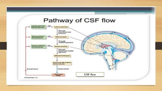
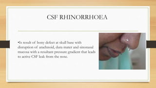
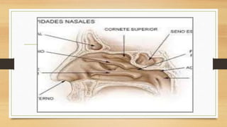
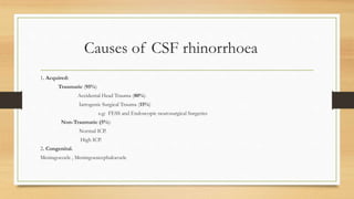
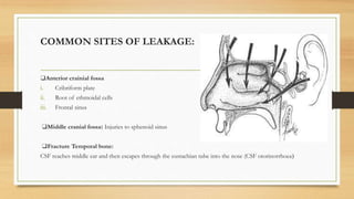
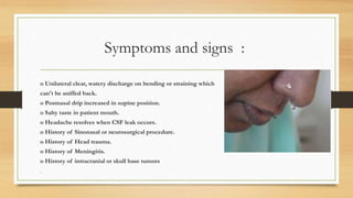
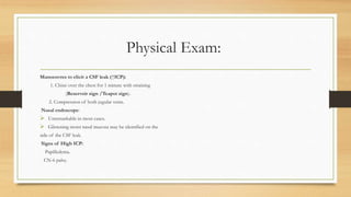
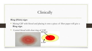
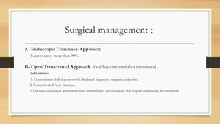
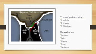
Dr. Ali Alrashidi
ENT Resident-KSH- SA
CSF RHINORRHOEA
In this lecture will discus the following :
• Basics information about CSF .
• Difintion of CSF rhinorrhea .
• causes of CSF rhinorrhea .
• most common sites of leaks .
• Symptoms and signs of CSF rhinorrhea .
• Physical Examination for patients with CSF rhinorrhea.
• How to diagnose patient with CSF rhinorrhea.
• Management patient with CSF rhinorrhea.
CSF BASICS
Cerebrospinal fluid(CSF) is a clear, colorless body fluid found in the brain
and spine .
It is produced in the choroid plexuses of the ventricles of the brain.
 It acts as a cushion or buffer for the brain's cortex, providing basic
mechanical and immunological protection to the brain.
CSF BASICS
Total volume of CSF varies from 90 to 150 m.l.
 It is secreted at the rate about 20m1/h (500 ml/day)
 There fore total CSF is replaced 3-5 times a day.
 Normal CSF pressure at lumbar puncture is 50-150 mm H2O
 It rises on coughing, sneezing, nose blowing, straining or lifting heavy
weight.
CSF flow
CSF RHINORRHOEA
•Is result of bony defect at skull base with
disruption of arachnoid, dura mater and sinonasal
mucosa with a resultant pressure gradient that leads
to active CSF leak from the nose.
Causes of CSF rhinorrhoea
1. Acquired:
Traumatic (95%)
Accidental Head Trauma (80%).
Iatrogenic Surgical Trauma (15%)
e.g: FESS and Endoscopic neurosurgical Surgeries
Non-Traumatic (5%):
Normal ICP.
High ICP.
2. Congenital.
Meningocoele , Meningoencephalocoele
COMMON SITES OF LEAKAGE:
❑Anterior crainial fossa
i. Cribriform plate
ii. Root of ethmoidal cells
iii. Frontal sinus
❑Middle cranial fossa: Injuries to sphenoid sinus
❑Fracture Temporal bone:
CSF reaches middle ear and then escapes through the eustachian tube into the nose (CSF otorinorrhoea)
Symptoms and signs :
o Unilateral clear, watery discharge on bending or straining which
can't be sniffed back.
o Postnasal drip increased in supine position.
o Salty taste in patient mouth.
o Headache resolves when CSF leak occurs.
o History of Sinonasal or neurosurgical procedure.
o History of Head trauma.
o History of Meningitis.
o History of intracranial or skull base tumors
•
Physical Exam:
Manoeuvres to elicit a CSF leak (↑ICP):
1. Chine over the chest for 1 minute with straining
(Reservoir sign /Teapot sign).
2. Compression of both jugular veins.
Nasal endoscope:
 Unremarkable in most cases.
 Glistening moist nasal mucosa may be identified on the
side of the CSF leak.
Signs of High ICP:
Papilledema.
CN-6 palsy.
Clinically
Ring (Halo) sign:
• Mixing CSF with blood and placing it onto a piece of filter paper will give a
Ring sign.
• Central blood with clear ring of CSF.
LABORATORY TESTS
o Beta-2 transferrin: a
protein seen in CSF
and not in nasal
discharge: sensitive
and specific test .
o Beta trace protein:
also specific rof, FSC
detercesby meninges
and choroid plexus.
Imaging :
1. High Resolution CT Scan:
 Initial imaging study of choice.
 Should have 1mm cuts with axial,
sagittal and coronal views.
 Sensitivity and specificity > 90%.
Treatment of CSF rhinorrhoea:
Conservative
management
Surgical
intervention
conservative management:
The goal of conservative management is to reduce the CSF leak
flow by decompressing the intracranial pressure, in this way,
healing at the defect site may seal the leak without surgical
intervention.
o 70% of traumatic CSF leak will spontaneously resolve with
conservative measures.
Conservative management Consist of:
1. Bed Rest (5-7 days):
• Head of bed elevated 30 degrees.
• Maintain normal BP.
2. Avoid (6-8 weeks):
• Sneezing (Anti-histamine).
• Coughing (Anti-tussives).
• Vomiting (Anti-emetics).
• Straining (Stool softeners).
• Nose blowing.
• Heavy lifting (> 10lbs = 4.5kg).
3. Antibiotics:
• Controversial.
• Used to prevent intracranial infection (meningitis).
4. Diuretics:
• Indicated in patients with high ICP (benign intracranial hypertension).
• Acetazolamide (Diamox) is used to decrease ICP.
5. Lumbar Drain (5-7 days):
• Indicated if CSF leak fails to responds after 5-7 days of
conservative management.
• Function to lower ICP and reduce flow through defect.
Surgical management :
Indications:
1. Failed conservative management.
2. Intra-op recognition of CSF leak .
3. Large defects/leaks:
Especially in association with pneumocephalus.
4. Idiopathic (spontaneous) leaks.
5. Open traumatic head wounds with CSF leakage .
Surgical management :
A- Endoscopic Transnasal Approach:
Success rates more than 90%.
B- Open Transcranial Approach: it’s either extracranial or intracranial .
Indications:
1. Comminuted skull fractures with displaced fragments requiring reduction.
2. Extensive skull base fractures.
3. Fractures associated with intracranial hemorrhages or contusions that require craniotomy for treatment.
Types of graft technical :
•1- underlay
•2- Overlay
•3- Multilayers
The graft to be :
•fat tissue.
•facia .
•Mucosa .
•Bone .
•Cartilages.
Questions?
Thank you

Csf rhinorrhea

  • 1.
    Dr. Ali Alrashidi ENTResident-KSH- SA CSF RHINORRHOEA
  • 2.
    In this lecturewill discus the following : • Basics information about CSF . • Difintion of CSF rhinorrhea . • causes of CSF rhinorrhea . • most common sites of leaks . • Symptoms and signs of CSF rhinorrhea . • Physical Examination for patients with CSF rhinorrhea. • How to diagnose patient with CSF rhinorrhea. • Management patient with CSF rhinorrhea.
  • 3.
    CSF BASICS Cerebrospinal fluid(CSF)is a clear, colorless body fluid found in the brain and spine . It is produced in the choroid plexuses of the ventricles of the brain.  It acts as a cushion or buffer for the brain's cortex, providing basic mechanical and immunological protection to the brain.
  • 4.
    CSF BASICS Total volumeof CSF varies from 90 to 150 m.l.  It is secreted at the rate about 20m1/h (500 ml/day)  There fore total CSF is replaced 3-5 times a day.  Normal CSF pressure at lumbar puncture is 50-150 mm H2O  It rises on coughing, sneezing, nose blowing, straining or lifting heavy weight.
  • 5.
  • 6.
    CSF RHINORRHOEA •Is resultof bony defect at skull base with disruption of arachnoid, dura mater and sinonasal mucosa with a resultant pressure gradient that leads to active CSF leak from the nose.
  • 8.
    Causes of CSFrhinorrhoea 1. Acquired: Traumatic (95%) Accidental Head Trauma (80%). Iatrogenic Surgical Trauma (15%) e.g: FESS and Endoscopic neurosurgical Surgeries Non-Traumatic (5%): Normal ICP. High ICP. 2. Congenital. Meningocoele , Meningoencephalocoele
  • 9.
    COMMON SITES OFLEAKAGE: ❑Anterior crainial fossa i. Cribriform plate ii. Root of ethmoidal cells iii. Frontal sinus ❑Middle cranial fossa: Injuries to sphenoid sinus ❑Fracture Temporal bone: CSF reaches middle ear and then escapes through the eustachian tube into the nose (CSF otorinorrhoea)
  • 10.
    Symptoms and signs: o Unilateral clear, watery discharge on bending or straining which can't be sniffed back. o Postnasal drip increased in supine position. o Salty taste in patient mouth. o Headache resolves when CSF leak occurs. o History of Sinonasal or neurosurgical procedure. o History of Head trauma. o History of Meningitis. o History of intracranial or skull base tumors •
  • 11.
    Physical Exam: Manoeuvres toelicit a CSF leak (↑ICP): 1. Chine over the chest for 1 minute with straining (Reservoir sign /Teapot sign). 2. Compression of both jugular veins. Nasal endoscope:  Unremarkable in most cases.  Glistening moist nasal mucosa may be identified on the side of the CSF leak. Signs of High ICP: Papilledema. CN-6 palsy.
  • 12.
    Clinically Ring (Halo) sign: •Mixing CSF with blood and placing it onto a piece of filter paper will give a Ring sign. • Central blood with clear ring of CSF.
  • 13.
    LABORATORY TESTS o Beta-2transferrin: a protein seen in CSF and not in nasal discharge: sensitive and specific test . o Beta trace protein: also specific rof, FSC detercesby meninges and choroid plexus.
  • 14.
    Imaging : 1. HighResolution CT Scan:  Initial imaging study of choice.  Should have 1mm cuts with axial, sagittal and coronal views.  Sensitivity and specificity > 90%.
  • 17.
    Treatment of CSFrhinorrhoea: Conservative management Surgical intervention
  • 18.
    conservative management: The goalof conservative management is to reduce the CSF leak flow by decompressing the intracranial pressure, in this way, healing at the defect site may seal the leak without surgical intervention. o 70% of traumatic CSF leak will spontaneously resolve with conservative measures.
  • 19.
    Conservative management Consistof: 1. Bed Rest (5-7 days): • Head of bed elevated 30 degrees. • Maintain normal BP. 2. Avoid (6-8 weeks): • Sneezing (Anti-histamine). • Coughing (Anti-tussives). • Vomiting (Anti-emetics). • Straining (Stool softeners). • Nose blowing. • Heavy lifting (> 10lbs = 4.5kg). 3. Antibiotics: • Controversial. • Used to prevent intracranial infection (meningitis). 4. Diuretics: • Indicated in patients with high ICP (benign intracranial hypertension). • Acetazolamide (Diamox) is used to decrease ICP. 5. Lumbar Drain (5-7 days): • Indicated if CSF leak fails to responds after 5-7 days of conservative management. • Function to lower ICP and reduce flow through defect.
  • 20.
    Surgical management : Indications: 1.Failed conservative management. 2. Intra-op recognition of CSF leak . 3. Large defects/leaks: Especially in association with pneumocephalus. 4. Idiopathic (spontaneous) leaks. 5. Open traumatic head wounds with CSF leakage .
  • 21.
    Surgical management : A-Endoscopic Transnasal Approach: Success rates more than 90%. B- Open Transcranial Approach: it’s either extracranial or intracranial . Indications: 1. Comminuted skull fractures with displaced fragments requiring reduction. 2. Extensive skull base fractures. 3. Fractures associated with intracranial hemorrhages or contusions that require craniotomy for treatment.
  • 22.
    Types of grafttechnical : •1- underlay •2- Overlay •3- Multilayers The graft to be : •fat tissue. •facia . •Mucosa . •Bone . •Cartilages.
  • 23.
  • 24.