Downloaded 137 times


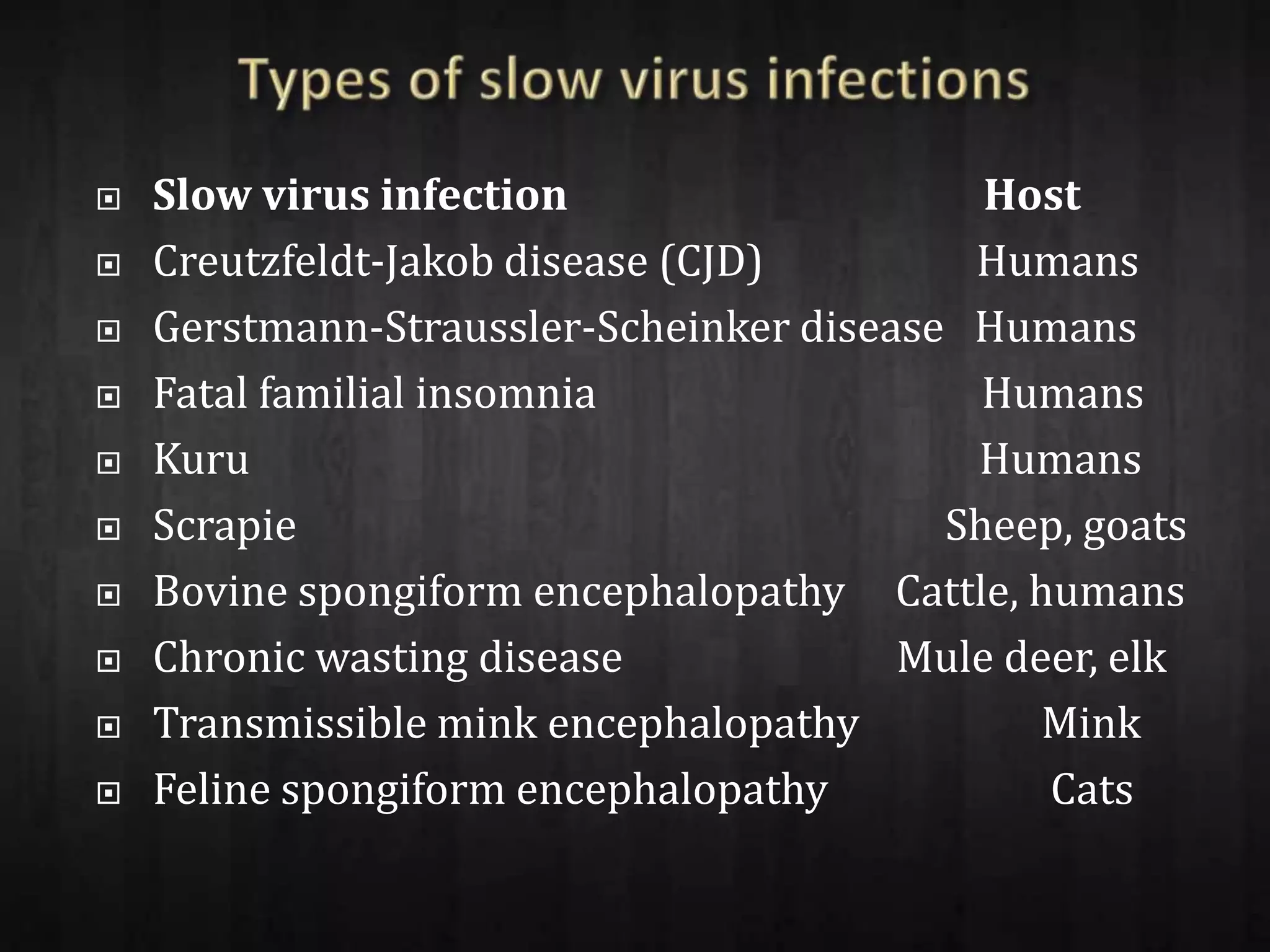
![ Prion diseases are categorized into three groups:
sporadic, (85%), hereditary &acquired(15%)
15% consist of hereditary forms (hereditary
Creutzfeldt-Jakob disease [CJD], Gerstmann-
Straussler-Scheinker disease, fatal familial insomnia)
and acquired forms.
Acquired forms may be transmitted iatrogenically
(through human growth hormone therapy, dura
mater grafts, or other neurosurgical procedures) or
through cannibalism (kuru) , variant CJD (vCJD) in
humans, scrapie in sheep, and bovine spongiform
encephalopathy (BSE) in cattle.](https://image.slidesharecdn.com/cjd-120908225251-phpapp02/75/Creutzfeldt-Jakob-disease-30-2048.jpg)


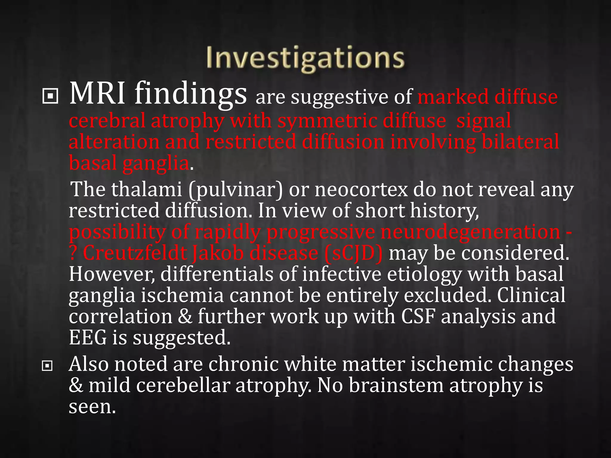
This patient, a 65-year-old male, presented with 6 months of gradually worsening gait difficulties and incoordination. Over the past 4 months, he developed forgetfulness, hallucinations, and sensorium changes. Exams found rigidity, myoclonic jerks, and a non-reactive semi-conscious state. Imaging and tests were suggestive of Creutzfeldt-Jakob disease (CJD), a prion disease characterized by spongiform changes in the brain. A definitive diagnosis requires brain biopsy or autopsy.


