
More Related Content
What's hot
What's hot (19)
Similar to Cranial nerves presentation
Similar to Cranial nerves presentation (20)
Recently uploaded
Recently uploaded (20)
Cranial nerves presentation
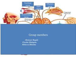
- 1. Trochlear nerve Optic nerve Olfactory nerve Oculomotor nerve Group members Abenezer Bogale Fisseha Mulugeta Rebecca Shisema
- 2. Outline • Origin • Course • Special features • Clinical Significance
- 3. Origin -Arises from olfactory epithelium of the upper part of the nasal cavity as olfactory fascicles where it takes the olfactory information from the environment. Olfactory Nerve
- 4. Course -After arising as olfactory fascicles travel up in to the the cribriform plate of the ethimoid bone -Come together as olfactory bulb. In the olfactory bulb the fibers from the nasal mucosa will join with mitral dendrites found on the bulb. -After they come together in the olfactory bulb they will become olfactory tract -It goes to anterior perforated substance and divide to medial and lateral olfactory striae.
- 6. Special features • It is purely sensory nerve (special visceral afferent • is one of the two nerves that do not arise from the brain stem • Shortest of the 12 cranial nerves. • As it originates from olfactory placode it is the only cranial nerve that has ability to regenerate
- 7. Clinical Significance Anosmia • Damage to this nerve may lead to the state of inability to smell or anosmia. • But inability to smell is not only caused by nerve damage but may be due to blunt trauma, tumor of the frontal lobe, Menningitis,Covid-19
- 8. Optic Nerve It is a paired nerve arising from the ganglion layer of the retina. Origin
- 9. Course • It leaves the eye after converging in the optic disc • Goes to the optic chiasm then ascends as optic tract to the lateral geniculate nucleus. • Then to the pretectal nucleus and superior colliculus. • In the optic chiasm it will cross with the contra lateral optic nerve fiber.
- 10. Special features • It is purely sensory nerve and its nucleus do not arise from the brain stem. • Unlike any other cranial nerve its is lined by oligodendrocytes rather than Schwann cells which makes it part of the central nerves system and susceptible to diseases affected the central nervous system such as multiple sclerosis. • The optic nerve is ensheathed in all three meningeal layers (dura, arachnoid, and pia mater) rather than the epineurium, perineurium, and endoneurium found in peripheral nerves
- 11. Clinical Significance -Visual loss from the nerve damage depends on the location of the damage.
- 12. Occulomotor Nerve Origin -It starts from the midbrain in the anterior part from its nucleus -Goes to the eye orbit via superior orbital fissure -Innervates extrinsic eye muscles that enable most movements of the eye and that raise the eyelid.
- 13. Course • After arising from its nucleus from the mid brain it goes lateral to the cavernous sinus. In the cavernous sinus it divides into:- -Superior:- for sup. rectus & levator palpebrae superioris -Inferior:- for inferior & medial rectus, inferior oblique
- 15. Special features -It is purely motor nerve and it is one of the nerves that arises from the brain stem -Has characteristics of both general somatic efferent and general visceral efferent. -Contain parasympathetic fibers arising from Edinger Westephal nuclei to sphincter pupilae and ciliary muscles
- 16. Clinical Significance Caused by DM Hypertension Demyelinating diseases
- 17. Trochlear nerve Origin -It arises from the back of the mid brain and comes in front of the brain stem to supply the superior oblique muscle Course • Has same path with the cranial nerve III and it goes to the orbit via superior orbital fissure.
- 18. Special features The smallest cranial nerve in route. It runs the longest intracranial route. Emerges from the posterior wall of midbrain
- 19. Clinical Significance Results in diplopia and inability to rotate the eye inferolateral. So the eye deviates upward and slightly inward. Person has difficulty in walking downstairs.
- 20. Sources and reference Snell’s Neuroanatomy 7th edition Netters atlas anatomy
Editor's Notes
- You will ask students to read the title and when they read it you will say they have optic nerve
- Read about this and explain
- Or use this