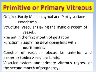
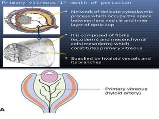
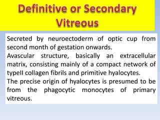
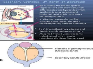
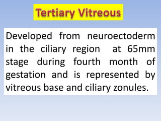
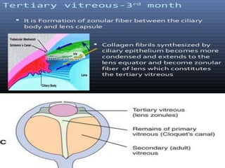
Vitreous humor is an inert, transparent,
colourless, jelly-like, hydrophilic gel that
serves the optical functions and also acts as
important supporting structure for eyeball.
Primitive or Primary
vitreous
Definitive or
Secondary vitreous
Tertiary vitreous
Origin : Partly Mesenchymal and Partly surface
ectodermal.
Structure: Vascular Having the Hyaloid system of
vessels.
Present in the first month of gestation.
Function: Supply the developing lens with
nourishment.
Consists of vascular plexus i.e anterior and
posterior tunica vasculosa lentis.
Vascular system and primary vitreous regress at
the second month of pregnancy.
Secreted by neuroectoderm of optic cup from
second month of gestation onwards.
Avascular structure, basically an extracellular
matrix, consisting mainly of a compact network of
typeII collagen fibrils and primitive hyalocytes.
The precise origin of hyalocytes is presumed to be
from the phagocytic monocytes of primary
vitreous.
Content of hyaluronic acid is very low during
the prenatal period, but increases after birth.
When this vitreous fills the cavity by fifth to
sixth month of gestation, primitive vitreous is
reduced to a small central space , Cloquet’s
canal, which lies between the optic nerve
head and the posterior lens surface.
The development may continue as late as 65-
70mm stage.
Developed from neuroectoderm
in the ciliary region at 65mm
stage during fourth month of
gestation and is represented by
vitreous base and ciliary zonules.
1.Name the stages of vitreous development?
2.Definitive vitreous consist of………………
3.Which stage of vitreous development is
vascular?
4. In the postnatal period the density of ………
increases and ……….decreases.
5. Cloquet’s canal lies between ……….. and
………………
6.Function of Primitive vitreous?
7. Tertiary vitreous is represented by……………..
8.When does primitive vitreous regress?
1.Primitive or Primary vitreous, Definitive or
secondary vitreous and tertiary vitreous.
2. TypeII collagen fibrils and primitive
hyalocytes.
3. Primitive vitreous.
4. Hyaluronic acid and Collagen fibrils.
5. Optic nerve head and Posterior lens surface.
6. Supply nutrition for the developing lens.
7. Vitreous base and ciliary zonules.
8. At the second month of pregnancy.
Vitreous

Vitreous

  • 2.
    Vitreous humor isan inert, transparent, colourless, jelly-like, hydrophilic gel that serves the optical functions and also acts as important supporting structure for eyeball.
  • 3.
    Primitive or Primary vitreous Definitiveor Secondary vitreous Tertiary vitreous
  • 4.
    Origin : PartlyMesenchymal and Partly surface ectodermal. Structure: Vascular Having the Hyaloid system of vessels. Present in the first month of gestation. Function: Supply the developing lens with nourishment. Consists of vascular plexus i.e anterior and posterior tunica vasculosa lentis. Vascular system and primary vitreous regress at the second month of pregnancy.
  • 6.
    Secreted by neuroectodermof optic cup from second month of gestation onwards. Avascular structure, basically an extracellular matrix, consisting mainly of a compact network of typeII collagen fibrils and primitive hyalocytes. The precise origin of hyalocytes is presumed to be from the phagocytic monocytes of primary vitreous.
  • 7.
    Content of hyaluronicacid is very low during the prenatal period, but increases after birth. When this vitreous fills the cavity by fifth to sixth month of gestation, primitive vitreous is reduced to a small central space , Cloquet’s canal, which lies between the optic nerve head and the posterior lens surface. The development may continue as late as 65- 70mm stage.
  • 9.
    Developed from neuroectoderm inthe ciliary region at 65mm stage during fourth month of gestation and is represented by vitreous base and ciliary zonules.
  • 12.
    1.Name the stagesof vitreous development? 2.Definitive vitreous consist of……………… 3.Which stage of vitreous development is vascular? 4. In the postnatal period the density of ……… increases and ……….decreases. 5. Cloquet’s canal lies between ……….. and ……………… 6.Function of Primitive vitreous? 7. Tertiary vitreous is represented by…………….. 8.When does primitive vitreous regress?
  • 14.
    1.Primitive or Primaryvitreous, Definitive or secondary vitreous and tertiary vitreous. 2. TypeII collagen fibrils and primitive hyalocytes. 3. Primitive vitreous. 4. Hyaluronic acid and Collagen fibrils. 5. Optic nerve head and Posterior lens surface. 6. Supply nutrition for the developing lens. 7. Vitreous base and ciliary zonules. 8. At the second month of pregnancy.