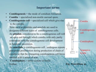
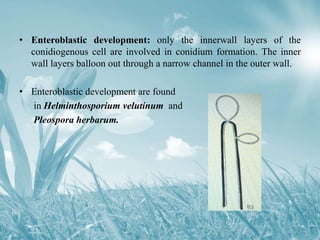
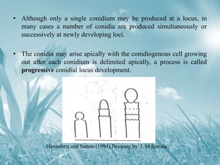
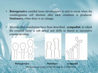
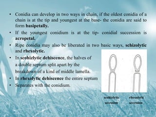
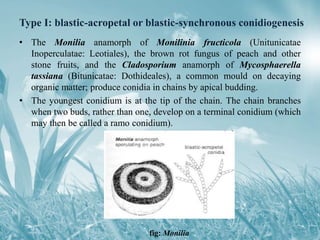
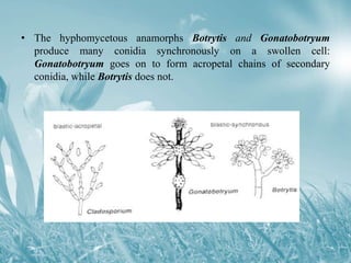
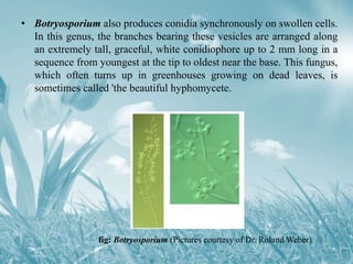
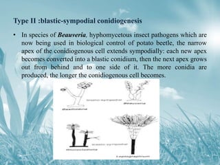
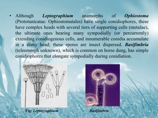
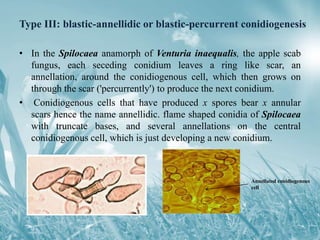
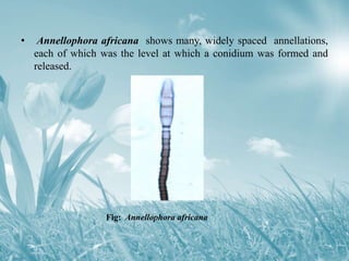
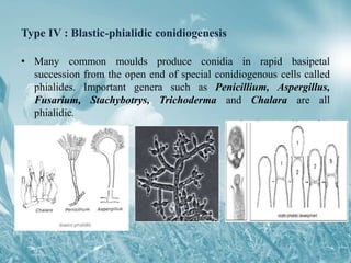
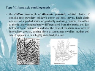
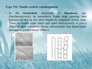
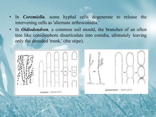
Conidial ontogeny refers to the modes of formation and development of asexual spores known as conidia. There are eight types of conidial ontogeny: six involve blastic or enlargement development and two involve thallic or non-enlargement development. Examples are provided to illustrate the different types including phialidic, annellidic, synchronous, and arthroconidial development. Key terms are also defined such as conidiogenous cell, conidiophore, and schizolytic versus rhexolytic dehiscence.
There is a tremendous variety of morphologically different conidia produced. The study of development of conidia based on its origin is referred to as “Conidial Ontogeny”.
Heterothallic species have sexes that reside in different individuals. . The term is applied particularly to distinguish heterothallic fungi, which require two compatible partners to produce sexual spores, from homothallic ones, which are capable of sexual reproduction from a single organism.
A chemical substance that is produced in one portion of an organism and moves by diffusion or transport to another portion of same individual or to other individual of same species where it induce specific response is called a hormone.
Introduction,In some fungi ,true sexual cycle comprising of nuclear fusion and meiosis is absent.
These fungi derive the benefits of sexuality through a cycle know as parasexuaL cycle.
First Reported by- Gudio Pontecorvo and J.A.Roper(1952)
Parasexual cycle was reported in
Aspergillus nidulans,the imperfect stage of Emericella nidulans.
Since then parasexual cycle has been discovered not only in several members of Deutromycetes but also in fungi belonging to Ascomycetes and Basidiomycetes.
DEFINETION - Parasexuality is defined as a cycle in which Plasmogamy, Karyogamy and Meiosis [Haploidization] take place in sequence but not at a specified time or at specified points in the life cycle of an organism.
Generally parasexual cycle occurs in those fungi in which true sexual cycle does not take place.
Parasexualcycle also know as Somatic recombination. PASEXUALITY ALSO REPORTED IN SOME ORGANISMS- Aspergillus nigar, Penicillium crysogenum, STEPS OF PARASEXUAL CYCLE - 1) ESTABLISHMENT OF HETEROKARYOSIS, 2) Formation of Heterozygous DIPLOIDS, 3) occasional mitotic crossing-over during multiplication of diploid nuclei, 4)occasional haplodization through aneuploidy , COMPARISION BETWEEN SEXUAL AND PARASEXUAL CYCLE, IMPORTANCE OF PARASEXUALITY, C0NCLUSION
As part of our project on educational technology 3/ field study 3
I am asking for your help with regards to the ppt that i made kindly leave a comment on my presentation thanks alot
Detail about Basidiomycetes.In this detail about its Ecosystem Relationship,Symbiotic Relationships,General characters,Basidiospores,Life cycle and its Fruiting body.
Contact Email: mzeeshan_93@yahoo.com
FUNGI - ROLE IN AGRICULTURE,MEDICINE & COMMERCIAL PRODUCTS. ChhanKumarkalita
TOPIC: FUNGI - ROLE IN AGRICULTURE,MEDICINE & COMMERCIAL PRODUCTS.
Presented By: Chhan kumar kalita
B.Sc. in Botany, Dept of Botany, B.P. Chaliha College, Nagarbera
M.Sc. in Botany, Dept of Botany, Nowgown College (Autonomous)
Molecular basis of plant resistance and defense responses to pathogensSenthil Natesan
In response to pathogen attack, plants have evolved sophisticated defense mechanisms to delay or arrest pathogen growth.Unlike animals, plants lack a circulating immune system recognizing microbial pathogens. Plant cells are more autonomous in their defense mechanisms and rely on the innate immune capacity of each cell and systemic signals that disseminate from infection sites (Jones and Dangl, 2006). Plant innate immunity consists of preformed physical and chemical barriers (such as leaf hairs, rigid cell walls, pre-existing antimicrobial compounds) and induced defenses. Should an invading microbe successfully breach the pre-formed barriers, it may be recognized by the plant, resulting in the activation of cellular defense responses that stop or restrict further development of the invader.
Plant - Pathogen Interaction and Disease DevelopmentKK CHANDEL
Plant diseases are the result of infection by any living organisms that adversely affect the growth, development, physiological functioning and productivity of a plant, manifesting outwardly as visible symptoms.
The SlideShare 101 is a quick start guide if you want to walk through the main features that the platform offers. This will keep getting updated as new features are launched.
The SlideShare 101 replaces the earlier "SlideShare Quick Tour".
Each month, join us as we highlight and discuss hot topics ranging from the future of higher education to wearable technology, best productivity hacks and secrets to hiring top talent. Upload your SlideShares, and share your expertise with the world!
Not sure what to share on SlideShare?
SlideShares that inform, inspire and educate attract the most views. Beyond that, ideas for what you can upload are limitless. We’ve selected a few popular examples to get your creative juices flowing.
Deuteromycotina is a polyphyletic group of fungi that reproduce asexually by the generation of conidia (asexual spores). Because these fungi lack a sexual reproductive cycle, they do not have a known sexual stage in their life cycle. The categorization of Deuteromycotina has been debated, as the lack of a documented sexual stage has made determining their evolutionary links with other fungal taxa problematic. With the introduction of molecular biology tools in recent years, several Deuteromycotina species have been reassigned into other fungal phyla based on genetic similarities. Aspergillus, Penicillium, and Trichoderma are examples of Deuteromycotina that are commonly used in the biotechnology and pharmaceutical industries for the synthesis of antibiotics and other chemicals. However, genetic analysis has led to the reclassification of many of these fungi into different phyla.
PHARMACEUTICAL MICROBIOLOGY (BP303T) Unit-III Part-1 Study of morphology, cla...Ms. Pooja Bhandare
PHARMACEUTICAL MICROBIOLOGY (BP303T)Unit-IIIPart-1Study of morphology, classification, reproduction/replication and cultivation of fungi, Introduction fungi. Morphological Characteristics of fungi, CLASSIFICATION: Depending on cell morphology, fungi can be divided into 4 classes:
Moulds Yeasts ,Yeast like fungi and
Dimorphic fungi
Depending on their sexual spores formation fungi are divided into 4 classes:
Zygomycetes Ascomycetes
Basidiomycetes Dueteromycetes
Reproduction and sporulation;Vegetative, Asexual
and Sexual
Vegetative reproduction: Fragmentation ,Fission, budding, Sclerotia Rhizomorphs
Asexual reproduction: Zoospores
Sporangiospore, Conidia
Oidia Uredospores ,Basidiospores
Sexual reproduction:Planogametic copulation: Isogamy Heterogamy
Gametangial contact
Gametangial copulation Spermatization Somatogamy CULTIVATION OF FUNGI: Brain Heart Infusion (BHT) agar
Czapek’s agar
Mycobiotic agar Inhibitory mold agar (IMA)
Potato dextrose agar
Sabouraud’s dextrose agar (SDA):
Sabouraud’s heart infusion (SABHI) agar
Potato Flake agar
Potato dextrose-yeast extract agar (PDYA)
. Cornmeal agar
Malt extract agar (MEA)
Structure and reproduction of Aspergillus niger ,with picture of different reproduction methods in detail ,also called sac fungi,large groupof true fungi ,saprophyte,it also known to cause food contaminations or food spoilage ,also cause black mold in fruits and vegetables like grapes, apricote ,onions and peanuts .Aspergillus niger is common group of Aspergillus.reproduction by sexual ,asexual or vegetative methods. vegetative mthods by fragmentation ,sclerotia
Penicillium (/ˌpɛnɪˈsɪliəm/) is a genus of ascomycetous fungi that is part of the mycobiome of many species and is of major importance in the natural environment, in food spoilage, and in food and drug production.
Penicillium is a genus of ascomycetous fungi that is part of the mycobiome of many species and is of major importance in the natural environment, in food spoilage, and in food and drug production.
Seminar of U.V. Spectroscopy by SAMIR PANDASAMIR PANDA
Spectroscopy is a branch of science dealing the study of interaction of electromagnetic radiation with matter.
Ultraviolet-visible spectroscopy refers to absorption spectroscopy or reflect spectroscopy in the UV-VIS spectral region.
Ultraviolet-visible spectroscopy is an analytical method that can measure the amount of light received by the analyte.
This pdf is about the Schizophrenia.
For more details visit on YouTube; @SELF-EXPLANATORY;
https://www.youtube.com/channel/UCAiarMZDNhe1A3Rnpr_WkzA/videos
Thanks...!
A brief information about the SCOP protein database used in bioinformatics.
The Structural Classification of Proteins (SCOP) database is a comprehensive and authoritative resource for the structural and evolutionary relationships of proteins. It provides a detailed and curated classification of protein structures, grouping them into families, superfamilies, and folds based on their structural and sequence similarities.
This presentation explores a brief idea about the structural and functional attributes of nucleotides, the structure and function of genetic materials along with the impact of UV rays and pH upon them.
Professional air quality monitoring systems provide immediate, on-site data for analysis, compliance, and decision-making.
Monitor common gases, weather parameters, particulates.
Cancer cell metabolism: special Reference to Lactate PathwayAADYARAJPANDEY1
Normal Cell Metabolism:
Cellular respiration describes the series of steps that cells use to break down sugar and other chemicals to get the energy we need to function.
Energy is stored in the bonds of glucose and when glucose is broken down, much of that energy is released.
Cell utilize energy in the form of ATP.
The first step of respiration is called glycolysis. In a series of steps, glycolysis breaks glucose into two smaller molecules - a chemical called pyruvate. A small amount of ATP is formed during this process.
Most healthy cells continue the breakdown in a second process, called the Kreb's cycle. The Kreb's cycle allows cells to “burn” the pyruvates made in glycolysis to get more ATP.
The last step in the breakdown of glucose is called oxidative phosphorylation (Ox-Phos).
It takes place in specialized cell structures called mitochondria. This process produces a large amount of ATP. Importantly, cells need oxygen to complete oxidative phosphorylation.
If a cell completes only glycolysis, only 2 molecules of ATP are made per glucose. However, if the cell completes the entire respiration process (glycolysis - Kreb's - oxidative phosphorylation), about 36 molecules of ATP are created, giving it much more energy to use.
IN CANCER CELL:
Unlike healthy cells that "burn" the entire molecule of sugar to capture a large amount of energy as ATP, cancer cells are wasteful.
Cancer cells only partially break down sugar molecules. They overuse the first step of respiration, glycolysis. They frequently do not complete the second step, oxidative phosphorylation.
This results in only 2 molecules of ATP per each glucose molecule instead of the 36 or so ATPs healthy cells gain. As a result, cancer cells need to use a lot more sugar molecules to get enough energy to survive.
introduction to WARBERG PHENOMENA:
WARBURG EFFECT Usually, cancer cells are highly glycolytic (glucose addiction) and take up more glucose than do normal cells from outside.
Otto Heinrich Warburg (; 8 October 1883 – 1 August 1970) In 1931 was awarded the Nobel Prize in Physiology for his "discovery of the nature and mode of action of the respiratory enzyme.
WARNBURG EFFECT : cancer cells under aerobic (well-oxygenated) conditions to metabolize glucose to lactate (aerobic glycolysis) is known as the Warburg effect. Warburg made the observation that tumor slices consume glucose and secrete lactate at a higher rate than normal tissues.
THE IMPORTANCE OF MARTIAN ATMOSPHERE SAMPLE RETURN.Sérgio Sacani
The return of a sample of near-surface atmosphere from Mars would facilitate answers to several first-order science questions surrounding the formation and evolution of the planet. One of the important aspects of terrestrial planet formation in general is the role that primary atmospheres played in influencing the chemistry and structure of the planets and their antecedents. Studies of the martian atmosphere can be used to investigate the role of a primary atmosphere in its history. Atmosphere samples would also inform our understanding of the near-surface chemistry of the planet, and ultimately the prospects for life. High-precision isotopic analyses of constituent gases are needed to address these questions, requiring that the analyses are made on returned samples rather than in situ.
2. Introduction
• Deuteromycetes or Fungi imperfecti are fungi without a teleomorph.
It is believed to be the primitive condition of Ascomycetes and
Basidiomycetes.
• Most food- and airborne fungi belong to this group: Penicillium,
Aspergillus, Fusarium and Cladosporium etc.
• This group of fungi is artificial and is characterized by their way of
sporulation, and produced conidia-asexual spore .
• Besides true conidia, some fungi, especially animal and human
pathogens, produce other types of asexual spores: microconidia,
blastospores, and arthrospores.
3. • Microconidia are very small conidia. Blastospores (Gr. Blastos- bud,
shoot, + sporos- seed, spore) are asexual spores formed by budding
either directly from a hypha, or from any other cell.
• Arthrospores (Gr. Arthron- joint, + sporos- seed, spore) are formed
by the breaking up of the hyphae into their component cells. They
are no different from oidia. All three of germinate to form mycelium
and function the same as conidia.
4. Important terms
• Conidiogenesis = the mode of conidium formation.
• Conidia = specialized non-motile asexual spore.
• Conidiogenous cell = specialized cell which give rise
to the conidia.
Term such as phialides and annelide are used to
designate different types of conidiogenous cells.
An phialide considered to be a conidiogenous cell with
an open end through which conidia with only partly
continuous with the conidiogenous cell development
in basipetal succession.
An annellide is conidiogenous cell , undergoes repeated
percurrent proliferation during production of chain of
conidia so that the elongating conidiogenous cell become
marked with a series of scar.
• Conidiophore = entire system of fertile
hyphae. Fig: Penicillium sp.
Conidia
Conidiogenous
cell
Conidiophore
Vegetative
hypha
5. Conidia and Conidial Ontogeny
• Conidiospores, commonly known as conidia, are asexual
reproductive structures. The word is derived from the
Greek konis ‘dust’ + the diminutive suffix -idium (Sutton, 1986).
• Conidia are found in many different groups of fungi, but especially
within Deuteromycotina. The term conidium has, unfortunately,
been used in a number of different ways, so that it no longer has any
precise meaning.
• In many fungi conidia represent a means of rapid spread and
colonization from an initial focus of infection. Conidia may
originated from conidiogenous cell in several ways.
6. • There is great variation in conidial ontogeny, which may be either
thallic or
blastic.
1. Thallic- is used to describe development where there is no
enlargement of the conidium initial, i.e. the conidium arises by
conversion of a pre-existing segment of the fungal thallus. An
example : Galactomyces candidus, in which the conidia are
formed by dissolution of septa along a hypha.
Fig: Galactomyces candidus
7. 2. In most conidia, development is blastic, i.e. there is enlargement of
the conidium initial before it is delimited by a septum.
• Two main kinds of blastic development have been distinguished:
i. Holoblastic,
ii. Enteroblastic,
Holoblastic development- All the wall layers of the conidiogenous cell
balloon out to form a conidium initial recognizably larger than the
conidiogenous cell. Conidia of Sclerotinia fructigena.
Fig: Sclerotinia fructigena
8. • Enteroblastic development: only the innerwall layers of the
conidiogenous cell are involved in conidium formation. The inner
wall layers balloon out through a narrow channel in the outer wall.
• Enteroblastic development are found
in Helminthosporium velutinum and
Pleospora herbarum.
9. • Although only a single conidium may be produced at a locus, in
many cases a number of conidia are produced simultaneously or
successively at newly developing loci.
• The conidia may arise apically with the conidiogenous cell growing
out after each conidium is delimited apically, a process is called
progressive conidial locus development.
Hennebert and Sutton (1994) Drawing by L M Barona.
10. • Retrogressive conidial locus development is said to occur when the
conidiogenous cell shortens after each conidium is produced.
Stationary, when there is no change.
• Several other conidiation have been described , sympodial; in which
the conidial locus is sub apical and shifts to lateral as successive
conidia develop.
Retrogressive Stationary sympodial
(Hennebert and Sutton (1994) Drawing by L M Barona.)
11. • Conidia can develop in two ways in chain, if the oldest conidia of a
chain is at the tip and youngest at the base- the conidia are said to
form basipetally.
• If the youngest conidium is at the tip- conidial succession is
acropetal.
• Ripe conidia may also be liberated in two basic ways, schizolytic
and rhexolytic.
• In scohizlytic dehiscence, the halves of
a double septum split apart by the
breakdown of a kind of middle lamella.
• In rhexolytic dehiscence the entire septum
• Separates with the conidium.
scohizlytic rhexolytic
secession secession
12. • There are eight different kinds of conidium development: six are
blastic, two thallic development.
i. Blastic-acropetal or blastic-synchronous conidiogenesis.
ii. blastic-sympodial conidiogenesis.
iii. blastic-annellidic or blastic-percurrent conidiogenesis.
iv. Blastic-phialidic conidiogenesis.
v. Blastic-retrogressive conidiogenesis.
vi. Basauxic conidiogenesis.
vii. Thallic-arthric conidiogenesis.
viii. Thallic-solitary conidiogenesis
13. Type I: blastic-acropetal or blastic-synchronous conidiogenesis
• The Monilia anamorph of Monilinia fructicola (Unitunicatae
Inoperculatae: Leotiales), the brown rot fungus of peach and other
stone fruits, and the Cladosporium anamorph of Mycosphaerella
tassiana (Bitunicatae: Dothideales), a common mould on decaying
organic matter; produce conidia in chains by apical budding.
• The youngest conidium is at the tip of the chain. The chain branches
when two buds, rather than one, develop on a terminal conidium (which
may then be called a ramo conidium).
fig: Monilia
14. • The hyphomycetous anamorphs Botrytis and Gonatobotryum
produce many conidia synchronously on a swollen cell:
Gonatobotryum goes on to form acropetal chains of secondary
conidia, while Botrytis does not.
15. • Botryosporium also produces conidia synchronously on swollen cells.
In this genus, the branches bearing these vesicles are arranged along
an extremely tall, graceful, white conidiophore up to 2 mm long in a
sequence from youngest at the tip to oldest near the base. This fungus,
which often turns up in greenhouses growing on dead leaves, is
sometimes called 'the beautiful hyphomycete.
fig: Botryosporium (Pictures courtesy of Dr. Roland Weber)
16. Type II :blastic-sympodial conidiogenesis
• In species of Beauveria, hyphomycetous insect pathogens which are
now being used in biological control of potato beetle, the narrow
apex of the conidiogenous cell extends sympodially: each new apex
becomes converted into a blastic conidium, then the next apex grows
out from behind and to one side of it. The more conidia are
produced, the longer the conidiogenous cell becomes.
17. • Although Leptographium anamorphs of Ophiostoma
(Prototunicatae: Ophiostomatales) have single conidiophores, these
have complex heads with several tiers of supporting cells (metulae),
the ultimate ones bearing many sympodially (or percurrently)
extending conidiogenous cells, and innumerable conidia accumulate
in a slimy head; these spores are insect dispersed. Basifimbria
(teleomorph unknown), which is common on horse dung, has simple
conidiophores that elongate sympodially during conidiation.
Fig: Leptographium Basifimbria
18. Type III: blastic-annellidic or blastic-percurrent conidiogenesis
• In the Spilocaea anamorph of Venturia inaequalis, the apple scab
fungus, each seceding conidium leaves a ring like scar, an
annellation, around the conidiogenous cell, which then grows on
through the scar ('percurrently') to produce the next conidium.
• Conidiogenous cells that have produced x spores bear x annular
scars hence the name annellidic. flame shaped conidia of Spilocaea
with truncate bases, and several annellations on the central
conidiogenous cell, which is just developing a new conidium.
Annellated conidiogenous
cell
19. • Annellophora africana shows many, widely spaced annellations,
each of which was the level at which a conidium was formed and
released.
Fig: Annellophora africana
20. Type IV : Blastic-phialidic conidiogenesis
• Many common moulds produce conidia in rapid basipetal
succession from the open end of special conidiogenous cells called
phialides. Important genera such as Penicillium, Aspergillus,
Fusarium, Stachybotrys, Trichoderma and Chalara are all
phialidic.
21. Type V: Blastic-retrogressive conidiogenesis
• In Basipetospora (thermotolerant fungus used in Indonesia in the
preparation of a red food colouring), a conidium forms at the tip of
the relatively undifferentiated conidiogenous hypha and is delimited
by a crosswall; then a short zone of the hypha just below the
conidium balloons out to produce the second conidium.
• After this has been delimited by a septum, the next segment of the
hypha plasticizes and blows out, and so on. As the chain of conidia
elongates, the conidiogenous hypha becomes shorter.
22. Type VI: basauxic conidiogenesis
• the Oidium anamorph of Blumeria graminis, whitish chains of
conidia (the 'powdery mildew') cover the host leaves. Each chain
consists of a graded series of gradually maturing conidia, the oldest
at the tip, the youngest barely differentiated from the hyphal cell just
below it. New material is added at the base of the chain in a form of
intercalary growth, arising from a sometimes swollen mother cell
which appears to be a highly modified phialide.
23. Type VII: Thallic-arthric conidiogenesis
• In the Geotrichum anamorphs of Dipodascus spp.
(Saccharomycetes), an assimilative hypha stops growing, then
becomes divided up into short lengths by irregularly arising septa.
These are double septa which split apart schizolytically to give a
'chain' of short cylindrical 'fission arthroconidia' that disarticulates
and appears jointed (hence 'arthric').
24. • In Coremiella, some hyphal cells degenerate to release the
intervening cells as 'alternate arthroconidia.‘
• In Oidiodendron, a common soil mould, the branches of an often
tree like conidiophore disarticulate into conidia, ultimately leaving
only the denuded 'trunk,' (the stipe).
25. Type VIII: Thallic-solitary conidiogenesis
• The Microsporum anamorphs of Nannizzia (Prototunicatae:
Onygenales), which can digest keratin, and cause skin diseases in
humans, develop large thallic phragmospores at the ends of hyphae.
These conidia are liberated rhexolytically.