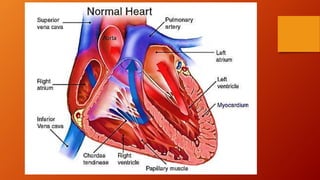
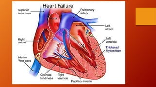
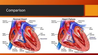
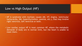
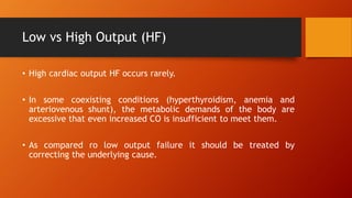
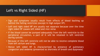
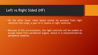
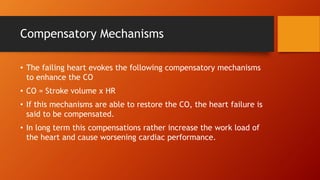
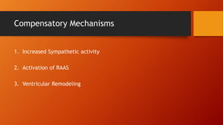
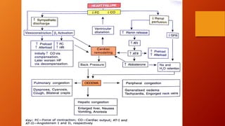
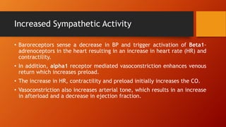
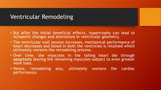
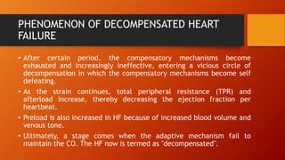
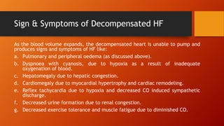
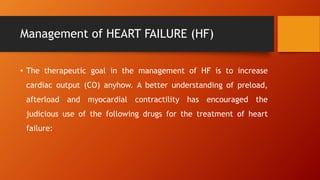
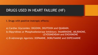
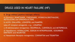
2. Common Terms related to Heart
ā¢ Cardiac output
ā¢ Heart Rate
ā¢ Force of Contraction
ā¢ Peripheral vascular resistance
ā¢ Preload
ā¢ Afterload
ā¢ Stroke volume
ā¢ Systole
ā¢ Diastole
ā¢ EDV
ā¢ ESV
ā¢ Congestion
3. ā¢ Cardiac Output The amount of blood the heart pumps through the
circulatory system in a minute.
ā¢ Heart rate is the speed of the heartbeat measured by the number
of contractions of the heart per minute.
ā¢ Cardiac contractility can be defined as the tension developed and
velocity of shortening (i.e., the āstrengthā of contraction) of
myocardial fibers at a given preload and afterload.
ā¢ Vascular resistance is the resistance that must be overcome to push
blood through the circulatory system and create flow.
ā¢ Preload is the amount of sarcomere stretch experienced by cardiac
muscle cells, called cardiomyocytes, at the end of ventricular
filling during diastole
ā¢ Afterload is the pressure that the heart must work against to eject
blood during systole.
4. ā¢ Stroke volume is the volume of blood pumped from the left
ventricle per beat.
ā¢ Systole is contraction of heart to pump the blood out of heart
ā¢ Diastole is the relaxation of heart after a contraction (atrium
emptying blood into ventricles)
ā¢ EDV: End diastolic volume is volume of blood inside heart after a
diastole
ā¢ ESV: End systolic volume is volume of blood inside heart after a
Systole
ā¢ Congestion: The blockage of something or passage of something is
called congestion.
5. Calculations
Stroke Volume (ml/beat) = EDV ā ESV
Cardiac output (CO) (ml/min) = Stroke volume * Heart Rate (bpm)
CO = SV x HR
EDV and ESV are measured by Echocardiograph in which sound
waves depicts the image of heart.
7. Heart Failure
ā¢ The term heart failure literally means a condition in which the
body is unable to pump the sufficient amount of blood to meet the
metabolic demands of the body and also unable to receive it back,
because every time after a systole, some residual blood remains in
its ventricles.
ā¢ In past, it was termed as congestive heart failure because of fluid
retention and edematous state leading to pulmonary and
peripheral congestion, but now precisely it is called āheart failureā
because all patients do not show fluid overload initially.
11. Low vs High Output (HF)
ā¢ HF is syndrome with multiple causes (MI, HT, Angina, ventricular
tachycardia, DM, hyperthyroidism, anemia, etc.), that may involve
right ventricle, left ventricle or both.
ā¢ Low cardiac output HF is most common HF, where the metabolic
demands of body are in normal limit, but the heart is unable to
meet them.
12. Low vs High Output (HF)
ā¢ High cardiac output HF occurs rarely.
ā¢ In some coexisting conditions (hyperthyroidism, anemia and
arteriovenous shunt), the metabolic demands of the body are
excessive that even increased CO is insufficient to meet them.
ā¢ As compared ro low output failure it should be treated by
correcting the underlying cause.
13. Left vs Right Sided (HF)
ā¢ Sign and symptoms usually result from effects of blood backing up
behind the failing ventricles (except in high output HF)
ā¢ Left and right sided HF are usually not separate because over the time
right sided HF causes left sided and vice versa
ā¢ If the blood cannot be pumped adequately from the left ventricle to the
peripheral circulation, a part of it will be retained in left ventricle
during systole.
ā¢ Because of this left ventricle will not be able to receive complete blood
from left atrium and lungs.
ā¢ Hence left sided HF is characterized by presence of pulmonary
congestion and oedema (presented as shortness of breath and dyspnoea)
14. Left vs Right Sided (HF)
ā¢ On the other hand, when blood cannot be pumped from right
ventricle into lungs, a part of it retains in right ventricle.
ā¢ Because of this accumulation, the right ventricle will be unable to
accept blood from peripheral organs, hence it is characterized by
peripheral oedema.
15. Compensatory Mechanisms
ā¢ The failing heart evokes the following compensatory mechanisms
to enhance the CO
ā¢ CO = Stroke volume x HR
ā¢ If this mechanisms are able to restore the CO, the heart failure is
said to be compensated.
ā¢ In long term this compensations rather increase the work load of
the heart and cause worsening cardiac performance.
18. Increased Sympathetic Activity
ā¢ Baroreceptors sense a decrease in BP and trigger activation of Beta1-
adrenoceptors in the heart resulting in an increase in heart rate (HR) and
contractility.
ā¢ In addition, alpha1 receptor mediated vasoconstriction enhances venous
return which increases preload.
ā¢ The increase in HR, contractility and preload initially increases the CO.
ā¢ Vasoconstriction also increases arterial tone, which results in an increase
in afterload and a decrease in ejection fraction.
19. Increased Sympathetic Activity
ā¢ CO finally decreases, which reduces renal perfusion.
ā¢ After sometime, there is a down-regulation of Beta1 receptors,
which negates the stimulatory effect of sympathetic discharge.
ā¢ Sustained increase in sympathetic activity, however, increases the
work load on the failing heart and therefore contributes adversely
to the cardiac performance ultimately.
20. Activation of Renin-Angiotensin Aldosterone
System (RAAS)
ā¢ A fall in CO decreases blood flow to kidneys.
ā¢ This prompts renin release, synthesis of angiotensin II, and release
of aldosterone.
ā¢ This results in an increase in PVR with Na+ and water retention.
ā¢ Thus blood volume increases and more blood reaches the heart (in
preload). Since afterload also increases with increase in PVR, the
heart is unable to pump this extra volume.
21. Activation of Renin-Angiotensin Aldosterone
System (RAAS)
ā¢ The resulting fluid back-up between the left ventricle and the lungs,
and up to right ventricle from peripheral circulation, causes
pulmonary and peripheral oedema.
ā¢ Although atrial natriuretic peptide is also increased in HF, owing to
increased arterial pressure, paradoxically it does not produce
natriuretic or vasodilatory effects in patients of HF.
ā¢ Excessive compensatory neurohormonal activity, therefore, ultimately
depresses cardiac functions.
22. Ventricular Remodeling
ā¢ The most important intrinsic compensatory mechanism is
myocardial hypertrophy with a resultant increase in a more
spherical shape of the heart referred to as "cardiac remodeling"
which means a type of dilatation other than that due to passive
stretch of heart muscle.
ā¢ During remodeling there is proliferation of the connective tissue
cells as well as of abnormal myocardial cells under the influence of
Ang-II.
ā¢ Initially, the increase in muscle mass helps to maintain cardiac
performance.
23. Ventricular Remodeling
ā¢ But after the initial beneficial effects, hypertrophy can lead to
ischaemic changes and alterations in ventricular geometry.
ā¢ The ventricular wall tension increases, mechanical performance of
heart decreases and blood in both the ventricles is retained which
ultimately worsens the remodeling process.
ā¢ Over time, the myocytes in the failing heart die through
apoptosis leaving the remaining myocytes subject to even greater
work load.
ā¢ Hence, remodeling also, ultimately worsens the cardiac
performance.
24. PHENOMENON OF DECOMPENSATED HEART
FAILURE
ā¢ After certain period, the compensatory mechanisms become
exhausted and increasingly ineffective, entering a vicious circle of
decompensation in which the compensatory mechanisms become self
defeating.
ā¢ As the strain continues, total peripheral resistance (TPR) and
afterload increase, thereby decreasing the ejection fraction per
heartbeat.
ā¢ Preload is also increased in HF because of increased blood volume and
venous tone.
ā¢ Ultimately, a stage comes when the adaptive mechanism fail to
maintain the CO. The HF now is termed as "decompensated".
25. Sign & Symptoms of Decompensated HF
As the blood volume expands, the decompensated heart is unable to pump and
produces signs and symptoms of HF like:
a. Pulmonary and peripheral oedema (as discussed above).
b. Dyspnoea with cyanosis, due to hypoxia as a result of inadequate
oxygenation of blood.
c. Hepatomegaly due to hepatic congestion.
d. Cardiomegaly due to myocardial hypertrophy and cardiac remodeling.
e. Reflex tachycardia due to hypoxia and decreased CO induced sympathetic
discharge.
f. Decreased urine formation due to renal congestion.
g. Decreased exercise tolerance and muscle fatigue due to diminished CO.
26. Management of HEART FAILURE (HF)
ā¢ The therapeutic goal in the management of HF is to increase
cardiac output (CO) anyhow. A better understanding of preload,
afterload and myocardial contractility has encouraged the
judicious use of the following drugs for the treatment of heart
failure:
27. DRUGS USED IN HEART FAILURE (HF)
I. Drugs with positive inotropic effects:
a) Cardiac Glycosides: DIGOXIN, DIGITOXIN and QUABAIN.
b) Bipyridines or Phosphodiesterase Inhibitors: INAMRINONE, MILRINONE,
LEVOSIMENDAN and ENOXIMONE
c) B-Adrenergic Agonists: DOPAMINE, DOBUTAMINE and DOPEXAMINE
28. DRUGS USED IN HEART FAILURE (HF)
II. Drugs without positive inotropic effects:
a) Diuretics: BUMETANIDE, FUROSEMIDE, HYDROCHLOROTHIAZIDE,
METOLAZONE and SPIRONOLACTONE
b) ACEIS: ENALAPRIL, LISINOPRIL and RAMIPRIL
(also AT receptor antagonist, e.g., LOSARTAN
c) B-Adrenoceptor Antagonists: BISOPROLOL, CARVEDILOL and METOPROLOL
d) Vasodilators: HYDRALAZINE, SODIUM NI NITROPRUSSIDE, ISOSORBIDE
DINITRATE and NESIRITIDE
e) Vasopressin Receptor Antagonists: CONIVAPTAN and TOLVAPTAN