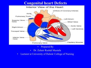
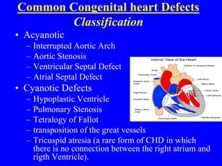
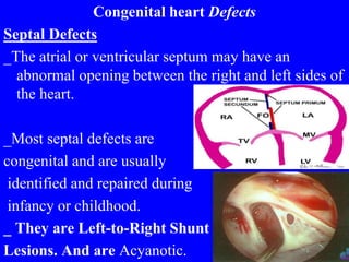
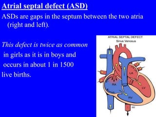
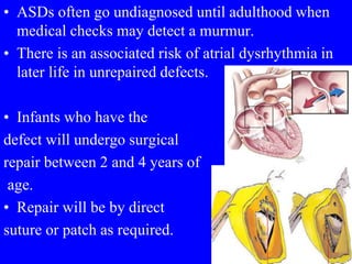
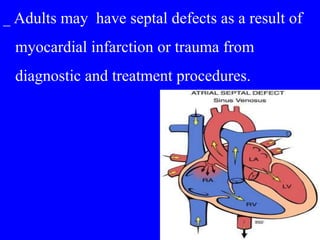
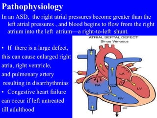
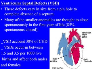
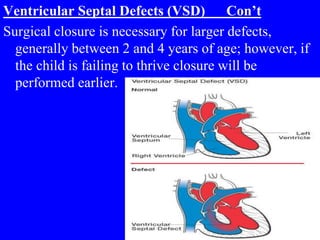
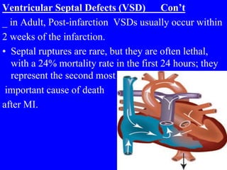
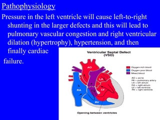
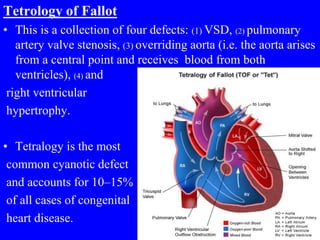
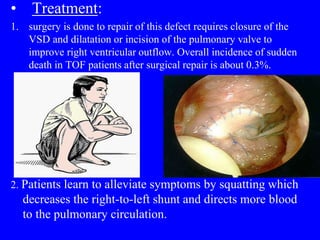
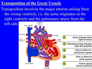
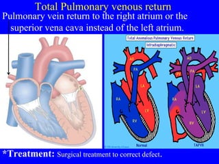
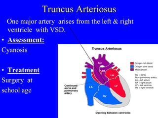
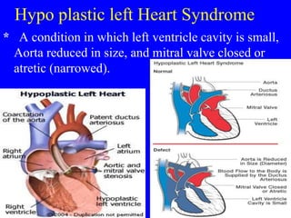
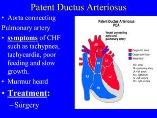
Congenital heart Defects
• Prepared By
• Dr. Zuhair Rushdi Mustafa
• Lecturer at University of Duhok/ College of Nursing
Common Congenital heart Defects
Classification
• Acyanotic
– Interrupted Aortic Arch
– Aortic Stenosis
– Ventricular Septal Defect
– Atrial Septal Defect
• Cyanotic Defects
– Hypoplastic Ventricle
– Pulmonary Stenosis
– Tetralogy of Fallot
– transposition of the great vessels
– Tricuspid atresia (a rare form of CHD in which
there is no connection between the right atrium and
rigth Ventricle).
Congenital heart Defects
Septal Defects
_The atrial or ventricular septum may have an
abnormal opening between the right and left sides of
the heart.
_Most septal defects are
congenital and are usually
identified and repaired during
infancy or childhood.
_ They are Left-to-Right Shunt
Lesions. And are Acyanotic.
Atrial septal defect (ASD)
ASDs are gaps in the septum between the two atria
(right and left).
This defect is twice as common
in girls as it is in boys and
occurs in about 1 in 1500
live births.
• ASDs often go undiagnosed until adulthood when
medical checks may detect a murmur.
• There is an associated risk of atrial dysrhythmia in
later life in unrepaired defects.
• Infants who have the
defect will undergo surgical
repair between 2 and 4 years of
age.
• Repair will be by direct
suture or patch as required.
_ Adults may have septal defects as a result of
myocardial infarction or trauma from
diagnostic and treatment procedures.
Pathophysiology
In an ASD, the right atrial pressures become greater than the
left atrial pressures , and blood begins to flow from the right
atrium into the left atrium—a right-to-left shunt.
• If there is a large defect,
this can cause enlarged right
atria, right ventricle,
and pulmonary artery
resulting in disarrhythmias
• Congestive heart failure
can occur if left untreated
till adulthood
Clinical Manifestation
Patients with septal defects may be asymptomatic or
have:
1. Decreased exercise tolerance.
2. dyspnea on exertion
3. Palpitations
4. Syncope
5. Symptoms of right ventricular or congestive heart
failure.
6. Cyanosis may result.
7. strokes (cerebrovascular accidents, brain attacks)
may occur.
Treatment of Atrial Septal Defects
• Most ASDs should be closed. Many are now closed
percutaneously (patient must weigh ≥10 kg).
• Infants who have the defect will undergo surgical
repair between 2 and 4 years of age.
• If defect is small (less than 2mm), will usually
resolves spontaneously.
• If defect is large, surgical correction is needed.
• Transcatheter devices, such as a septal occluder may
be used.
• Surgical closure by suturing.
• patch.
An Amplatzer
septal
occluder
Ventricular Septal Defects (VSD)
• These defects vary in size from a pin hole to
complete absence of a septum.
• Many of the smaller anomalies are thought to close
spontaneously in the first year of life (85%
spontaneous closed).
_VSD account 30% of CHD
_ VSDs occur in between
1.5 and 3.5 per 1000 live
births and affect both males
and females
Ventricular Septal Defects (VSD) Con’t
Surgical closure is necessary for larger defects,
generally between 2 and 4 years of age; however, if
the child is failing to thrive closure will be
performed earlier.
Ventricular Septal Defects (VSD) Con’t
_ in Adult, Post-infarction VSDs usually occur within
2 weeks of the infarction.
• Septal ruptures are rare, but they are often lethal,
with a 24% mortality rate in the first 24 hours; they
represent the second most
important cause of death
after MI.
Pathophysiology
Pressure in the left ventricle will cause left-to-right
shunting in the larger defects and this will lead to
pulmonary vascular congestion and right ventricular
dilation (hypertrophy), hypertension, and then
finally cardiac
failure.
• Clinical Manifestation
• Patients with VSDs who gradually develop
symptoms describe to have:
1. shortness of breath
2. Syncope
3. chest pain
4. symptoms of left ventricular failure.
Treatments for Ventricular Septal
Defects
• Lasix and Aldactone to decrease symptoms of
CHF
• Digoxin to increase effectiveness of myocardial
function
• If surgery needed, patching or suturing the
defect can be done
• Mortality from surgery is low
Tetrology of Fallot
• This is a collection of four defects: (1) VSD, (2) pulmonary
artery valve stenosis, (3) overriding aorta (i.e. the aorta arises
from a central point and receives blood from both
ventricles), (4) and
right ventricular
hypertrophy.
• Tetralogy is the most
common cyanotic defect
and accounts for 10–15%
of all cases of congenital
heart disease.
Tetralogy of Fallot
• Symptoms:
– Dyspnea on exertion or when crying
– irritability
– Cyanosis especially when crying or feeding
– Hyperventilation
– sometimes syncope
– convulsions due to cerebral hypoxemia.
• Treatment:
1. surgery is done to repair of this defect requires closure of the
VSD and dilatation or incision of the pulmonary valve to
improve right ventricular outflow. Overall incidence of sudden
death in TOF patients after surgical repair is about 0.3%.
2. Patients learn to alleviate symptoms by squatting which
decreases the right-to-left shunt and directs more blood
to the pulmonary circulation.
Nursing Management of septal defects
1. postprocedure cardiac catheterization care. After
undergoing percutaneous septal repair, the patient
usually remains in the hospital for 24 to 48 hours.
2. Care for a patient recovering from a surgical septal
defect repair is the same as other cardiac surgeries.
Mixed blood flow
1. Transposition of the great Arteries
2. Total pulmonary venous return
3. Truncus Arteriosus
4. Hypo plastic left Heart Syndrome
Transposition of the Great Vessels
Transposition involves the major arteries arising from
the wrong ventricle, i.e. the aorta originates in the
right ventricle and the pulmonary artery from the
left side.
• This is the most commonly diagnosed heart defect in
the newborn period.
• The affected infant will present
with cyanosis either on feeding
or on crying because oxygenation
is inadequate for activity.
Treatment
• Surgery to correct the defect must be done at 1 week
to 3months.
Total Pulmonary venous return
Pulmonary vein return to the right atrium or the
superior vena cava instead of the left atrium.
*Treatment: Surgical treatment to correct defect.
Truncus Arteriosus
One major artery arises from the left & right
ventricle with VSD.
• Assessment:
Cyanosis
• Treatment
Surgery at
school age
Hypo plastic left Heart Syndrome
* A condition in which left ventricle cavity is small,
Aorta reduced in size, and mitral valve closed or
atretic (narrowed).
Assessment:
1. Cyanosis
2. Heart Murmur
3.Enlargment of the heart (as seen in an Echo)
4.Noticeably troubled breathing
5.Apparent weakness
6.Inability to feed normally
7.Cold extremities
• Treatment:
• Surgery to correct the defect
Patent Ductus Arteriosus
• Aorta connecting
Pulmonary artery
• symptoms of CHF
such as tachypnea,
tachycardia, poor
feeding and slow
growth.
• Murmur heard
• Treatment:
–Surgery

Congenital hear defects

  • 1.
    Congenital heart Defects •Prepared By • Dr. Zuhair Rushdi Mustafa • Lecturer at University of Duhok/ College of Nursing
  • 2.
    Common Congenital heartDefects Classification • Acyanotic – Interrupted Aortic Arch – Aortic Stenosis – Ventricular Septal Defect – Atrial Septal Defect • Cyanotic Defects – Hypoplastic Ventricle – Pulmonary Stenosis – Tetralogy of Fallot – transposition of the great vessels – Tricuspid atresia (a rare form of CHD in which there is no connection between the right atrium and rigth Ventricle).
  • 3.
    Congenital heart Defects SeptalDefects _The atrial or ventricular septum may have an abnormal opening between the right and left sides of the heart. _Most septal defects are congenital and are usually identified and repaired during infancy or childhood. _ They are Left-to-Right Shunt Lesions. And are Acyanotic.
  • 4.
    Atrial septal defect(ASD) ASDs are gaps in the septum between the two atria (right and left). This defect is twice as common in girls as it is in boys and occurs in about 1 in 1500 live births.
  • 5.
    • ASDs oftengo undiagnosed until adulthood when medical checks may detect a murmur. • There is an associated risk of atrial dysrhythmia in later life in unrepaired defects. • Infants who have the defect will undergo surgical repair between 2 and 4 years of age. • Repair will be by direct suture or patch as required.
  • 6.
    _ Adults mayhave septal defects as a result of myocardial infarction or trauma from diagnostic and treatment procedures.
  • 7.
    Pathophysiology In an ASD,the right atrial pressures become greater than the left atrial pressures , and blood begins to flow from the right atrium into the left atrium—a right-to-left shunt. • If there is a large defect, this can cause enlarged right atria, right ventricle, and pulmonary artery resulting in disarrhythmias • Congestive heart failure can occur if left untreated till adulthood
  • 8.
    Clinical Manifestation Patients withseptal defects may be asymptomatic or have: 1. Decreased exercise tolerance. 2. dyspnea on exertion 3. Palpitations 4. Syncope 5. Symptoms of right ventricular or congestive heart failure. 6. Cyanosis may result. 7. strokes (cerebrovascular accidents, brain attacks) may occur.
  • 9.
    Treatment of AtrialSeptal Defects • Most ASDs should be closed. Many are now closed percutaneously (patient must weigh ≥10 kg). • Infants who have the defect will undergo surgical repair between 2 and 4 years of age. • If defect is small (less than 2mm), will usually resolves spontaneously. • If defect is large, surgical correction is needed. • Transcatheter devices, such as a septal occluder may be used.
  • 10.
    • Surgical closureby suturing. • patch. An Amplatzer septal occluder
  • 11.
    Ventricular Septal Defects(VSD) • These defects vary in size from a pin hole to complete absence of a septum. • Many of the smaller anomalies are thought to close spontaneously in the first year of life (85% spontaneous closed). _VSD account 30% of CHD _ VSDs occur in between 1.5 and 3.5 per 1000 live births and affect both males and females
  • 12.
    Ventricular Septal Defects(VSD) Con’t Surgical closure is necessary for larger defects, generally between 2 and 4 years of age; however, if the child is failing to thrive closure will be performed earlier.
  • 13.
    Ventricular Septal Defects(VSD) Con’t _ in Adult, Post-infarction VSDs usually occur within 2 weeks of the infarction. • Septal ruptures are rare, but they are often lethal, with a 24% mortality rate in the first 24 hours; they represent the second most important cause of death after MI.
  • 14.
    Pathophysiology Pressure in theleft ventricle will cause left-to-right shunting in the larger defects and this will lead to pulmonary vascular congestion and right ventricular dilation (hypertrophy), hypertension, and then finally cardiac failure.
  • 15.
    • Clinical Manifestation •Patients with VSDs who gradually develop symptoms describe to have: 1. shortness of breath 2. Syncope 3. chest pain 4. symptoms of left ventricular failure.
  • 16.
    Treatments for VentricularSeptal Defects • Lasix and Aldactone to decrease symptoms of CHF • Digoxin to increase effectiveness of myocardial function • If surgery needed, patching or suturing the defect can be done • Mortality from surgery is low
  • 17.
    Tetrology of Fallot •This is a collection of four defects: (1) VSD, (2) pulmonary artery valve stenosis, (3) overriding aorta (i.e. the aorta arises from a central point and receives blood from both ventricles), (4) and right ventricular hypertrophy. • Tetralogy is the most common cyanotic defect and accounts for 10–15% of all cases of congenital heart disease.
  • 18.
    Tetralogy of Fallot •Symptoms: – Dyspnea on exertion or when crying – irritability – Cyanosis especially when crying or feeding – Hyperventilation – sometimes syncope – convulsions due to cerebral hypoxemia.
  • 19.
    • Treatment: 1. surgeryis done to repair of this defect requires closure of the VSD and dilatation or incision of the pulmonary valve to improve right ventricular outflow. Overall incidence of sudden death in TOF patients after surgical repair is about 0.3%. 2. Patients learn to alleviate symptoms by squatting which decreases the right-to-left shunt and directs more blood to the pulmonary circulation.
  • 20.
    Nursing Management ofseptal defects 1. postprocedure cardiac catheterization care. After undergoing percutaneous septal repair, the patient usually remains in the hospital for 24 to 48 hours. 2. Care for a patient recovering from a surgical septal defect repair is the same as other cardiac surgeries.
  • 21.
    Mixed blood flow 1.Transposition of the great Arteries 2. Total pulmonary venous return 3. Truncus Arteriosus 4. Hypo plastic left Heart Syndrome
  • 22.
    Transposition of theGreat Vessels Transposition involves the major arteries arising from the wrong ventricle, i.e. the aorta originates in the right ventricle and the pulmonary artery from the left side.
  • 23.
    • This isthe most commonly diagnosed heart defect in the newborn period. • The affected infant will present with cyanosis either on feeding or on crying because oxygenation is inadequate for activity. Treatment • Surgery to correct the defect must be done at 1 week to 3months.
  • 24.
    Total Pulmonary venousreturn Pulmonary vein return to the right atrium or the superior vena cava instead of the left atrium. *Treatment: Surgical treatment to correct defect.
  • 25.
    Truncus Arteriosus One majorartery arises from the left & right ventricle with VSD. • Assessment: Cyanosis • Treatment Surgery at school age
  • 26.
    Hypo plastic leftHeart Syndrome * A condition in which left ventricle cavity is small, Aorta reduced in size, and mitral valve closed or atretic (narrowed).
  • 27.
    Assessment: 1. Cyanosis 2. HeartMurmur 3.Enlargment of the heart (as seen in an Echo) 4.Noticeably troubled breathing 5.Apparent weakness 6.Inability to feed normally 7.Cold extremities • Treatment: • Surgery to correct the defect
  • 28.
    Patent Ductus Arteriosus •Aorta connecting Pulmonary artery • symptoms of CHF such as tachypnea, tachycardia, poor feeding and slow growth. • Murmur heard • Treatment: –Surgery