Cyanotic heart diseases
•
3 likes•217 views

A congenital heart anomaly is any structure or functional abnormalities or defects of the heart or great vessels existing from birth

Recommended
More Related Content
What's hot
What's hot (20)
Similar to Cyanotic heart diseases
Similar to Cyanotic heart diseases (20)
Recently uploaded
Recently uploaded (20)
Cyanotic heart diseases
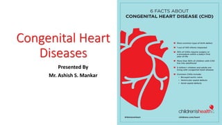
- 1. Congenital Heart Diseases Presented By Mr. Ashish S. Mankar
- 2. Classification of congenital heart disease Acyanotic Increased pulmonary blood flow Atrial septal Defects Ventricular septal defects Patent ductus arteriosus Atrioventricular canal Obstruction to blood flow from ventricles Coarction of aorta Aortic stenosis Pulmonic stenosis Cyanotic Decreased blood flow Tetrology of fallot Tricuspid atresia Mixed blood flow Transposition of great arteries Total anomalies venous return Hypoplastic left heart syndrome
- 3. CYANOTIC HEART DISEASES (DEFECTS WITH DECREASED BLOOD FLOW)
- 5. TETRALOGY OF FALLOT • It is the most common cause of cyanosis beyond one year of age. • It constitutes 10% of all congenital heart defects. • Fallot defined it as a constellation of four abnormalities to include : • Ventricular septal defect (VSD), • Pulmonary stenosis (PS), • Right ventricular hypertrophy • Dextroposition of the aorta or An overriding aorta is a congenital heart defect where the aorta is positioned directly over a ventricular septal defect (VSD), instead of over the left ventricle
- 7. Clinical Manifestation • Become symptomatic any time after birth • Paroxysmal attack of dyspnea • Cyanosis may be present from birth or make its appearance some years after birth • Commonest symptoms are dyspnea on exertion and exercise intolerance • Patients assume a sitting posture – squatting – as soon as they get dyspneic. Although squatting is not specific for TOF, it is the commonest congenital lesion in which squatting is noted • Anoxic spells occur predominantly after waking up or following exertion.
- 8. • Normal growth and development depend on a normal workload for the heart and normal flow of oxygen-rich blood to all parts of the body. Babies who have tetralogy of Fallot may not gain weight or grow as quickly as children who have healthy hearts because they tire easily while feeding. • Squatting (a compensatory mechanism) is uniquely characteristic of a right-to-left shunt that presents in the exercising child. Squatting increases the peripheral vascular resistance, which diminishes the right-to-left shunt and increases pulmonary blood flow. –Child becomes more cyanosed while crying, these are called ‘cyanotic spells’or Fallot’s spells.
- 9. What causes a spell ? • Due to "spasm" or contraction of a band of muscle in the right ventricle just under the pulmonary valve. When this muscle contracts, it further narrows the channel for blood flow into the lungs. As a result, oxygen delivery becomes further reduced. This causes a spell
- 10. Investigations Physical Examination • Cyanosis , clubbing, slightly prominent `a ‘ wave in the jugular venous pulse, normal sized heart with parasternal impulse, a systolic thrill in less than 30 % patients. • Normal first sound, • Single second sound and • An ejection systolic murmur at 3rd ICS
- 11. Chest X – ray : • Boot shaped heart (it means apex is lifted up & there is a concavity in the region of pulmonary artery)
- 12. Treatment • Management of complications and correction of anemia • Treatment of Anoxic spells : • Knee chest position to increase aortic resistance. The increased aortic and left ventricular pressure reduces the rush of blood through the septal hole from the right ventricle and improves blood circulation to the lungs, decreasing the right to left shunt thus decreasing the amount of deoxygenated blood entering the systemic circulation. • Oxygen through a face mask to increase the amount of oxygen in the blood.
- 13. • beta-blockers such as propranolol • acute episodes may require rapid intervention with morphine to reduce ventilatory drive and a vasopressor such as epinephrine, phenylephrine, or norepinephrine to increase blood pressure. • Correction of anemia • Consider operation
- 14. Surgical Management • Blalock-Taussig operation : connection between the right subclavian artery, and the right pulmonary artery, which increases the amount of red oxygenated blood reaching the lungs, relieving cyanosis. • Pott`s shunt : descending aorta is anastomosed to the pulmonary artery • Waterstont`s shunt : ascending aorta right pulmonary artery anastomosis • Total correction: The hole in the ventricular septum is closed with a patch and the obstruction to right ventricular outflow, pulmonic stenosis, is opened. • These corrections allow blood flow to the lungs for oxygenation before being pumped out into the body.
- 15. Surgical Management • If oxygen levels are critically low soon after birth, a prostaglandin E1 infusion is usually initiated to keep the ductus arteriosus open which will provide additional pulmonary blood flow and increase the child's oxygen level. • Complete repair is usually done electively when children are about 6 months of age, as long as the oxygen levels remain adequate. • Surgical correction of the defect is always necessary. Occasionally, patients will require a surgical palliative procedure prior to the final correction. • Corrective repair of tetralogy of Fallot involves closure of the ventricular septal defect with a synthetic Dacron patch so that the blood can flow normally from the left ventricle to the aorta.
- 16. • The narrowing of the pulmonary valve and right ventricular outflow tract is then augmented (enlarged) by a combination of cutting away (resecting) obstructive muscle tissue in the right ventricle and by enlarging the outflow pathway with a patch. • In some babies, however, the coronary arteries will branch across the right ventricular outflow tract where the patch would normally be placed. In these babies an incision in this area to place the patch would damage the coronary artery so this cannot safely be done. • When this occurs, a hole is made in the front surface of the right ventricle (avoiding the coronary artery) and a conduit (tube) is sewn from the right ventricle to the bifurcation of the pulmonary arteries to provide unobstructed blood flow from the right ventricle to the lungs.
- 17. • Surgical repair is more difficult when the pulmonary arteries are critically small or when the lung blood flow is supplied predominantly by aortopulmonary collaterals. • Most babies are fairly sick in the first few days after surgery, since the right ventricle is "stiff" from the previous hypertrophy (thickness) and because an incision is made into the muscle of the ventricle, making the muscle temporarily weaker. • This right ventricular dysfunction usually improves significantly in the days following surgery. Patients may also have rhythm problems after surgery.
- 18. • An abnormally fast rhythm (called junctional tachycardia) may occur and may require treatment with medication or the use of a temporary pacemaker. This abnormal rhythm is usually temporary and the rhythm generally will return to normal as the right ventricle recovers. • Patients are also at risk for slow heart rates after surgery due to heart block. Heart block may be caused by injury to or inflammation of the conduction system in the heart. In many patients, the conduction improves and normal rhythm returns. Rarely, a permanent pacemaker may be necessary. • Since a normal circulation is produced by the tetralogy of Fallot repair procedure, long-term cardiac function is usually excellent.
- 19. • However, the repair does usually leave the child with a leaky (insufficient) pulmonary valve. In this situation, after the right ventricle pumps blood out to the pulmonary arteries, some of the blood will flow back into the right ventricle. This creates extra volume in the right ventricle forcing it to work harder and become dilated. • In a small percentage of children, this pulmonary insufficiency can lead to diminished function of the right ventricle. Symptoms of fatigue, especially with exercise, may develop. In these cases, replacement of the pulmonary valve is often recommended. • Patients who have had repair of tetralogy of Fallot can also redevelop a narrowing at the outflow area or in the branch (left or right) pulmonary arteries, which will cause the right ventricle to pump at abnormally high pressures.
- 20. • If these problems occur, surgical intervention to further widen the outflow tract or pulmonary arteries may be necessary. Narrowing the pulmonary arteries can sometimes be treated without surgery, with balloon dilation of the vessels during cardiac catheterization. • Long-term follow-up with a cardiologist to detect recurrent or new problems as early as possible is essential. Follow-up visits in the cardiology clinic usually consist of a physical examination, electrocardiogram and periodic echocardiography. In addition, these visits will also include occasional cardiac MRI scans, exercise stress tests and Holter evaluations as a child reaches the teenage and adult years.
- 21. Palliative Management • It was common in the past to do temporary surgery during infancy in babies who had tetralogy of Fallot. This surgery improved blood flow to the lungs. ... In the temporary surgery, the surgeon places a tube called a shunt between a large artery branching off the aorta and the pulmonary artery. • The Blalock–Thomas–Taussig shunt (commonly called the Blalock–Taussig shunt) is a surgical procedure used to increase pulmonary blood flow for palliation in duct dependent cyanotic heart defects like pulmonary atresia, which are common causes of blue baby syndrome.
- 22. i.Classic BT shunt: The subclavian artery is anastomosed to the ipsilateral pulmonary artery (PA). This procedure is usually performed in infants older than 3- months; a right-sided shunt is performed in patients with left aortic arch; a left sided shunt is performed for right aortic arch. ii.Modified Blalock-Taussig shunt: A Gore-Tex interposition graft is placed between the subclavian artery and the ipsilateral PA. This is the most popular procedure for any age, especially for small infants younger than 3 months of age. A left-sided shunt is preferred for patients with a right aortic arch
- 23. TRICUSPID ATRESIA • Tricuspid atresia is a type of heart disease that is present at birth (congenital heart disease), in which the tricuspid heart valve is missing or abnormally developed. The defect blocks blood flow from the right atrium to the right ventricle. • Incidence: 0.06/1000 live births
- 24. Types • Muscular (89%) • Membranous (6.6%) • Valvar (fused cusp) • Ebsteins Anomaly • The atrioventricular canal (extremely rare 0.2%)
- 25. Risk factors • A mother who had German measles (rubella) or another viral illness during early pregnancy • A parent who has a congenital heart defect • Older parental age at conception • Mother's obesity • Drinking alcohol during pregnancy • Smoking before or during pregnancy • A mother who has poorly controlled diabetes • Use of some types of medications during pregnancy, such as the acne drug isotretinoin (Claravis, Amnesteem, others), some anti-seizure medications and some bipolar disorder medications • The presence of Down syndrome, a genetic condition that results from an extra 21st chromosome
- 26. Pathophysiology Atresia of tricuspid valve No communica tion between RA and RV RV is underdevel -oped Systemic venous blood received by RA Enters LA through ASD Mixing of systemic and pulmonary blood
- 27. Enters LV Blood enters RV through VSD From RV blood enters Pulmonary trunk Blood enters enters pul trunk via PDA Increased pulmonary blood flow LA and LV hypertrophy CHF
- 28. Clinical Manifestation • The clinical feature of tricuspid atresia largely depend on the quantity of pulmonary blood flow • Decreased pulm flow: - Severe cyanosis - Hypoxemia - Acidosis - Pulmonary oligemia - May have central cyanosis - Tachypnoea
- 29. Increased pulm flow • Difficult to diagnose • May not appear cyanotic but may present with signs of heart failure later in infancy • Pulmonary plethora present with symptoms of dyspnoea, fatigue, difficulty in feeding, and perspiration which are suggestive of congestive heart failure. • Cyanosis is minimal
- 30. Other features • Holosystolic murmur at the lower sternal border • Problems related to chronic cyanosis like • Clubbing • Polycythemia, relative anemia • Stroke • Coagulation abnormalities • Some babies or older people with tricuspid atresia also develop symptoms of heart failure, including: • Fatigue and weakness • Shortness of breath • Swelling (edema) in the legs, ankles and feet • Swelling of the abdomen (ascites) • Sudden weight gain from fluid retention
- 31. Investigation • Physical examination • Pulse oximetry • ABG • Hb and hematocrit • Echo • Chest X-ray • Angiography
- 32. Therapeutic Management • For the neonate, whose pulmonary blood flow depends on the patency of the ductus arteriosus, a continuous infusion of prostaglandin E1 (0.03-0.1 mcg/kg/min) is started until surgical intervention can be arranged. • Digoxin • Diuretics
- 34. Palliation for decreased pulm blood flow • Systemic to pulmonary artery shunt: increases pulmonary blood flow through surgically created left to right shunt at the great vessels.
- 37. Palliation for increased pulm blood flow • Control amount of pulmonary blood flow to prevent CHF and pulmonary vascular disease from pulmonary over circulation
- 38. Palliation for tricuspid atresia
- 39. Fontan Procedure • Between the ages of 2 and 5 years, children with tricuspid atresia will be ready for the third operation required to optimize their circulation. • This operation is called the Fontan procedure, and involves connection of the inferior vena cava directly to the pulmonary artery, which forces all blood returning from the body to pass through the lungs and pick up oxygen before being pumped to the body. • This allows a more normal color in the skin and lips as well due to a more normal oxygen saturation in the blood.
- 40. Modified Fontan Procedure • Systemic venous return is directed to the lungs without a ventricular pump through surgical connections between the right atrium and the pulmonary artery • A fenestration (opening) in the right arterial baffle is sometimes done to relieve pressure. • The patient must have normal ventricular function and a low pulmonary vascular resistance for the procedure to be successful
- 41. Bidirectional Glenn Procedure • The operation at 3 to 6 months is called a bidirectional Glenn. The superior vena cava is detached from the heart and connected directly to the pulmonary artery. • This allows blood from the upper body to flow directly to the lungs to pick up oxygen without having to be pumped by the heart. • It also prevents blood that already has oxygen from returning to the lungs, and, thereby, keeps the heart from doing unnecessary work. • After this operation, however, there is still blood returning from the body through the inferior vena cava going directly back to the body without first passing through the lungs. Because of this, some level of cyanosis will persist.
- 43. Complications later in life • Although treatment greatly improves the outcome for babies with tricuspid atresia, complications can develop later in life, including: • Formation of blood clots that can lead to a clot blocking an artery in the lungs (pulmonary embolism) or cause a stroke • Easy tiring when participating in activity or exercise • Heart rhythm abnormalities (arrhythmias) • Kidney or liver disease
- 44. CYANOTIC HEART DISEASES (DEFECTS WITH MIXED BLOOD FLOW)
- 46. Transposition of Great Arteries (TGA) • Second most common form (5-7%) of congenital cardiac anomalies. • Aorta arises from RV and Pulmonary Arteries from LV. • Most common in males. • Without an abnormality, life would not be possible. • ASD • VSD (30-40%) • PDA
- 47. Transposition of Great Arteries (TGA) • The transposition of the great arteries is ventriculo arterial discordance, in which the aorta arises from the morphologic right ventricle and the pulmonary artery arises from the morphologic left artery.
- 48. Etiology • Idiopathic • Genetic causes • Rubella infection to mother during pregnancy • Maternal age over 40years • Incidence is more in infants of diabetic mothers
- 49. Pathophysiology Pumped to the systemic circulation effectively bypassing the lungs Deoxygenated systemic venous blood returns to the right atrium and right ventricle Recirculated to the pulmonary vascular bed via the abdominal pulmonary arterial connection to the left ventricle Oxygenated pulmonary venous blood returns to the left atrium and left ventricle Transposition of great arteries
- 50. If remain untreated, Heart Failure ASD,VSD,PDA It is incompatible with prolonged survival unless mixing of oxygenated and deoxygenated blood occurs at some anatomic level like
- 51. Clinical Manifestation • Depends on the type and size of associated defects. • Cyanosis • Tachypnoea • Tachycardia • Diaphoresis • Failure to gain wight • Signs and symptoms of heart failure. • Cardiomegaly
- 52. Investigations • Auscultation- a single or narrowly split, diminished second heart sound. Systolic ejection murmur may be present. • ABG analysis • Chest X-Ray: “Egg on a String” • Echocardiography • Cardiac Catheterization
- 53. Medical Management • To provide interacting mixing: The administration of intravenous prostaglandin E1 may be initiated to temporarily increase blood mixing if systemic and pulmonary mixing is inadequate to provide an oxygen saturation of 75% or to maintain cardiac output. • Bicarbonate administration for Acidosis • During cardiac catheterization, a balloon atrial septostomy (Rashkind procedure) may also be performed to increase mixing and maintain cardiac output over a longer period.
- 54. Surgical Treatment Atrial Switch Procedure- • Procedure of choice performed in first week of life involves transecting the great arteries and anastomosing the main pulmonary artery to the proximal aorta (just above the aortic valve) and anastomosing the ascending aorta to the proximal pulmonary artery. • The coronary arteries are switched from the proximal aorta to the proximal aorta to the proximal pulmonary artery creating a new aorta. • Potential complication of the arterial switch includes narrowing at the great artery anastomosis or coronary artery insufficiency
- 55. Intra-atrial baffle repairs • It is created to divert venous blood to the mitral valve and pulmonary venous blood to the tricuspid valve using the septum (Stenning procedure) or a prosthetic material (mustard procedure) • These are performed in the first year of life. A disadvantage is the continuing role of the right ventricle as the systemic pump and the late development of right ventricular failure and rhythm disturbances
- 56. Rastelli Procedure • It involves closure of the VSD with a baffle directing left ventricular blood through the VSD in to the aorta. • The pulmonic valve is then closed and a conduit is placed from the right ventricle to the pulmonary artery creating a physiologically normal circulation. • Unfortunately this procedure requires multiple conduit replacement as the child grows.
- 58. Hypoplastic Left Heart Syndrome • Hypoplastic left heart syndrome occurs when parts of the left side of the heart (mitral valve, left ventricle, aortic valve and aorta) do not develop completely. • The left ventricle is non-functional • Thus the left side of the heart is completely unable to supply blood to the systemic circulation
- 59. Defects 1. Patent foramen ovale 2. Coarctation of the aorta 3. Patent ductus arteriosus 4. Narrowed aorta 5. Hypo plastic left ventricle 6. Aortic atresia • It is the 4th most common congenital heart defect. • Occurs in up to 4% of cases of CHD
- 60. Pathophysiology The right ventricle must then pump blood to the lungs and also to the systemic circulation through PDA Blood returning from the lungs into the left atrium must pass through an ASD to the right side of the heart. As a result, the right side of the heart must maintain the circulation for both the lungs and the body. In patient with HLHS, the left side of the heart is unable to send enough blood to the body.
- 61. The right ventricle can support the circulation to the both lungs and the body for a while, but this extra workload eventually causes the right side of the heart to fail. A few days after birth when the ductus arteriosus closes, the heart cannot pump blood into the systemic circulation, causing poor perfusion of the vital organs & shock.
- 62. Clinical Manifestation • The neonates are born healthy, no cyanosis, no murmur. • But after some hours to a day or two, the infant become critically ill and may die due to closure of ductus. • Cyanosis • Irritability • Low volume pulse with hypotension • Single heart sound
- 63. Investigations • Health History: Onset of cyanosis, poor feeding, history of tiring easily • Physical examination: Evaluate vital signs, noting tachycardia, tachypnea, hypothermia. Observe for increased work of breathing and gradually increasing cyanosis. Note pallor of the extremities. • Pulse oximetry- shows decreased oxygen saturation. • Auscultation- Adventitious breath sound, a gallop rhythm, a single second heart sound, and a soft systolic ejection or holosystolic murmur • Echocardiogam- can diagnose this syndrome in prenatal period • USG – for prenatal diagnosis
- 64. Medical Management • After diagnosis, baby will be admitted in neonatal intensive care unit. • A ventilator may be needed for breathing support • Prostaglandin E1 is used to keep ductus arteriosus open
- 65. Surgical treatment of hypo plastic left heart syndrome • Three separate surgeries. • Norwood procedure • First few days after birth. • Glenn Shunt (Cavo Pulmonary Connection) • 3-9 months of age • Fontan Procedure • 2 years of age • Less wait because of damage from pulmonary hypertension
- 66. Norwood procedure (Stage I) • It consist of building a new aorta by using the pulmonary valve and artery • Connecting the hypoplastic old aorta and coronary arteries to the new aorta • Removing the wall between the atrial septum • Making an artificial connection from right ventricle to the pulmonary artery to maintain blood flow to the lungs (called a shunt)
- 67. Glenn Shunt (Stage II) • The operation at 3 to 6 months is called a bidirectional Glenn. The superior vena cava is detached from the heart and connected directly to the pulmonary artery. • This allows blood from the upper body to flow directly to the lungs to pick up oxygen without having to be pumped by the heart. • It also prevents blood that already has oxygen from returning to the lungs, and, thereby, keeps the heart from doing unnecessary work.
- 68. Fontan Procedure (Stage III) • IVC is connected directly to the pulmonary arteries. • The right ventricle now serves only as the pumping chamber for the body. • This surgery is usually performed when the baby is 18months to 3years old. • After this final step baby is no longer blue.
- 70. Heart Transplantation • In some cases, heart transplantation is considered a better choice than the three step surgery process. • However there are few donated hearts available for small infants.
- 72. Total Anomalies Venous Return • TAPVR is a congenital heart defect in which the pulmonary veins do not connect normally to the left atrium. Instead they connect to the right atrium, often by way of the superior venacava. • Relatively rare, it occurs in about 1 in 17,000 live birth. • ASD or foramen ovale is always present.
- 73. TYPES OF TAPVR
- 74. Supra cardiac TAPVR • The pulmonary veins drain into the right atrium through the superior vena cava. • The superior vena cava is a large vein that normally carries only deoxygenated, or “blue,” blood into the right atrium from the upper half of the body.
- 75. Infra cardiac TAPVR • The pulmonary veins drain into the right atrium through the liver (hepatic) veins and the inferior vena cava. • The inferior vena cava is another large vein that normally carries only deoxygenated blood into the right atrium from the lower half of the body.
- 76. Cardiac TAPVR • There are two types. In one, the pulmonary veins can directly enter into the right side of the heart, into the right atrium. • Alternatively, the pulmonary veins can drain into the coronary sinus. The coronary sinus is a vein that normally carries deoxygenated blood from the heart muscle into the right atrium. • This vein is usually very small, but becomes quite large with this abnormal amount of blood. Mixed TAPVR • The pulmonary veins split up and drain partially to more than one of these options.
- 77. Pathophysiology • Oxygenated blood that would normally enter the left atrium now enters the right ventricle. • As a result the pressure on the right side of the heart increases, leading to hypertrophy. • Since none of the pulmonary veins connect normally to the left atrium, the only source of blood to the left atrium is blood that is shunted from the right atrium across the defect to the left side of the heart. • The highly oxygenated blood from the lungs completely mixes with the poorly oxygenated blood returning from the systemic circulation. • This can cause an overload of the right atrium and right ventricle. • The increase blood volume going into the lungs can lead to pulmonary hypertension and pulmonary edema
- 78. Clinical Manifestations • In some cases, newborns with TAPVR have difficulty breathing and quickly become very ill. This occurs when the pulmonary veins are too narrow or are obstructed at some point, and blood can’t flow from the lungs as quickly as it should. This is called TAPVR with pulmonary obstruction. These children are typically transported to the Cardiac Center. • In other cases, TAPVR is diagnosed in the first few months of life, after a child demonstrates milder symptoms such as a heart murmur or cyanosis (blue tint to skin). • Blue or purple tint to lips, skin and nails (cyanosis) • Rapid breathing or working harder while breathing, especially while eating • Heart murmur, S2 wide and split. • The severity of TAPVR symptoms varies.
- 79. • Chest X-ray: this will show right heart enlargement and increased blood flow through the pulmonary artery. If the veins are obstructed, there will be pulmonary edema (buildup of fluid in the lungs). • Electrocardiogram (ECG): This usually shows evidence of right heart hypertrophy, right axis deviation. • Echocardiogram • Pulse oximetry • Cardiac catheterization- to visualize the abnormal connection of pulmonary veins particularly if an obstruction is present. • Cardiac MRI Investigations
- 80. Management • TAPVR require open-heart surgery in all cases. • Critically ill newborn will have surgery immediately. • If the child is not critically ill, Doctors may wait up to two months to perform surgery, depending on the strength of the child and on the heart anatomy.
- 81. • To understand the surgery, one important thing to know about TAPVR is that the pulmonary veins, despite their abnormal connections to other veins, all end in a collection (called a “confluence”) at the back of the left atrium. • The surgeon opens the confluence so that the veins can drain into the left atrium. Then he or she ties off all the abnormal connections between the pulmonary veins and other veins, so that blood can follow only the path to the left atrium. • The surgeon also closes septal defects (the abnormal holes) with tiny patches or stitches, and closes the patent ductus arteriosus. As the child ages, the lining of the heart will grow over the stitches Surgical Management
- 83. LIST OF NURSING DIAGNOSIS (PRE-OPERATIVE) • Ineffective breathing pattern related to pulmonary congestion • Decreased cardiac output related to structural defect secondary to atrial septal defect • Activity intolerance related to imbalance between oxygen supply and demand • Fluid volume excess related to compromised regulatory mechanism secondary to heart failure • Altered family process related to a child with life threatening illness
- 84. LIST OF NURSING DIAGNOSIS (POST-OPERATIVE) • Decreased cardiac output related to increased demand of myocardium • Ineffective breastfeeding related to generalized weakness secondary to postoperative status • Risk for hyperthermia related to the infectious process secondary to presence of IV lines, etc. • Risk for infection related to presence of chest tube drainage • Deficient knowledge related to the postoperative care