
Recommended
More Related Content
Similar to Congenital heart diesease- presenatation.pdf
Similar to Congenital heart diesease- presenatation.pdf (20)
Recently uploaded
Recently uploaded (20)
Congenital heart diesease- presenatation.pdf
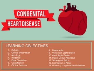
- 1. LEARNING OBJECTIVES 1. Definition 2. Clinical presentation 3. Aetiology 4. Incidence 5. Fetal Circulation 6. Classification 7. Clinical Features 8. Dextocardia 9. Ventricular Septal Defect 10. Atrial Septal Defect 11. Patent Ductus Arteriosus 12. Tetralogy of Fallot 13. Coarctation of Aorta 14. Grown-up congenital heart disease
- 2. CONGENITAL HEART DISEASE? Congenital heart disease is the abnormality of the heart present from birth. Congenital heart disease usually manifests in childhood but may pass unrecognized and not present until adult life. Defects that are well tolerated, such as atrial septal defect may cause no symptoms until adult life or may be detected incidentally on routine examination or chest x-ray.
- 3. CONGENITAL HEART DISEASE? Congenital heart disease is the abnormality of the heart present from birth. Congenital heart disease usually manifests in childhood but may pass unrecognized and not present until adult life. Defects that are well tolerated, such as atrial septal defect may cause no symptoms until adult life or may be detected incidentally on routine examination or chest x-ray.
- 4. CONGENITAL HEART DISEASE? Congenital defects that were previously fatal in childhood can now be (completely or partially) corrected so that survival to adult life is the norm. Such patient remain well for many years but subsequently re-present in later life with related problems such as arrhythmia or ventricular dysfunction.
- 5. PRESENTATION BIRTH AND NEONATAL PERIOD INFANCY AND CHILDHOOD ADOLESCENCE AND ADULTHOOD Cyanosis Heart failure Cyanosis Arrhythmia Heart Failure Murmur Failure to thrive Arrhythmia Heart Failure Cyanosis (Eisenmenger’s Syndrome) Hypertension Late consequences of previous surgery
- 6. AETIOLOGY 1. Maternal infection (E.g. Rubella) 1. Maternal alcohol misuse 1. Maternal lupus erythematosus 1. Genetic or chromosomal abnormalities 5. Gene defects 6. Exposure to drugs or toxins Associated with atrial septal defect, patent ductus arteriosus and pulmonary valvular or/and artery stenosis. Associated with septal defects Associated with congenital complete heart block Associated with septal defects
- 7. INCIDENCE LESION % OF ALL CONGENITAL HEART DEFECTS Ventricular Septal Defect 30 Atrial Septal Defect 10 Patent Ductus Arteriosus 10 Pulmonary Stenosis 7 Coarctation of Aorta 7 Aortic Stenosis 6 Tetralogy of Fallot 6 Transposition of Great Arteries 4 Others 20 The incidence of hemodynamically significant congenital cardiac abnormalities is about 0.8% of live birth
- 9. AFTER BIRTH
- 10. FETAL CIRCULATION In the fetus, oxygenated blood comes through the (1) umbilical vein where it enters the inferior vena cava via the (2) ductus venosus (red). The oxygenated blood streams from the right atrium through the open (3) foramen ovale to the left atrium and via the left ventricle into the aorta. Venous blood from the superior vena cava (blue) crosses under the main blood stream into the right atrium and then, partly mixed with oxygenated blood (purple), into the right ventricle and pulmonary artery. The pulmonary vasculature has a high resistance and so little blood passes to the lungs; most blood passes through the (4) ductus arteriosus to the descending aorta. The aortic isthmus is a constriction in the aorta that lies in the aortic arch before the junction with the ductus arteriosus and limits the flow of oxygen-rich blood to the descending aorta. Then blood passes throught the (5) umbilical artery and back to the placenta. BEFORE BIRTH
- 11. FETAL CIRCULATION At birth, the lungs expand with air and pulmonary vascular resistance falls, so that blood now flows to the lungs and back to the left atrium. The left atrial pressure rises above right atrial pressure and the flap valve of the foramen ovale closes. The umbilical arteries and the ductus venosus close. In the next few days, the ductus arteriosus closes under the influence of hormonal changes (particularly prostaglandins) and the aortic isthmus expands AT BIRTH
- 12. CLASSIFICATION MALPOSITIONS OF THE HEART Dextocardia SHUNTS Coarctation of Aorta Aortic Stenosis and Atresia Pulmonary Stenosis and Atresia OBSTRUCTION ACYANOTIC / LATE CYANOTIC CYANOTIC Ventricular Septal Defect (VSD) Atrial Septal Defect (ASD) Patent Ductus Arteriosus (PDA) Tetralogy of Fallot (TOF) Transposition of Great Arteries Persistent Trunchus Arteriosus Tricuspid Atresia and stenosis
- 13. CLINICAL FEATURES Asymptomatic Breathless Cyanosis Failure to attain normal growth and development Growth restriction and learning difficulties Murmurs Thrills Signs of cardiomegaly Radiofemoral delay (Coarctation of Aorta) Syncope Digital clubbing CLINICAL SIGNS SYMPTOMS
- 14. DEXTOCARDIA MALPOSITION OF THE HEART
- 15. Dextrocardia is a rare condition whereby the apex of the heart points to the right side of the chest. It may be accompanied by situs inversus so that all other organs of the body are also transposed in similar way and thus heart is in normal position in relation to them. However, isolated dextrocardia is associated with major anomalies of the heart such as transposition of the atria in relation to ventricles or transposition of the great arteries. WHAT IS DEXTOCARDIA?
- 16. DEXTOCARDIA
- 17. SYMPTOMS There are no symptoms of dextrocardia if the heart is normal. Conditions that may include dextrocardia may cause the following symptoms: Bluish skin (cyanosis) Difficulty breathing Failure to grow and gain weight Fatigue Jaundice (yellow skin and eyes) Pale skin (pallor) Repeated sinus or lung infections
- 18. Chest x-ray CT Scan of the heart Electrocardiogram MRI of the heart Echocardiogram DIAGNOSTIC TESTS
- 19. TREATMENT A complete mirror image Dextrocardia with no heart defects requires no treatment. The type of treatment needed depends on the heart or physical problems the infant may have in addition to dextrocardia. If heart defects are present with dextrocardia, the baby will most likely need surgery. Water pills (diuretics) Drugs that help the heart muscle pump more forcefully (inotropic agents) Drugs that lower blood pressure and ease the workload on the heart (ACE inhibitors) MEDICATIONS
- 21. VSD is the most common congenital anomaly of the heart and comprises about 30% of all congenital heart diseases. Occurs as a result of incomplete septation of the ventricles. The smaller defects often close spontaneously, while larger defects remain patent and produce significant effects. Acquired ventricular septal defect may result from rupture as a complication of acute MI or rarely from trauma. VENTRICULAR SEPTAL DEFECT
- 23. CLINICAL FEATURES 1. Pansystolic murmur 2. Cardiac failure (in infants) 3. Eisenmenger’s Syndrome 4. Parasternal pulsation 5. Tachypnea 6. Indrawing of the lower ribs on inspiration CHEST X-RAY Chest x-ray shows pulmonary plethora ELECTROCARDIOGRAM (ECG) Bilateral ventricular hypertrophy
- 24. COMPLICATIONS 1. Pulmonary hypertension 1. Right ventricular hypertrophy 1. At a later stage, the pressure on the right side is higher than on the left side creating late cyanotic heart disease.
- 25. MANAGEMENT Small ventricular septal defects require no specific treatment. Cardiac failure in infancy is initially treated medically with digoxin and diuretics. Persisting failure is an indication for surgical repair of the defect. Eisenmenger’s syndrome is avoided by monitoring for signs of rising pulmonary resistance (serial ECG and echocardiography) and carrying out surgical repair, when appropriate. Surgical closure is contraindicated in fully developed Eisenmenger’s syndrome when heart–lung transplantation may be the only effective treatment. Except in Eisenmenger’s syndrome, long-term prognosis is very good in congenital ventricular septal defect. Many patients with Eisenmenger’s syndrome die in the second or third decade of life.
- 27. Isolated ASD comprises about 10% of congenital heart diseases. The condition remains unnoticed in infancy and childhood till pulmonary hypertension is induced causing late cyanotic heart disease and right- sided heart failure. ATRIAL SEPTAL DEFECT
- 28. Depending upon the location of the defect, there are 3 types of ASD: A. Fossa Ovalis type or Ostium Secundum type is the most common form comprising about 90% cases of ASD. The defect is situated in the region of the fossa ovalis that, in utero, was the foramen ovale A. Ostium Primum type comprises about 5% cases of ASD. The defect lies low in the interatrial septum adjacent to atrioventricular valves. There may be cleft in the aortic leaflet of the mitral valve producing mitral insufficiency. A. Sinus venosus type accounts for about 5% cases of ASD. The defect is located high in the interatrial septum near the entry of the superior vena cava. TYPES
- 29. TYPES
- 30. TYPES
- 32. CLINICAL FEATURES CHEST X-RAY Enlargement of the heart and pulmonary artery as well as pulmonary plethora ELECTROCARDIOGRAM (ECG) Incomplete right bundle branch block 1. Asymptomatic 2. Dyspnea 3. Chest infections 4. Cardiac failure 5. Arrhythmias (atrial fibrillation) CHARACTERISITC PHYSICAL SIGN 1. Wide, fixed splitting second heart sound 2. Systolic flow murmur over the pulmonary valve
- 33. INVESTIGATIONS 1. Chest x-ray 2. Electrocardiogram 3. Transoesophageal echocardiogram Directly demonstrate the defect and typically shows RV dilatation, RV hypertrophy and pulmocary artery dilatation, as well as the precise size and location of defects.
- 34. COMPLICATIONS 1. Pulmonary hypertension 1. Enlargement of the right side of the heart 1. At a later stage, the pressure on the right side is higher than on the left side creating late cyanotic heart disease.
- 35. MANAGEMENT Pulmonary flow in Atrial Septal Defects will be increased by 50% than the systemic flow. For example 1.5 : 1 ratio of flow. This problem should be corrected surgically. Closure can be accomplished by using cardiac catheterization of implantable closure device. Long term prognosis is excellent except if there is pulmonary hypertension as it is contradict for surgery.
- 37. Normally, the ductus closes soon after birth but sometimes fails to do so. Persistence of the ductus is associated with other abnormalities and is more common in females. Since the pressure in the aorta is higher than that in the pulmonary artery, there will be a continuous arteriovenous shunt, the volume of which depends on the size of the ductus. If pulmonary vascular resistance increases, pulmonary artery pressure may rise until it equals or exceeds aortic pressure. The shunt through the defect may then reverse, causing Eisenmenger’s Syndrome. PDA
- 38. PDA
- 39. CLINICAL FEATURES ELECTROCARDIOGRAM (ECG) Evidence of right ventricular hypertrophy in Eisenmenger’s syndrome 1. Asymptomatic (small shunts) 2. Growth and development retardation 3. Dyspnea 4. Cardiac failure CHARACTERISITC SIGN 1. Continuous machinery murmur 2. Thrill 3. Increase in pulse volume
- 40. MANAGEMENT 1. Patent ductus is closed with cardiac catheterisation of an implantable occlusive device. 2. Closure should be undertaken in infancy if the shunt is significant and pulmonary resistance not elevated 3. When the ductus is structurally intact, a prostaglandin synthetase inhibitor (Indometacin or Ibuprofen) may be used in the first week of life to induce closure. 4. However, in the presence of a congenital defect with impaired lung perfusion (E.g. severe pulmonary stenosis), it may be advisable to improve oxygenation by keeping the ductus open with prostaglandin treatment.
- 42. Tetralogy of Fallot is the most common cyanotic congenital heart disease, found in about 1 in 2000 births. Classically there are 4 defects: 1. Ventricular septal defect 2. Pulmonary stenosis 3. Right ventricular hypertrophy 4. Overriding aorta TOF
- 43. TOF
- 44. Cyanosis ( or Tets Spell) Stunting of growth Digital clubbing Polycythemia CLINICAL FEATURES PHYSICAL EXAMINATION Loud ejection systolic murmur
- 45. Chest x-ray Abnormally small pulmonary artery Boot-shaped heart Echocardiogram Demonstrate that the aorta is not continuous with the anterior ventricular septum INVESTIGATIONS
- 46. MANAGEMENT The definitive management is total correction of the defect by surgical relief of the pulmonary stenosis and closure of the ventricular septal defect. Primary surgical correction may be undertaken prior to the age of 5 years. If the pulmonary arteries are too hypoplastic, then palliation in the form of a Blalock–Taussig shunt may be performed, with an anastomosis created between the pulmonary artery and subclavian artery. The prognosis after total correction is good, especially if the operation is performed in childhood. Follow-up is needed to identify residual shunting, recurrent pulmonary stenosis and arrhythmias.
- 48. Narrowing of the aorta occurs in the region where the ductus arteriosus joins the aorta, i.e. at the isthmus just below the origin of the left subclavian artery. The condition is twice as common in males and occurs in 1 in 4000 children. Associated with other abnormalities, most frequently bicuspid aortic valve and ‘berry’ aneurysms of the cerebral circulation Acquired coarctation of the aorta is rare but may follow trauma or occur as a complication of a progressive arteritis (Takayasu’s disease) COA
- 49. COA
- 50. Often asymptomatic when detected in older children or adults Headache (due to hypertension proximal to coarctation) Weakness or cramps in legs (due to decreased circulation in the lower part of the body) Weak and delayed femoral pulse as compared to radial pulse Systolic murmur CLINICAL FEATURES CHEST X-RAY Normal in early childhood May show contour of the aorta (indentation of descending aorta) Notching of under-surfaces of the ribs from collaterals MRI Normal in early childhood May show contour of the aorta (indentation of descending aorta) Notching of under-surfaces of the ribs from collaterals
- 51. Chest x-ray Normal in early childhood May show contour of the aorta (indentation of descending aorta) Notching of under-surfaces of the ribs from collaterals Electrocardiogram Left ventricular hypertrophy Echocardiogram MRI INVESTIGATIONS
- 52. MANAGEMENT In untreated cases, death may occur from left ventricular failure, dissection of the aorta or cerebral haemorrhage Surgical correction is advisable in all but the mildest cases. Patients repaired in late childhood or adult life often remain hypertensive or develop recurrent hypertension later on. Recurrence of stenosis may occur as the child grows and this may be managed by balloon dilatation and sometimes stenting.
- 53. MANAGEMENT
- 55. There are increasing number of children who have had surgical repair of defects and who may have further problems as adult. The transition period between paediatric and adult care needs to be managed carefully. Those who have undergone correction of COA may develop hypertension in adult life. Those who have had surgery involving the atria may develop atrial arrhythmias and those who have ventricular scars may develop ventricular arrhythmias Such patient require careful follow-up from teenage years througout adult life so that peoblems can be identified early and appropriate medical or surgical management can be done.