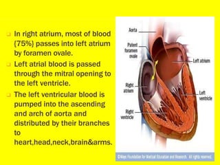
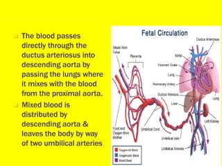
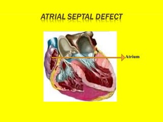
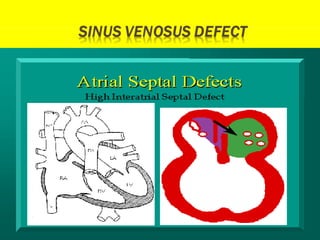
Acyanotic heart
disease
โ‘ The heart is a roughly cone shaped, hallow
muscular organ
โ‘ Heart is about 10 cm long
โ‘ size is ownerโ€™s fist
โ‘ weighs about 225gms in women and heavier in
men
โ‘ Heart lies in thoracic cavity in mediastinum
between lungs
โ‘ The umbilical vein
carrying the oxygenated
blood from the placenta.
โ‘ Enters the fetus at
umbilicus and the blood
goes to liver.
โ‘ The blood from the liver
enter inferior vena cava
through ductus venosus
and then to right atrium
of heart.
โ‘ In right atrium, most of blood
{75%} passes into left atrium
by foramen ovale.
โ‘ Left atrial blood is passed
through the mitral opening to
the left ventricle.
โ‘ The left ventricular blood is
pumped into the ascending
and arch of aorta and
distributed by their branches
to
heart,head,neck,brain&arms.
โ‘ The blood passes
directly through the
ductus arteriosus into
descending aorta by
passing the lungs where
it mixes with the blood
from the proximal aorta.
โ‘ Mixed blood is
distributed by
descending aorta &
leaves the body by way
of two umbilical arteries
โ‘ The mean CARDIAC OUTPUT 225ml/kg/min. in
uterine life.
โ‘ Following BIRTH:- cardiac output 500/min.
โ‘ Heart rate:- 120-140/min.
โ‘ Acyanotic Heart Disease is the common
congenital heart disease present at birth.
โ‘ Itโ€™s prevalence is mostly unknown.
โ‘ In this mostly the cyanosis is not seen hence
known as ACYANOTIC HEART DISEASE
โ‘ It is the one of the major cause of death in the 1st
year of life.
ACYANOTIC HEART DISEASE is a congenital
heart disease with structural malformation of the
heart or great vessels.
โ‘ Idiopathic {90%} cases.
โ‘ Hereditary & Consangious marriage.
โ‘ Genetic disorder & Chromosomal abnormalities.
โ‘ Other associated factors-
fetal & maternal teratrogenic infection {Rubella}.
Teratogenic drug {Thalidomide} intake
Alcohol intake by mother.
Fetal hypoxia.
Birth asphyxia.
There is diminished pulmonary blood flow due to left to
right shut.
It includes:
โ‘ ATRIAL SEPTAL DEFECT
โ‘ VENTRICULAR SEPTAL DEFECT
โ‘ PATENT DUCTUS ARTERIOSUS
โ‘ CO-ARCTATION OF AORTA
Atrium
Atrial septal defect is an abnormal opening
between right to left atria resulting left to right
shunting of blood.
Atrial septal defect are of 3 types
โ‘ Ostium primum.
โ‘ Ostium secundum
โ‘ Sinus venosus defect
โ‘ Asymptomatic
โ‘ Dyspnea
โ‘ Bulging of chest
โ‘ Easy fatigability
โ‘ Poor weight gain
โ‘ Congestive cardiac failure
โ‘ Cardiac enlargement
โ‘ History collection
โ‘ Physical examination
โ‘ Chest x ray
โ‘ ECG
โ‘ 2D Echo
โ‘ Cardiac catheterization
โ‘ Infective endocarditis
โ‘ Pulmonary arterial hypertension
โ‘ Growth retardation
โ‘ Congestive cardiac failure
โ‘ Post- operative complications
โ‘ Atrial fibrillation
โ‘ Chest infection :- Antibiotics
Nebulization
Physiotherapy
โ‘ Fever:-
โ‘ Poor weight gain :-
Antipyretic
IV infusion
โ‘ Open heart surgery :-
Done on affected children before
they enter school even if no
symptoms are present.
โ‘ Purse string closure :-
For small defects a stitching
around the opening and pulling it
closed
โ‘ Knitted Dacron patch :-
If the defect is larger.
Gortex patch
ASD
VENTRICULAR SEPTUM
ventricular septal defect is an abnormal
opening in the septum between right and left
ventricles.
โ‘ Pansystolic murmur
โ‘ Breathlessness
โ‘ Poor feeding
โ‘ Growth failure
โ‘ Dyspnea
โ‘ Sweating
โ‘ Recurrent respiratory infection
โ‘ Tachycardia
โ‘ Poor weight gain
โ‘ Failure to thrive
โ‘ Biventricular hypertrophy
โ‘ History collection
โ‘ Physical examination
โ‘ Chest x ray
โ‘ ECG
โ‘ 2D Echo
โ‘ Cardiac catheterization
โ‘ Doppler
โ‘ Pulmonary atrial hypertension
โ‘ Prolapse of aortic valve
โ‘ Infective endocarditis
โ‘ Control of congestive cardiac failure
โ‘ Treatment of repeated chest infection
โ‘ Prevention and treatment of anemia and infective
endocarditis
Surgical treatment is indicated if :-
โ‘ CCF occurs in infancy
โ‘ Left to right shunt
โ‘ Pulmonary stenosis, pulmonary atrial
hypertension , aortic regurgitation
Closure of the ventricular septal
defect with the use of a DACRON
patch
The operation can be done
BELOW the age of one year.
If congestive cardiac failure
cannot be controlled with medical
management with evidence of
pulmonary hypertension the
operation should be performed by
the age of two years
Dacron graft
VSD
The three major complication are
โ‘ Complete heart block
โ‘ Bifasciular block
โ‘ Re-opened or Residual ventricular septal defect
patent ductus arteriosus is the failure of the
fetal ductus arteriosus {artery connecting the
aorta and pulmonary artery} to close within the
first weeks of the life.
โ‘ 11% of all congenital heart disease
โ‘ Tachypnea
โ‘ Bounding pulse
โ‘ Dyspnea
โ‘ Frequent respiratory infection
โ‘ Increased systolic pressure
โ‘ Low diastolic pressure
โ‘ Pre cordial pain
โ‘ Feeding difficulty
โ‘ Weight gain
โ‘ Growth failure
โ‘ CCF
โ‘ History collection
โ‘ Physical examination
โ‘ ECG
โ‘ 2D Echo
โ‘ Congestive cardiac failure
โ‘ Pulmonary atrial hypertension
โ‘ Cyanosis
โ‘ Endocarditis
Supportive medical management
โ‘ Fluid restriction
โ‘ Ligation of the
ductus
Co-arctation of aorta is the narrowing near the
insertion of the ductus arterious, resulting in the
increased pressure proximal to the defect {head
and upper extremities} and decreased pressure
distal to the obstruction {body and lower
extremities}
โ‘ The condition is more commonly found in male.
โ‘ Accompanies with other defects like VSD,PDA.
โ‘ Blood pressure is higher than normal in upper
part of body
โ‘ In leg, it is relatively low resulting in absence or
diminution of the femoral pulses
โ‘ Legs may be cooler than the arms
โ‘ If childโ€™s exercise muscle cramps in legs
โ‘ A murmur may or may not be present
โ‘ Hypertension
โ‘ Tachypnea
โ‘ Endocarditis
โ‘ Congestive cardiac failure
โ‘ Older children asymptomatic
โ‘ History collection
โ‘ Physical examination
โ‘ Chest x-ray
โ‘ Barium swallow
โ‘ Thoracic roentgenogram
โ‘ 2D Echo
โ‘ Doppler
โ‘ Cardiac catheterization
โ‘ Intracranial hemorrhage
โ‘ Aortic aneurysm
โ‘ Infective endocarditis
โ‘ Congestive heart failure
โ‘ Antibiotics
โ‘ Prevention and treatment of complication
โ‘ Removal of narrowed portion of aorta with
anastomosis of ends
โ‘ In some cases, a graft of transplanted aorta
โ‘ Surgery is postponed until the preferred age 3-6
years
โ‘ Balloon angioplasty
โ‘ Relieving respiratory distress
โ‘ Improving cardiac output
โ‘ Providing adequate nutrition
โ‘ Preventing infection
โ‘ Completion of immunization schedule
โ‘ Reducing fear and anxiety
โ‘ Follow up teaching
โ‘ Avoidance of over protection and rejection
โ‘ Promoting growth and development
โ‘ Referring social service agency
โ‘ Admission history
โ‘ Physical examination
โ‘ Pre-operative studies
โ‘ Baseline vital signs
โ‘ Fluid replacement
โ‘ Pre-operative teaching
โ‘ Transfer the child to recovery room or intensive
care unit .
โ‘ Continues monitoring
โ‘ Restoring optimal function
โ‘ Adequate rest
โ‘ Warm bed
โ‘ Auscultated apically for a full minute
โ‘ Asculte the lungs
โ‘ Check central venous pressure
โ‘ Close observation of system functioning
โ‘ Give fluids gradually
โ‘ Cardiovascular
โ‘ Respiratory
โ‘ Renal
โ‘ Central nervous infection
โ‘ Hyperthermia related to the chest infection
โ‘ Ineffective breathing pattern related to the
dyspnea, septal defect
โ‘ Ineffective airway clearance related to the
retained secretions
โ‘ Nutritional requirement less than the body
demand
โ‘ Fear and anxiety related to the long hospital stay
โ‘ Risk for infection related to the congenital heart
disease or IV cannula
โ‘ Knowledge deficient regarding the care of the
child at home
The acynotic heart diseases are asymptomatic
initially and shows symptoms in later months or
year. So at the birth the child thorough systemic
examination is done for early detection and
treatment
The risk for operation is less in the infancy or
up to 3 years in some cases once the vascular
changes takes place as the years passes then it
may lead to complication like HTN, CCF, surgical
intervention is at greater risk.
ACYANOTIC HEART DISEASE.pptx

ACYANOTIC HEART DISEASE.pptx

  • 1.
  • 3.
    โ‘ The heartis a roughly cone shaped, hallow muscular organ โ‘ Heart is about 10 cm long โ‘ size is ownerโ€™s fist โ‘ weighs about 225gms in women and heavier in men โ‘ Heart lies in thoracic cavity in mediastinum between lungs
  • 4.
    โ‘ The umbilicalvein carrying the oxygenated blood from the placenta. โ‘ Enters the fetus at umbilicus and the blood goes to liver. โ‘ The blood from the liver enter inferior vena cava through ductus venosus and then to right atrium of heart.
  • 5.
    โ‘ In rightatrium, most of blood {75%} passes into left atrium by foramen ovale. โ‘ Left atrial blood is passed through the mitral opening to the left ventricle. โ‘ The left ventricular blood is pumped into the ascending and arch of aorta and distributed by their branches to heart,head,neck,brain&arms.
  • 6.
    โ‘ The bloodpasses directly through the ductus arteriosus into descending aorta by passing the lungs where it mixes with the blood from the proximal aorta. โ‘ Mixed blood is distributed by descending aorta & leaves the body by way of two umbilical arteries
  • 7.
    โ‘ The meanCARDIAC OUTPUT 225ml/kg/min. in uterine life. โ‘ Following BIRTH:- cardiac output 500/min. โ‘ Heart rate:- 120-140/min.
  • 9.
    โ‘ Acyanotic HeartDisease is the common congenital heart disease present at birth. โ‘ Itโ€™s prevalence is mostly unknown. โ‘ In this mostly the cyanosis is not seen hence known as ACYANOTIC HEART DISEASE โ‘ It is the one of the major cause of death in the 1st year of life.
  • 10.
    ACYANOTIC HEART DISEASEis a congenital heart disease with structural malformation of the heart or great vessels.
  • 11.
    โ‘ Idiopathic {90%}cases. โ‘ Hereditary & Consangious marriage. โ‘ Genetic disorder & Chromosomal abnormalities. โ‘ Other associated factors- fetal & maternal teratrogenic infection {Rubella}. Teratogenic drug {Thalidomide} intake Alcohol intake by mother. Fetal hypoxia. Birth asphyxia.
  • 12.
    There is diminishedpulmonary blood flow due to left to right shut. It includes: โ‘ ATRIAL SEPTAL DEFECT โ‘ VENTRICULAR SEPTAL DEFECT โ‘ PATENT DUCTUS ARTERIOSUS โ‘ CO-ARCTATION OF AORTA
  • 13.
  • 14.
    Atrial septal defectis an abnormal opening between right to left atria resulting left to right shunting of blood.
  • 15.
    Atrial septal defectare of 3 types โ‘ Ostium primum. โ‘ Ostium secundum โ‘ Sinus venosus defect
  • 20.
    โ‘ Asymptomatic โ‘ Dyspnea โ‘Bulging of chest โ‘ Easy fatigability โ‘ Poor weight gain โ‘ Congestive cardiac failure โ‘ Cardiac enlargement
  • 21.
    โ‘ History collection โ‘Physical examination โ‘ Chest x ray โ‘ ECG โ‘ 2D Echo โ‘ Cardiac catheterization
  • 22.
    โ‘ Infective endocarditis โ‘Pulmonary arterial hypertension โ‘ Growth retardation โ‘ Congestive cardiac failure โ‘ Post- operative complications โ‘ Atrial fibrillation
  • 23.
    โ‘ Chest infection:- Antibiotics Nebulization Physiotherapy โ‘ Fever:- โ‘ Poor weight gain :- Antipyretic IV infusion
  • 24.
    โ‘ Open heartsurgery :- Done on affected children before they enter school even if no symptoms are present. โ‘ Purse string closure :- For small defects a stitching around the opening and pulling it closed
  • 25.
    โ‘ Knitted Dacronpatch :- If the defect is larger. Gortex patch ASD
  • 26.
  • 27.
    ventricular septal defectis an abnormal opening in the septum between right and left ventricles.
  • 29.
    โ‘ Pansystolic murmur โ‘Breathlessness โ‘ Poor feeding โ‘ Growth failure โ‘ Dyspnea โ‘ Sweating
  • 30.
    โ‘ Recurrent respiratoryinfection โ‘ Tachycardia โ‘ Poor weight gain โ‘ Failure to thrive โ‘ Biventricular hypertrophy
  • 31.
    โ‘ History collection โ‘Physical examination โ‘ Chest x ray โ‘ ECG โ‘ 2D Echo โ‘ Cardiac catheterization โ‘ Doppler
  • 32.
    โ‘ Pulmonary atrialhypertension โ‘ Prolapse of aortic valve โ‘ Infective endocarditis
  • 33.
    โ‘ Control ofcongestive cardiac failure โ‘ Treatment of repeated chest infection โ‘ Prevention and treatment of anemia and infective endocarditis
  • 34.
    Surgical treatment isindicated if :- โ‘ CCF occurs in infancy โ‘ Left to right shunt โ‘ Pulmonary stenosis, pulmonary atrial hypertension , aortic regurgitation
  • 35.
    Closure of theventricular septal defect with the use of a DACRON patch The operation can be done BELOW the age of one year. If congestive cardiac failure cannot be controlled with medical management with evidence of pulmonary hypertension the operation should be performed by the age of two years Dacron graft VSD
  • 36.
    The three majorcomplication are โ‘ Complete heart block โ‘ Bifasciular block โ‘ Re-opened or Residual ventricular septal defect
  • 38.
    patent ductus arteriosusis the failure of the fetal ductus arteriosus {artery connecting the aorta and pulmonary artery} to close within the first weeks of the life.
  • 39.
    โ‘ 11% ofall congenital heart disease
  • 41.
    โ‘ Tachypnea โ‘ Boundingpulse โ‘ Dyspnea โ‘ Frequent respiratory infection โ‘ Increased systolic pressure โ‘ Low diastolic pressure
  • 42.
    โ‘ Pre cordialpain โ‘ Feeding difficulty โ‘ Weight gain โ‘ Growth failure โ‘ CCF
  • 43.
    โ‘ History collection โ‘Physical examination โ‘ ECG โ‘ 2D Echo
  • 44.
    โ‘ Congestive cardiacfailure โ‘ Pulmonary atrial hypertension โ‘ Cyanosis โ‘ Endocarditis
  • 45.
  • 46.
  • 48.
    Co-arctation of aortais the narrowing near the insertion of the ductus arterious, resulting in the increased pressure proximal to the defect {head and upper extremities} and decreased pressure distal to the obstruction {body and lower extremities}
  • 49.
    โ‘ The conditionis more commonly found in male. โ‘ Accompanies with other defects like VSD,PDA.
  • 51.
    โ‘ Blood pressureis higher than normal in upper part of body โ‘ In leg, it is relatively low resulting in absence or diminution of the femoral pulses โ‘ Legs may be cooler than the arms โ‘ If childโ€™s exercise muscle cramps in legs โ‘ A murmur may or may not be present
  • 52.
    โ‘ Hypertension โ‘ Tachypnea โ‘Endocarditis โ‘ Congestive cardiac failure โ‘ Older children asymptomatic
  • 53.
    โ‘ History collection โ‘Physical examination โ‘ Chest x-ray โ‘ Barium swallow โ‘ Thoracic roentgenogram โ‘ 2D Echo
  • 54.
  • 55.
    โ‘ Intracranial hemorrhage โ‘Aortic aneurysm โ‘ Infective endocarditis โ‘ Congestive heart failure
  • 56.
    โ‘ Antibiotics โ‘ Preventionand treatment of complication
  • 57.
    โ‘ Removal ofnarrowed portion of aorta with anastomosis of ends โ‘ In some cases, a graft of transplanted aorta โ‘ Surgery is postponed until the preferred age 3-6 years โ‘ Balloon angioplasty
  • 59.
    โ‘ Relieving respiratorydistress โ‘ Improving cardiac output โ‘ Providing adequate nutrition โ‘ Preventing infection โ‘ Completion of immunization schedule
  • 60.
    โ‘ Reducing fearand anxiety โ‘ Follow up teaching โ‘ Avoidance of over protection and rejection โ‘ Promoting growth and development โ‘ Referring social service agency
  • 61.
    โ‘ Admission history โ‘Physical examination โ‘ Pre-operative studies โ‘ Baseline vital signs โ‘ Fluid replacement โ‘ Pre-operative teaching
  • 62.
    โ‘ Transfer thechild to recovery room or intensive care unit . โ‘ Continues monitoring โ‘ Restoring optimal function โ‘ Adequate rest โ‘ Warm bed
  • 63.
    โ‘ Auscultated apicallyfor a full minute โ‘ Asculte the lungs โ‘ Check central venous pressure โ‘ Close observation of system functioning โ‘ Give fluids gradually
  • 64.
    โ‘ Cardiovascular โ‘ Respiratory โ‘Renal โ‘ Central nervous infection
  • 65.
    โ‘ Hyperthermia relatedto the chest infection โ‘ Ineffective breathing pattern related to the dyspnea, septal defect โ‘ Ineffective airway clearance related to the retained secretions โ‘ Nutritional requirement less than the body demand
  • 66.
    โ‘ Fear andanxiety related to the long hospital stay โ‘ Risk for infection related to the congenital heart disease or IV cannula โ‘ Knowledge deficient regarding the care of the child at home
  • 67.
    The acynotic heartdiseases are asymptomatic initially and shows symptoms in later months or year. So at the birth the child thorough systemic examination is done for early detection and treatment The risk for operation is less in the infancy or up to 3 years in some cases once the vascular changes takes place as the years passes then it may lead to complication like HTN, CCF, surgical intervention is at greater risk.