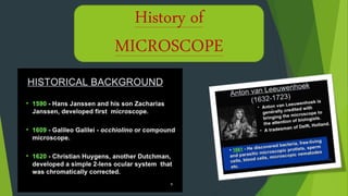
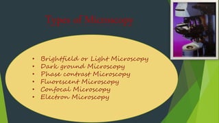
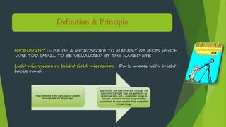
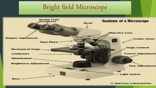
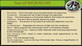
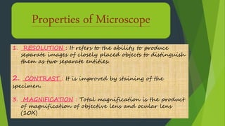
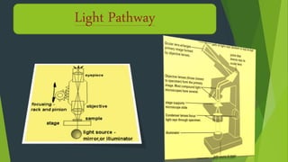
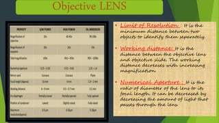
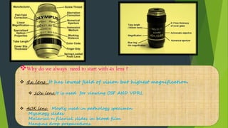
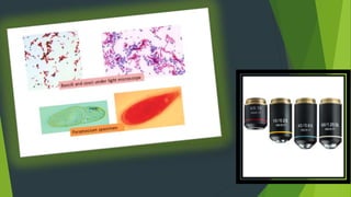
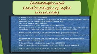
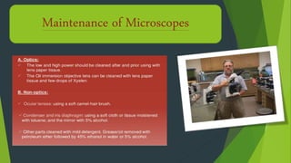
3. Types of Microscopy
• Brightfield or Light Microscopy
• Dark ground Microscopy
• Phase contrast Microscopy
• Fluorescent Microscopy
• Confocal Microscopy
• Electron Microscopy
4. PRINCIPLEDefinition & Principle
MICROSCOPY :-USE OF A MICROSCOPE TO MAGNIFY OBJECTS WHICH
ARE TOO SMALL TO BE VISUALIZED BY THE NAKED EYE
Light microscopy or bright field microscopy : Dark images with bright
background
Rays emitted from light source passes
through the iris diaphragm
and fall on the specimen and through the
specimen the light rays are gathered by
objective lens and a magnified image is
formed ,which is further magnified by
ocular lens to produce the final magnified
virtual image
7. Properties of Microscope
1. RESOLUTION : It refers to the ability to produce
separate images of closely placed objects to distinguish
them as two separate entities.
2. CONTRAST : It is improved by staining of the
specimen.
3. MAGNIFICATION : Total magnification is the product
of magnification of objective lens and ocular lens
(10X)
9. i
Objective LENS
• Limit of Resolution : It is the
minimum distance between two
objects to identify them separately.
• Working distance: It is the
distance between the objective lens
and objective slide. The working
distance decreases with increasing
magnification.
• Numerical Aperture : It is the
ratio of diameter of the lens to its
focal length. It can be decreased by
decreasing the amount of light that
passes through the lens.
10. Why do we always need to start with 4x lens ?
4x lens It has lowest field of vision but highest magnification.
10x lens It is used for viewing CSF AND VDRL
40X lens Mostly used in pathology specimen
Mycology slides
Malarial n filarial slides in blood film
Hanging drop preparations