Coats Disease: An
Overview
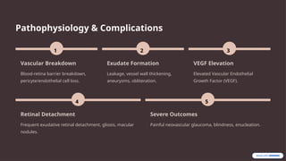
Firstdescribed in 1908, Coats disease is characterized by retinal vascular
abnormalities, exudates, and hemorrhage.
2.
Key Characteristics
Onset
Typically beforeage 15.
Leukocoria and strabismus
present earlier.
Laterality
Tends to be unilateral.
Demographics
Preferentially affects males;
no specific racial/ethnic
associations.
3.
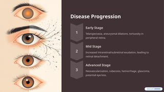
Disease Progression
Early Stage
Telangiectasia,aneurysmal dilations, tortuosity in
peripheral retina.
Mid Stage
Increased intraretinal/subretinal exudation, leading to
retinal detachment.
Advanced Stage
Neovascularization, rubecosis, hemorrhage, glaucoma,
potential eye loss.
4.
Etiology: Unraveling theCause
Genetics
Nonhereditary ocular disease.
Proposed somatic missense
mutation of NDP gene (Xp11.2).
High male-to-female ratio (3:1)
due to X inactivation.
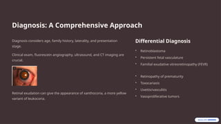
Diagnosis: A ComprehensiveApproach
Diagnosis considers age, family history, laterality, and presentation
stage.
Clinical exam, fluorescein angiography, ultrasound, and CT imaging are
crucial.
Retinal exudation can give the appearance of xanthocoria, a more yellow
variant of leukocoria.
Differential Diagnosis
• Retinoblastoma
• Persistent fetal vasculature
• Familial exudative vitreoretinopathy (FEVR)
• Retinopathy of prematurity
• Toxocariasis
• Uveitis/vasculitis
• Vasoproliferative tumors
7.
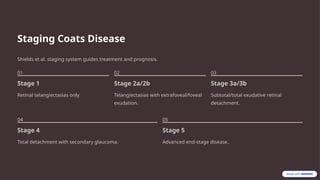
Staging Coats Disease
Shieldset al. staging system guides treatment and prognosis.
01
Stage 1
Retinal telangiectasias only.
02
Stage 2a/2b
Telangiectasias with extrafoveal/foveal
exudation.
03
Stage 3a/3b
Subtotal/total exudative retinal
detachment.
04
Stage 4
Total detachment with secondary glaucoma.
05
Stage 5
Advanced end-stage disease.
8.
Treatment Approaches
Laser Ablation
Obliteratesabnormal
vasculature, eliminates
hyperpermeability.
Cryotherapy
For peripheral telangiectasia
with significant exudative
detachment.
Surgery
For total detachment, vitreous hemorrhage, or secondary
complications.
9.
Adjuvant Therapy &Case Study
Anti-VEGF Agents
Effective in reducing subretinal fluid and exudation.
Example: Bevacizumab treatment showed complete
resolution in 24 children.
Caution: May lead to vitreoretinal fibrosis and tractional
retinal detachment.
Case: Young Adult
24-year-old with blurry vision, photopsias, floaters.
Diagnosed Stage 2A, received sectoral photoagulation.
Stable with 20/20 vision at 6-month follow-up.
10.
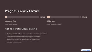
Prognosis & RiskFactors
<5 yrs
Younger Age
More rapid decline.
>10 yrs
Older Age
More insidious course.
Risk Factors for Visual Decline
• Postequatorial, diffuse, or superior telangiectasia/exudation.
• Failed resolution of subretinal fluid post-treatment.
• Retinal macrocysts or detachment at presentation.
• Macular involvement.