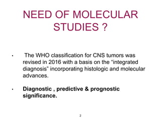
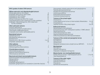
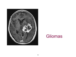
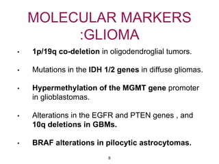
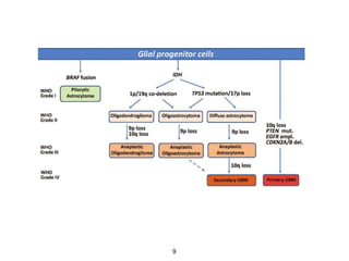
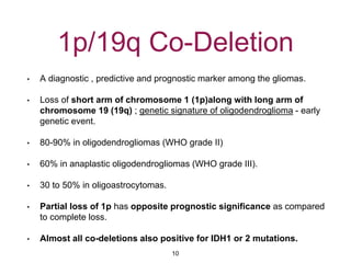
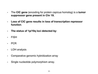
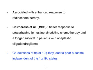
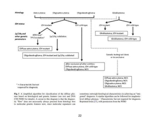
MOLECULAR STUDIES
IN CNS TUMORS
DR. AMRITA RAKESH
(DNB RESIDENT)
Bhagwan Mahaveer Cancer Hospital And Research Centre
Jaipur.
1
NEED OF MOLECULAR
STUDIES ?
• The WHO classification for CNS tumors was
revised in 2016 with a basis on the “integrated
diagnosis” incorporating histologic and molecular
advances.
• Diagnostic , predictive & prognostic
significance.
2
3
Cell of Origin For CNS
tumors
• Glial Cells
• Neural Stem Cells (NSCs)
• Genes that are expressed in NSCs are Nestin,
EGFR , PTEN , Hedgehog etc.
• Aberrant activation of developmental genetic
programs in NSC gives rise to CNS tumors.
4
Gliomas
5
• Gliomas are the most common intracranial
neoplasm.
• Astrocytoma , glioblastomas and oligodendrogliomas
accounting for more than 80%.
• Classified as grade I to IV.
• Grade I - JPA
• Grade II - fibrillary astrocytoma
• Grade III - anaplastic astrocytoma
• Grade IV - glioblastoma (most aggressive).
6
• 1985 : Epidermal growth factor receptor (EGFR) gene
amplification in glioblastoma.
• Subsequent discoveries :
• Phosphate and tensin homolog (PTEN) gene.
• Mutations in the Tp53 gene.
• BRAF mutation
• MGMT gene
• IDH mutations.
7
MOLECULAR MARKERS
:GLIOMA
• 1p/19q co-deletion in oligodendroglial tumors.
• Mutations in the IDH 1/2 genes in diffuse gliomas.
• Hypermethylation of the MGMT gene promoter
in glioblastomas.
• Alterations in the EGFR and PTEN genes , and
10q deletions in GBMs.
• BRAF alterations in pilocytic astrocytomas.
8
9
1p/19q Co-Deletion
• A diagnostic , predictive and prognostic marker among the gliomas.
• Loss of short arm of chromosome 1 (1p)along with long arm of
chromosome 19 (19q) ; genetic signature of oligodendroglioma - early
genetic event.
• 80-90% in oligodendrogliomas (WHO grade II)
• 60% in anaplastic oligodendrogliomas (WHO grade III).
• 30 to 50% in oligoastrocytomas.
• Partial loss of 1p has opposite prognostic significance as compared
to complete loss.
• Almost all co-deletions also positive for IDH1 or 2 mutations.
10
• The CIC gene (encoding for protein capicua homolog) is a tumor
suppressor gene present in Chr 19.
• Loss of CIC gene results in loss of transcription repressor
function.
• The status of 1p/19q loci detected by:
• FISH
• PCR
• LOH analysis
• Comparative genomic hybridization array
• Single nucleotide polymorphism array.
11
• Associated with enhanced response to
radiochemotherapy.
• Cairncross et al.:(1998) : better response to
procarbazine-lomustine-vincristine chemotherapy and
a longer survival in patients with anaplastic
oligodendroglioma.
• Co-deletions of 9p or 10q may lead to poor outcome
independent of the 1p/19q status.
12
IDH1 & IDH2 Mutations
• Diagnostic and prognostic marker among gliomas.
• Mutations in IDH1 are frequent (70-80%) in WHO grade II & III
astrocytomas and a small subset (5-10%) of GBMs (WHO grade
IV).
• IDH1 are completely absent in pilocytic astrocytomas.
• IDH2 genes mutations were detected in a similar proportion of
gliomas (5%) , mostly in oligodendroglial tumors.
• Presence of the mutation is associated with young age , a
secondary type GBM (60-90%) , and increased overall
survival.
13
• IDH normally catalyses the action of isocitrate to a-
ketoglutarate, whereas the mutant forms of IDH1 & IDH2
lead to the production of the oncometabolite 2-
hydroxyglutarate, which inhibits function of numerous a-
ketoglutarate-dependent enzymes and leads to higher
levels of DNA methylation.
• IDH mutations identify a subset of gliomas with a slower rate
of growth and substantially improved prognosis, grade for
grade, compared with gliomas that are IDH wild type.
14
MGMT Methylation
• The MGMT gene is located at chromosome 10q26 and
encodes for a DNA repair protein.
• Prognostic and predictive marker.
• Epigenetic silencing of this gene by promoter
hypermethylation leads to reduced expression of the
MGMT protein.
• MGMT gene silencing improves survival in patients
with glioblastoma who are treated concurrently
with alkylating drug temozolamide and radiation
therapy.
15
• Hegi and colleagues (2005) : reported that 49%
of patients with glioblastoma and methylated
MGMT were alive at 2 yrs after treatment with
temozolamide and radiotherapy, as compared to
15% of patients with unmethylated MGMT.
16
BRAF Mutation
• In gliomas : BRAF activation is by gene duplication or
point mutation.
• Fusion between the KIAA1549 and BRAF genes.
• Identified in 60-80% pilocytic astrocytomas ; rare in
diffuse astrocytic gliomas.
• Interphase FISH -currently the best method for testing
for this fusion.
• Prognostic significance is still under investigation.
17
EGFR and PTEN
alterations
• EGFR gene : located on chromosome 7p12.
• Activation of EGFR signalling through gene amplification or
mutations is found in about 30 to 40% of primary glioblastomas.
• Mutant EGFR is indicative of high-grade glioma .
• PTEN gene located on long arm of chromosome 10.
• Counteracts PI3K-AKT signaling pathway.
• frequently found in high-grade gliomas.
• Poor prognostic marker for anaplastic astrocytomas and
glioblastomas.
18
Ki-67
• Prognostic marker among grade II & III diffuse gliomas.
• Ki-67 is a nuclear antigen expressed in cells actively engaged in
the cell cycle but not expressed in the resting phase G0.
• Results expressed as percentage of positive staining cells.
• Among grade II and III diffuse gliomas, the Ki-67 index provides
prognostic value, as there is strong inverse relationship to
survival on multivariat analysis but not in GBM.
• Helpful in determining grade in histologically borderline cases.
19
TP53
• Diagnostic marker among gliomas.
• TP53 mutation is a marker of astrocytoma lineage
in the setting of IDH mutation and occurs in
infiltrative astrocytomas,grade II;anaplastic
astrocytomas,grade III; and GBM,WHO grade IV.
• Extremely rare in Oligodendrogliomas with IDH
mutation and 1p/19q codeletion.
20
ATRX
• ATRX (alpha thalassemia/mental retardation
syndrome X-linked) - diagnostic marker among
gliomas.
• Strongly associated with IDH mutation and TP53
mutation in infiltrating gliomas.
• Marker of astrocytic lineage.
21
22
Medulloblastoma
23
• Origin : stem cells located in the subependymal
matrix and the external granular layer of the
cerebellum.
• Medulloblastomas are tumors of the cerebellum ,
arising more frequently in the midline , especially
in the posterior vermis , adjacent to the roof of the
fourth ventricle.
24
• Five histological subtypes :
• Classical type (CMB)
• Desmoplastic / Nodular type (DN)
• Medulloblastoma with extensive nodularity
(MBEN)
• Anaplastic type
• Large cell Medulloblastoma (LC)
25
• 4 - MOLECULAR SUBGROUPS:
• WNT subgroup
• Sonic hedgehog subgroup
• Group 3
• Group 4
26
• WNT subgroup - the WNT/b-catenin pathway
participates in the control of vertebrate development.
• Rarest subgroup of medulloblastoma, accounting for 11%
; includes mainly classic MB.
• Patients with Turcot Syndrome : predisposition to WNT
MB.
• Monosomy of chromosome 6 is present in about
100% of WNT tumors.
• Overall excellent long term prognosis (90% 5 yr
survival rate).
27
• IHC : monoclonal antibodies against the C-
terminal domain of b-catenin.
• CTNNB1 (b-catenin encoding gene) mutation
analysis by direct gene sequencing.
• Cantharidin and noncantharidin : drugs on trial
against Wnt associated medulloblastoma.
28
• SHH medulloblastomas - accounts for 28% of all
medulloblastomas ; intermediate prognosis.
• Dichotomous age distribution : <4 yrs and >16 yrs.
• Sonic Hedgehog (shh) pathway plays a key role in
normal cerebellar development, induces
proliferation of neuronal precursor cells in the
developing cerebellum and other tissues.
• Desmoplastic/nodular and MBEN are almost
exclusively associated with shh pathway
activation.
29
• Group 3 Medulloblastomas - 28% of all medulloblastomas.
• associated with worst prognosis and frequently metastatic.
• predominantly found in infants/children.
• associated with MYC amplification.
• 3a-tumors — increased MYC expression and worse
prognosis.
• 3b-tumors — normal MYC expression and better prognosis.
• Mostly associated with classic or large cell/anaplastic
morphology.
30
• Group 4 Medulloblastomas - most common “typical” subgroup
of medulloblastoma, accounting for around 34%.
• Rarely affect infants (0-3 yrs) and mainly affect children, with a
peak age of 10 years.
• intermediate prognosis.
• classic histology.
• 2/3rd cases associated with isochromosome 17q.
• associated with CDK6 & MYCN amplification but minimal MYC
over-expression.
• Chromosome X-loss is seen in 80% of females.
31
32
33
EPENDYMAL
TUMORS
34
• Taylor et al., aCGH profiles of 103
ependymomas, three molecularly distinct
subtypes of ependymomas depending on tumor
location :
• Supratentorial ependymomas : CDKN2A
deletion in >90% cases, poor prognosis.
• Spinal tumors : deletion of chromosome 22q12
• Posterior fossa ependymomas : chromosome
1q gain , good prognosis.
35
• RELA fusion positive ependymomas : subset of
supratentorial ependymomas.
• amenable to targeted therapy.
• Methylation status of HIC-1 putative tumor
suppressor gene : down regulation in 81% of
cases , correlated with non-spinal localization and
paediatric age.
36
MENINGIOMA
37
• Most common CNS tumors in adults.
• Associated risk factors - deletions of the neurofibromatosis
type 2 (NF2) gene & ionising radiations.
• Benign meningiomas - slow growing & 5-yr recurrence rate
of 5% following gross-total resection.
• Atypical meningiomas have 5-yr recurrence rate of
40%.
• Anaplastic meningiomas have recurrence rates of up to
80%.
• Surgical resection and radiotherapy- mainstay of treatment.
38
Cytogenetic Abnormality
• Monosomy 22 is the most frequent genetic
abnormality.
• Association between the long arm of chromosome
22 (22q) and meningiomas was first studied in
patients with NF-2.
• NF-2 codes for tumor suppressor merlin , which
has critical role in controlling cell growth and
motility.
39
• Chr 1p deletions comprise the second most
common chromosomal abnormality.
• Loss of 1p also associated with a 30% recurrence
rate.
40
PRIMARY CNS
LYMPHOMA
41
• Accounts for less than 5% of all primary brain
tumours.
• 95-98% diagnosed as high-grade DLBCL.
• 70% are supratentorial
• Periventricular location , facilitating
leptomeningeal seeding.
• Extends across the corpus-callosum and involve
both cerebral hemispheres.
42
• Gain on chromosome 12 — most frequent alteration.
• Gain in the region of 12q.
• MDM2 , CDK4 & GLI1 overexpression.
• EBV - immunocompromised individuals.
• BCA1 (CXCR13) - expressed at significant levels in PCNSL
tumors.
• Ectopic expression of Interleukin-4 : not expressed in the
vasculature of normal brain.
• Hypermethylation of CDKN2A gene - established molecular
event; produces p14ARF.
43
• TO CONCLUDE - not only a morphological
diagnosis , but also molecular data is necessary
for prognosis and response to treatment.
44
Thank You
45

Molecular studies in cns tumors

  • 1.
    MOLECULAR STUDIES IN CNSTUMORS DR. AMRITA RAKESH (DNB RESIDENT) Bhagwan Mahaveer Cancer Hospital And Research Centre Jaipur. 1
  • 2.
    NEED OF MOLECULAR STUDIES? • The WHO classification for CNS tumors was revised in 2016 with a basis on the “integrated diagnosis” incorporating histologic and molecular advances. • Diagnostic , predictive & prognostic significance. 2
  • 3.
  • 4.
    Cell of OriginFor CNS tumors • Glial Cells • Neural Stem Cells (NSCs) • Genes that are expressed in NSCs are Nestin, EGFR , PTEN , Hedgehog etc. • Aberrant activation of developmental genetic programs in NSC gives rise to CNS tumors. 4
  • 5.
  • 6.
    • Gliomas arethe most common intracranial neoplasm. • Astrocytoma , glioblastomas and oligodendrogliomas accounting for more than 80%. • Classified as grade I to IV. • Grade I - JPA • Grade II - fibrillary astrocytoma • Grade III - anaplastic astrocytoma • Grade IV - glioblastoma (most aggressive). 6
  • 7.
    • 1985 :Epidermal growth factor receptor (EGFR) gene amplification in glioblastoma. • Subsequent discoveries : • Phosphate and tensin homolog (PTEN) gene. • Mutations in the Tp53 gene. • BRAF mutation • MGMT gene • IDH mutations. 7
  • 8.
    MOLECULAR MARKERS :GLIOMA • 1p/19qco-deletion in oligodendroglial tumors. • Mutations in the IDH 1/2 genes in diffuse gliomas. • Hypermethylation of the MGMT gene promoter in glioblastomas. • Alterations in the EGFR and PTEN genes , and 10q deletions in GBMs. • BRAF alterations in pilocytic astrocytomas. 8
  • 9.
  • 10.
    1p/19q Co-Deletion • Adiagnostic , predictive and prognostic marker among the gliomas. • Loss of short arm of chromosome 1 (1p)along with long arm of chromosome 19 (19q) ; genetic signature of oligodendroglioma - early genetic event. • 80-90% in oligodendrogliomas (WHO grade II) • 60% in anaplastic oligodendrogliomas (WHO grade III). • 30 to 50% in oligoastrocytomas. • Partial loss of 1p has opposite prognostic significance as compared to complete loss. • Almost all co-deletions also positive for IDH1 or 2 mutations. 10
  • 11.
    • The CICgene (encoding for protein capicua homolog) is a tumor suppressor gene present in Chr 19. • Loss of CIC gene results in loss of transcription repressor function. • The status of 1p/19q loci detected by: • FISH • PCR • LOH analysis • Comparative genomic hybridization array • Single nucleotide polymorphism array. 11
  • 12.
    • Associated withenhanced response to radiochemotherapy. • Cairncross et al.:(1998) : better response to procarbazine-lomustine-vincristine chemotherapy and a longer survival in patients with anaplastic oligodendroglioma. • Co-deletions of 9p or 10q may lead to poor outcome independent of the 1p/19q status. 12
  • 13.
    IDH1 & IDH2Mutations • Diagnostic and prognostic marker among gliomas. • Mutations in IDH1 are frequent (70-80%) in WHO grade II & III astrocytomas and a small subset (5-10%) of GBMs (WHO grade IV). • IDH1 are completely absent in pilocytic astrocytomas. • IDH2 genes mutations were detected in a similar proportion of gliomas (5%) , mostly in oligodendroglial tumors. • Presence of the mutation is associated with young age , a secondary type GBM (60-90%) , and increased overall survival. 13
  • 14.
    • IDH normallycatalyses the action of isocitrate to a- ketoglutarate, whereas the mutant forms of IDH1 & IDH2 lead to the production of the oncometabolite 2- hydroxyglutarate, which inhibits function of numerous a- ketoglutarate-dependent enzymes and leads to higher levels of DNA methylation. • IDH mutations identify a subset of gliomas with a slower rate of growth and substantially improved prognosis, grade for grade, compared with gliomas that are IDH wild type. 14
  • 15.
    MGMT Methylation • TheMGMT gene is located at chromosome 10q26 and encodes for a DNA repair protein. • Prognostic and predictive marker. • Epigenetic silencing of this gene by promoter hypermethylation leads to reduced expression of the MGMT protein. • MGMT gene silencing improves survival in patients with glioblastoma who are treated concurrently with alkylating drug temozolamide and radiation therapy. 15
  • 16.
    • Hegi andcolleagues (2005) : reported that 49% of patients with glioblastoma and methylated MGMT were alive at 2 yrs after treatment with temozolamide and radiotherapy, as compared to 15% of patients with unmethylated MGMT. 16
  • 17.
    BRAF Mutation • Ingliomas : BRAF activation is by gene duplication or point mutation. • Fusion between the KIAA1549 and BRAF genes. • Identified in 60-80% pilocytic astrocytomas ; rare in diffuse astrocytic gliomas. • Interphase FISH -currently the best method for testing for this fusion. • Prognostic significance is still under investigation. 17
  • 18.
    EGFR and PTEN alterations •EGFR gene : located on chromosome 7p12. • Activation of EGFR signalling through gene amplification or mutations is found in about 30 to 40% of primary glioblastomas. • Mutant EGFR is indicative of high-grade glioma . • PTEN gene located on long arm of chromosome 10. • Counteracts PI3K-AKT signaling pathway. • frequently found in high-grade gliomas. • Poor prognostic marker for anaplastic astrocytomas and glioblastomas. 18
  • 19.
    Ki-67 • Prognostic markeramong grade II & III diffuse gliomas. • Ki-67 is a nuclear antigen expressed in cells actively engaged in the cell cycle but not expressed in the resting phase G0. • Results expressed as percentage of positive staining cells. • Among grade II and III diffuse gliomas, the Ki-67 index provides prognostic value, as there is strong inverse relationship to survival on multivariat analysis but not in GBM. • Helpful in determining grade in histologically borderline cases. 19
  • 20.
    TP53 • Diagnostic markeramong gliomas. • TP53 mutation is a marker of astrocytoma lineage in the setting of IDH mutation and occurs in infiltrative astrocytomas,grade II;anaplastic astrocytomas,grade III; and GBM,WHO grade IV. • Extremely rare in Oligodendrogliomas with IDH mutation and 1p/19q codeletion. 20
  • 21.
    ATRX • ATRX (alphathalassemia/mental retardation syndrome X-linked) - diagnostic marker among gliomas. • Strongly associated with IDH mutation and TP53 mutation in infiltrating gliomas. • Marker of astrocytic lineage. 21
  • 22.
  • 23.
  • 24.
    • Origin :stem cells located in the subependymal matrix and the external granular layer of the cerebellum. • Medulloblastomas are tumors of the cerebellum , arising more frequently in the midline , especially in the posterior vermis , adjacent to the roof of the fourth ventricle. 24
  • 25.
    • Five histologicalsubtypes : • Classical type (CMB) • Desmoplastic / Nodular type (DN) • Medulloblastoma with extensive nodularity (MBEN) • Anaplastic type • Large cell Medulloblastoma (LC) 25
  • 26.
    • 4 -MOLECULAR SUBGROUPS: • WNT subgroup • Sonic hedgehog subgroup • Group 3 • Group 4 26
  • 27.
    • WNT subgroup- the WNT/b-catenin pathway participates in the control of vertebrate development. • Rarest subgroup of medulloblastoma, accounting for 11% ; includes mainly classic MB. • Patients with Turcot Syndrome : predisposition to WNT MB. • Monosomy of chromosome 6 is present in about 100% of WNT tumors. • Overall excellent long term prognosis (90% 5 yr survival rate). 27
  • 28.
    • IHC :monoclonal antibodies against the C- terminal domain of b-catenin. • CTNNB1 (b-catenin encoding gene) mutation analysis by direct gene sequencing. • Cantharidin and noncantharidin : drugs on trial against Wnt associated medulloblastoma. 28
  • 29.
    • SHH medulloblastomas- accounts for 28% of all medulloblastomas ; intermediate prognosis. • Dichotomous age distribution : <4 yrs and >16 yrs. • Sonic Hedgehog (shh) pathway plays a key role in normal cerebellar development, induces proliferation of neuronal precursor cells in the developing cerebellum and other tissues. • Desmoplastic/nodular and MBEN are almost exclusively associated with shh pathway activation. 29
  • 30.
    • Group 3Medulloblastomas - 28% of all medulloblastomas. • associated with worst prognosis and frequently metastatic. • predominantly found in infants/children. • associated with MYC amplification. • 3a-tumors — increased MYC expression and worse prognosis. • 3b-tumors — normal MYC expression and better prognosis. • Mostly associated with classic or large cell/anaplastic morphology. 30
  • 31.
    • Group 4Medulloblastomas - most common “typical” subgroup of medulloblastoma, accounting for around 34%. • Rarely affect infants (0-3 yrs) and mainly affect children, with a peak age of 10 years. • intermediate prognosis. • classic histology. • 2/3rd cases associated with isochromosome 17q. • associated with CDK6 & MYCN amplification but minimal MYC over-expression. • Chromosome X-loss is seen in 80% of females. 31
  • 32.
  • 33.
  • 34.
  • 35.
    • Taylor etal., aCGH profiles of 103 ependymomas, three molecularly distinct subtypes of ependymomas depending on tumor location : • Supratentorial ependymomas : CDKN2A deletion in >90% cases, poor prognosis. • Spinal tumors : deletion of chromosome 22q12 • Posterior fossa ependymomas : chromosome 1q gain , good prognosis. 35
  • 36.
    • RELA fusionpositive ependymomas : subset of supratentorial ependymomas. • amenable to targeted therapy. • Methylation status of HIC-1 putative tumor suppressor gene : down regulation in 81% of cases , correlated with non-spinal localization and paediatric age. 36
  • 37.
  • 38.
    • Most commonCNS tumors in adults. • Associated risk factors - deletions of the neurofibromatosis type 2 (NF2) gene & ionising radiations. • Benign meningiomas - slow growing & 5-yr recurrence rate of 5% following gross-total resection. • Atypical meningiomas have 5-yr recurrence rate of 40%. • Anaplastic meningiomas have recurrence rates of up to 80%. • Surgical resection and radiotherapy- mainstay of treatment. 38
  • 39.
    Cytogenetic Abnormality • Monosomy22 is the most frequent genetic abnormality. • Association between the long arm of chromosome 22 (22q) and meningiomas was first studied in patients with NF-2. • NF-2 codes for tumor suppressor merlin , which has critical role in controlling cell growth and motility. 39
  • 40.
    • Chr 1pdeletions comprise the second most common chromosomal abnormality. • Loss of 1p also associated with a 30% recurrence rate. 40
  • 41.
  • 42.
    • Accounts forless than 5% of all primary brain tumours. • 95-98% diagnosed as high-grade DLBCL. • 70% are supratentorial • Periventricular location , facilitating leptomeningeal seeding. • Extends across the corpus-callosum and involve both cerebral hemispheres. 42
  • 43.
    • Gain onchromosome 12 — most frequent alteration. • Gain in the region of 12q. • MDM2 , CDK4 & GLI1 overexpression. • EBV - immunocompromised individuals. • BCA1 (CXCR13) - expressed at significant levels in PCNSL tumors. • Ectopic expression of Interleukin-4 : not expressed in the vasculature of normal brain. • Hypermethylation of CDKN2A gene - established molecular event; produces p14ARF. 43
  • 44.
    • TO CONCLUDE- not only a morphological diagnosis , but also molecular data is necessary for prognosis and response to treatment. 44
  • 45.