SlideShare a Scribd company logo
BACHELOR OF SCIENCE IN NURSING:
HEALTH ASSESSMENT
COURSE MODULE COURSE UNIT WEEK
3 14 13
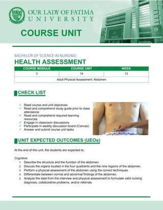
Adult Physical Assessment: Abdomen
✓ Read course and unit objectives
✓ Read and comprehend study guide prior to class
attendance
✓ Read and comprehend required learning
resources
✓ Engage in classroom discussions
✓ Participate in weekly discussion board (Canvas)
✓ Answer and submit course unit tasks
At the end of this unit, the students are expected to:
Cognitive:
1. Describe the structure and the function of the abdomen.
2. Discuss the organs located in the four quadrants and the nine regions of the abdomen.
3. Perform a physical assessment of the abdomen using the correct techniques.
4. Differentiate between normal and abnormal findings of the abdomen.
5. Analyze the data from the interview and physical assessment to formulate valid nursing
diagnosis, collaborative problems, and/or referrals.
Affective:
1. Listen attentively during class discussions
2. Demonstrate tact and respect when challenging other people’s opinions and ideas
3. Accept comments and reactions of classmates on one’s opinions openly and graciously.
Psychomotor:
1. Participate actively during class discussions
2. Confidently express personal opinion and thoughts in front of the class
Weber, J.R., and Kelley, J. H., (2018) Health Assessment in Nursing 6th Edition, Philadelphia:
Wolters Kluwer
ASSESSMENT OF THE ABDOMEN
Intraperitoneal Organs
• Organs covered with peritoneum and held in place by mesentery, it includes:
o spleen
o bile duct
o gallbladder
o small intestine
o stomach
o large intestine
o liver
Retroperitoneal Organs
• organs situated behind the peritoneum and without mesenteric attachment, it includes:
o pancreas
o ureters
o kidneys
o bladder
Anatomical Mapping
• 2 Methods of dividing the abdominal cavity:
o By quadrants
▪ most common assessment approach
▪ a vertical line from the xiphoid process to the pubic symphysis and a
horizontal line across the umbilicus
▪ The location of organs by quadrants
o By regions
▪ the vertical lines that extend superiorly from the midpoints of the inguinal
ligaments and 2 horizontal lines, one at the level of the edge of the lower ribs
and the other at the level of the iliac crest
THE ABDOMINAL VISCERA (ORGANS)
• Stomach
o J-shaped pouch like organ located in the left upper quadrant of the abdomen
beneath the diaphragm
o lies to the right of the spleen and is partially covered by the liver
o Functions
▪ reservoir where the complex mechanical and chemical processes of digestion
occurs
▪ breaksdown food particles into the molecular form of digestion
• Small Intestine
o tubular-shaped organ extending from the pyloric sphincter to the ileocecal valve at
the opening of the large intestine
o can measure from 10 feet to 30 feet
o Segments
▪ duodenum
• 1st and shortest section
• significant role in digestion
▪ jejunum
• composed of circular mucosal folds that provide surface area for
nutrient absorption
▪ ileum
• absorbs bile salts Vitamin B12
• Large Intestine
o tubular shaped organ extending from the ileocecal valve to the anus
o greater diameter than the small intestine
o Segments
▪ ascending
▪ transverse
▪ descending
• Sigmoid Colon
o Functions
▪ form stool from cellulose, indigestible fibers, fat, bacteria, cellular debris and
inorganic materials
▪ carry these intestinal contents to the end of the GIT
▪ absorption of water and electrolytes
• Liver
o largest solid organ in the body lies directly below the diaphragm
o located in the right upper quadrant but extends across the midline into the left upper
quadrant
o storage
▪ carbohydrates amino acids vitamins
▪ minerals
▪ blood
o detoxification and filtration
▪ drugs hormones
▪ bacteria
o metabolism
▪ carbohydrates
▪ proteins
▪ fats
▪ ammonia to urea
o synthesis and secretion
▪ bile production; 600 to 1000 ml/day
▪ formation of lymph bile salts
▪ plasma proteins
▪ fibrinogen
▪ blood-clotting substances
▪ antibodies
• Gallbladder
o pear-shaped sac located in the right upper quadrant of the abdomen
o Functions
▪ store and concentrate bile produced by the liver
▪ contributes to fat digestion and absorption
▪ stores approximately 30 to 50 ml of bile
• Pancreas
o elongated accessory organ of digestion lies in a transverse position along the
posterior abdominal wall
o Functions
▪ exocrine
• secretes bicarbonate and pancreatic enzymes which aid in digestion
▪ endocrine
• secretes the hormone insulin, glucagon and gastrin
• Spleen
o largest lymph of the body oval in shape found at the upper left quadrant of the
abdomen
o Functions
▪ serves the body as filter and reservoir for red blood cell mass
• Vermiform appendix
o finger-like shape that extends off the lower cecum in the right lower quadrant
• Kidneys
o bean-shaped organs that lie tucked against the posterior abdominal wall
o Functions
▪ get rid of the body waste products
▪ acid base balance fluid and electrolyte balance
▪ arterial blood pressure
• Ureters
o the passageway of urine from the kidneys
• Bladder
o stores urine can hold 200 to 400 ml of urine
ASSESSMENT PROPER
• Equipment
• Drapes
• Tape measure or small ruler with
centimeter markings
• Marking pen
• Stethoscope
Order of Assessment
• Inspection
• Auscultation
• Percussion
• Palpation
General Approach to Abdominal Assessment
1. Greet the patient and explain the assessment technique
2. Ensure that the room is at a warm, comfortable temperature to prevent patient chilling and
shivering
3. Use a quiet room that will be free from interruptions
4. Utilize an adequate light source. This includes a bright overhead light and a freestanding
lamp for tangential lightning
5. Ask the patient to urinate before the exam
6. Drape the patient from the xiphoid process to the symphisis pubis then expose the patient's
abdomen
7. Position the patient comfortably in a supine position with knees flexed over a pillow or
position the patient so that the arms are either folded across the chest or at the sides to
ensure abdominal relaxation
8. Stand to the right side of the patient for the examination
9. Visualize the underlying abdominal structures during the assessment process in order to
accurately describe the location of any pathology
10. Have the patient point to tender areas; assess these last. Mark these and other significant
findings on the body diagram in the patient's chart
11. Watch the patient's face closely for signs of discomfort or pain
12. Help the patient relax by using unhurried approach, diverting attention with questions and
so on
13. Ensure that your hands and the stethoscope are warm to promote patient comfort
Inspection
• Contour
o Normal
▪ the abdominal contour is flat (straight horizontal line from costal margin to
symphysis pubis)
▪ rounded (convexity of the abdomen from costal margin to symphysis pubis)
▪ abdomen should be evenly rounded
o Deviations from normal
▪ generalized protuberant or distended abdomen may be due to obesity, air or
fluid accumulation
▪ distended below umbilicus may be due to a full bladder, uterine enlargement,
or an ovarian tumor or cyst
▪ distended upper abdomen may be due to masses of pancreas or gastric
dilation
• Symmetry
o Normal
▪ abdomen should be symmetrical bilaterally
o Deviations from normal
▪ asymmetry may be seen with organ enlargement, large masses, hernia,
diastasis recti, or bowel obstruction
• Pigmentation and Color
o Normal
▪ abdominal skin may be paler than the general skin tone because it is seldom
exposed to the natural elements
o Deviations from normal
▪ purple discoloration at the flanks (Grey – Turner sign) indicates bleeding
within the abdominal wall, possibly from trauma of abdominal organs
(pancreas, kidneys, intestines, or from pancreatitis
▪ Pale, taut skin may be seen with ascites
▪ Redness may indicate inflammation
▪ Bruises or areas of discoloration
• Scar
o Normal
▪ Pale, smooth, minimally raised old scars may be seen
o Deviations from normal
▪ Nonhealing wounds, redness, inflammation
▪ Deep irregular scars may result from burns
• Striae
o Stretch Marks
o Due to past stretching of the reticular skin layers due to fast or prolonged stretching
o Normal
▪ Pink, bluish in color if new striae
▪ Silvery, white, linear if old striae
▪ Uneven stretch marks from past pregnancies or weight gain
o Deviations from normal
▪ Dark bluish – pink striae are associated with Cushing syndrome
▪ Stretch marks caused by ascites
• Masses / Nodules
o Normal
▪ No masses or nodules are present
• Umbilicus
o Normal
▪ Depressed and beneath the abdominal surface, protruding not more than
0.5cm
▪ Round or conical
▪ Skin tones are similar to surrounding abdominal skin tones
▪ Midline
o Deviations from normal
▪ Cullen sign
• Indicates intra – abdominal bleeding
▪ Deviated umbilicus
▪ Everted umbilicus
• Pulsation
o Normal
▪ Ripples of peristalsis may be observed in thin patient's
▪ Peristalsis movement slowly traverses the abdomen in a slanting downward
direction
Auscultation
• Bowel Sounds
o Heard as intermittent gurgling sounds throughout the abdominal quadrants at a rate
of 5 – 30 per minute
o Normally, bowel sounds are always present at the ileocecal valve area (RLQ)
o Normal hyperactive bowel sounds referred to as “borborygmus” may also be heard
o loud, prolonged gurgles characterized of one’s stomach growling
o Deviations from normal
▪ Hyperactive bowel sounds
• gastroenteritis
• diarrhea
• use of laxatives
▪ Hypoactive bowel sounds
• paralytic ileus
• peritonitis
• bowel obstruction
• Vascular Sounds
o Bruits
▪ use bell of stethoscope
▪ low – pitched, murmur- like sound
▪ whooshing sound, blood flows through a narrow vessel
▪ important if the client has hypertension or if you suspect arterial insufficiency
to the legs
▪ auscultated over the abdominal aorta and renal, iliac, and femoral arteries
best heard over these areas
▪ not normally heard
• may indicate aneurysm and or renal arterial stenosis
o Venous Hum
▪ sounds produced by abdominal organs that suggests increased collateral
circulation heard over the umbilicus and epigastric area cirrhosis of the liver
not normally heard
o Friction Rub
▪ high-pitched grating sound produced by large surface of an abdominal organ
(liver, spleen) rubs the peritoneum
▪ associated with respiration rare, and not normally heard
Percussion
• Patterns
o Abdominal percussion sequences may proceed
clockwise or up and down over the abdomen
o Cover all quadrants
• General Assessment
o Normal
▪ Tympany
• is predominant sound heard because air is present in the stomach and
in the intestines
▪ Dullness
• is normally heard over organs such as the liver or a distended bladder
• Liver Span
o Lower Border
▪ Begin liver percussion in the RLQ and percuss upward toward the chest
▪ Mark the percussion changes
o Upper Border
▪ Percuss over the upper right chest at the MCL and percuss downward
▪ Mark the percussion changes
o Measure the distance between the two marks:
▪ this is the span of the liver
o Normal liver span at the MCL is 8 – 12cm greater in men and taller clients, less in
shorter clients
o Normal liver span at the MSL is 4 – 8cm
o Deviations from normal
▪ Hepatomegaly, a liver span that exceeds normal limits (enlarged), is
characteristic of liver tumors, cirrhosis, abscess, and vascular engorgement
▪ ATROPHY of the liver is indicated by a decreased span
▪ A liver in a LOWER POSITION than normal may be caused by emphysema
▪ A liver in a HIGHER POSITION than normal may be caused by an
abdominal mass, ascites, or a paralyzed diaphragm
• Spleen
o Normal
▪ an oval area of dullness approximately 7 cm wide near the left tenth rib and
slightly posterior to the MAL
▪ tympany (or resonance) is heard at the last left interspace
o Deviations from normal
▪ Splenomegaly has an area of dullness greater than 7cm wide
▪ enlargement may result from traumatic injury, portal hypertension
mononucleusis
▪ Dullness at the anterior axillary line suggests enlargement of the spleen
• Kidney
o Blunt Percussion
▪ To assess for tenderness in difficult-to-palpate structures
▪ Normally, no tenderness or pain is elicited or reported by the client
▪ Tenderness or sharp pain elicited over the costovertebral angles (CVA)
suggests kidney infection (pyelonephritis), renal calculi, or hydronephrosis
Palpation
• Normally palpable structures in the abdomen
• Light Palpation
o Normal
▪ Abdomen should feel smooth and is nontender and soft
▪ There is no guarding
o Deviations from normal
▪ Involuntary reflex guarding reflects peritoneal irritation
▪ Right-side guarding may be due to cholecystitis
• Deep Palpation
o Normal
▪ (mild) tenderness is possible over the xiphoid, aorta, cecum, sigmoid colon,
and ovaries with deep palpation
▪ No organ enlargement should be palpable
▪ No abnormal masses, bulges, or swelling
▪ Only the aorta and the edge of the liver are palpable
o Deviations from normal
▪ Severe tenderness or pain may be related to trauma, peritonitis, infection,
tumors, or enlarged or diseased organs
• Liver
o Bimanual Technique
▪ Normal
• the liver is not palpable, although it may be felt in extremely thin adults
• liver edge presents as a firm, sharp, regular ridge with a smooth
surface
▪ Deviations from normal
• A hard, firm liver may indicate cancer
• Nodularity may occur with tumors, metastatic cancer, late cirrhosis, or
syphilis
• Tenderness may be from vascular engorgement (e.g., congestive
heart failure), acute hepatitis, or abscess
• Spleen
o Normal
▪ Seldom palpable at the left costal margin
▪ If palpable, it should be soft and nontender
o Deviations from normal
▪ Palpable spleen suggests enlargement
• infections
• trauma
• chronic blood disorders
▪ Palpating the spleen in side – lying position
• NOTE:
o Be gentle when palpating the spleen to avoid rupture and
trauma of the organ
• Kidneys
o Normal
▪ Usually not palpable
▪ If palpated, it should feel firm, smooth and rounded
o Deviations from normal
▪ An enlarged kidney may be due to a cyst, tumor, or hydronephrosis
• Urinary Bladder
o An empty bladder is neither palpable nor tender
o A moderately full bladder is smooth and round and it is palpable above the
symphysis pubis
o A full bladder is palpated above the symphysis pubis and it may be close to the
umbilicus
• Inguinal Lymph Nodes
o Normal
▪ Small, movable and palpable nodes are less than 1cm and are nontender
D’Amico, D., and Barbarito, C., (2019) Health & Physical Assessment in Nursing 3rd Edition,
Singapore: Pearson Education, Inc.
http://region9.dost.gov.ph/news/603-5-steps-of-a-breast-self-exam
https://journals.lww.com/nursingmadeincrediblyeasy/fulltext/2009/09000/making_sense_of_abdomi
nal_assessment.5.aspx#:~:text=With%20abdominal%20assessment%2C%20you%20inspect,perc
uss%2C%20palpate%2C%20and%20auscultate.
Weber, J.R., and Kelley, J. H., (2018) Health Assessment in Nursing
6th Edition, Philadelphia: Wolters Kluwer

More Related Content

What's hot

Diagnostic test for git
Diagnostic test for gitDiagnostic test for git
Diagnostic test for git
huma2012
 

What's hot (20)

Peptic ulcer disease 1
Peptic ulcer disease 1Peptic ulcer disease 1
Peptic ulcer disease 1
 
Hiatal hernia
Hiatal herniaHiatal hernia
Hiatal hernia
 
Hiatal Hernias
Hiatal HerniasHiatal Hernias
Hiatal Hernias
 
Esophageal motility disorders
Esophageal motility disordersEsophageal motility disorders
Esophageal motility disorders
 
Appendicitis
AppendicitisAppendicitis
Appendicitis
 
Achalasia
Achalasia Achalasia
Achalasia
 
Appendicitis
AppendicitisAppendicitis
Appendicitis
 
appendicitis.pptx
appendicitis.pptxappendicitis.pptx
appendicitis.pptx
 
Peptic Ulcer
Peptic Ulcer Peptic Ulcer
Peptic Ulcer
 
anatomy and physiology of small intestine
anatomy and physiology of small intestineanatomy and physiology of small intestine
anatomy and physiology of small intestine
 
Pancreas
PancreasPancreas
Pancreas
 
Guideline: Acute mesenteric ischemia
Guideline: Acute mesenteric ischemiaGuideline: Acute mesenteric ischemia
Guideline: Acute mesenteric ischemia
 
Inguinal Hernia .pptx
Inguinal Hernia .pptxInguinal Hernia .pptx
Inguinal Hernia .pptx
 
Peritoneum and retroperitoneum
Peritoneum and retroperitoneumPeritoneum and retroperitoneum
Peritoneum and retroperitoneum
 
Imaging modalities of intestinal obstruction Mithilesh Kumar Medical College ...
Imaging modalities of intestinal obstruction Mithilesh Kumar Medical College ...Imaging modalities of intestinal obstruction Mithilesh Kumar Medical College ...
Imaging modalities of intestinal obstruction Mithilesh Kumar Medical College ...
 
Management of ascites
Management of ascitesManagement of ascites
Management of ascites
 
Appendicitis
AppendicitisAppendicitis
Appendicitis
 
Inguinal Hernia Management, Presentation, by Dr. Shabir Ahmad
Inguinal Hernia Management, Presentation, by Dr. Shabir Ahmad Inguinal Hernia Management, Presentation, by Dr. Shabir Ahmad
Inguinal Hernia Management, Presentation, by Dr. Shabir Ahmad
 
Intestinal Obstruction
Intestinal ObstructionIntestinal Obstruction
Intestinal Obstruction
 
Diagnostic test for git
Diagnostic test for gitDiagnostic test for git
Diagnostic test for git
 

Similar to CM3 - CU14 ASSESSMENT OF ABDOMEN.pdf

Similar to CM3 - CU14 ASSESSMENT OF ABDOMEN.pdf (20)

GID.pptx
GID.pptxGID.pptx
GID.pptx
 
Abdominal assessment.pdf
Abdominal assessment.pdfAbdominal assessment.pdf
Abdominal assessment.pdf
 
abdominal assessment
abdominal assessmentabdominal assessment
abdominal assessment
 
ANATOMY PHYSIOLOGY AND COMMON DIAGNOSTIC TEST
ANATOMY PHYSIOLOGY AND COMMON DIAGNOSTIC TESTANATOMY PHYSIOLOGY AND COMMON DIAGNOSTIC TEST
ANATOMY PHYSIOLOGY AND COMMON DIAGNOSTIC TEST
 
Digestive System notes.pptx
Digestive System notes.pptxDigestive System notes.pptx
Digestive System notes.pptx
 
Clinical Examinations of the Abdomen with Images
Clinical Examinations of the Abdomen with ImagesClinical Examinations of the Abdomen with Images
Clinical Examinations of the Abdomen with Images
 
digestive system .pptx
 digestive system .pptx digestive system .pptx
digestive system .pptx
 
Lecture 1 elementary system
Lecture 1 elementary systemLecture 1 elementary system
Lecture 1 elementary system
 
FINALS.pptx
FINALS.pptxFINALS.pptx
FINALS.pptx
 
BILIARY AND GASTRO INTESTINAL CONDITIONS-1.pptx
BILIARY AND GASTRO INTESTINAL CONDITIONS-1.pptxBILIARY AND GASTRO INTESTINAL CONDITIONS-1.pptx
BILIARY AND GASTRO INTESTINAL CONDITIONS-1.pptx
 
11. Final Abdominal Examination.ppt
11. Final Abdominal Examination.ppt11. Final Abdominal Examination.ppt
11. Final Abdominal Examination.ppt
 
Gatrointestinal assessment
Gatrointestinal assessmentGatrointestinal assessment
Gatrointestinal assessment
 
SURGICAL ANATOMY OF STOMACH [Autosaved].pptx
SURGICAL ANATOMY OF STOMACH [Autosaved].pptxSURGICAL ANATOMY OF STOMACH [Autosaved].pptx
SURGICAL ANATOMY OF STOMACH [Autosaved].pptx
 
LIVER-STOMACH-INTESTINES.pptx
LIVER-STOMACH-INTESTINES.pptxLIVER-STOMACH-INTESTINES.pptx
LIVER-STOMACH-INTESTINES.pptx
 
Cathdgsys stomach-130716212423-phpapp01
Cathdgsys stomach-130716212423-phpapp01Cathdgsys stomach-130716212423-phpapp01
Cathdgsys stomach-130716212423-phpapp01
 
GI bleeding
GI bleeding GI bleeding
GI bleeding
 
abdominal assessment
abdominal assessmentabdominal assessment
abdominal assessment
 
Intestinal obstruction
Intestinal obstructionIntestinal obstruction
Intestinal obstruction
 
Mass in right hypochondrium.pptx
Mass in right hypochondrium.pptxMass in right hypochondrium.pptx
Mass in right hypochondrium.pptx
 
Mass in right hypochondrium.pptx
Mass in right hypochondrium.pptxMass in right hypochondrium.pptx
Mass in right hypochondrium.pptx
 

More from ZyraPascual1 (6)

CU13-assessment-of-perivascular-system.pptx
CU13-assessment-of-perivascular-system.pptxCU13-assessment-of-perivascular-system.pptx
CU13-assessment-of-perivascular-system.pptx
 
CM3 - CU16 ASSESSMENT OF NEUROLOGIC SYSTEM.pdf
CM3 - CU16 ASSESSMENT OF NEUROLOGIC SYSTEM.pdfCM3 - CU16 ASSESSMENT OF NEUROLOGIC SYSTEM.pdf
CM3 - CU16 ASSESSMENT OF NEUROLOGIC SYSTEM.pdf
 
CM3 - CU15 ASSESSMENT OF MUSCULOSKELETAL SYSTEM.pdf
CM3 - CU15 ASSESSMENT OF MUSCULOSKELETAL SYSTEM.pdfCM3 - CU15 ASSESSMENT OF MUSCULOSKELETAL SYSTEM.pdf
CM3 - CU15 ASSESSMENT OF MUSCULOSKELETAL SYSTEM.pdf
 
CM3 - CU13 ASSESSMENT OF PERIPHERAL VASCULAR SYSTEM.pdf
CM3 - CU13 ASSESSMENT OF PERIPHERAL VASCULAR SYSTEM.pdfCM3 - CU13 ASSESSMENT OF PERIPHERAL VASCULAR SYSTEM.pdf
CM3 - CU13 ASSESSMENT OF PERIPHERAL VASCULAR SYSTEM.pdf
 
CM3 - CU12 ASSESSMENT OF HEART & NECK VESSELS.pdf
CM3 - CU12 ASSESSMENT OF HEART & NECK VESSELS.pdfCM3 - CU12 ASSESSMENT OF HEART & NECK VESSELS.pdf
CM3 - CU12 ASSESSMENT OF HEART & NECK VESSELS.pdf
 
EUKARYOTIC-MICROBES.pptx
EUKARYOTIC-MICROBES.pptxEUKARYOTIC-MICROBES.pptx
EUKARYOTIC-MICROBES.pptx
 

Recently uploaded

GENERAL PHARMACOLOGY - INTRODUCTION DENTAL.ppt
GENERAL PHARMACOLOGY - INTRODUCTION DENTAL.pptGENERAL PHARMACOLOGY - INTRODUCTION DENTAL.ppt
GENERAL PHARMACOLOGY - INTRODUCTION DENTAL.ppt
Mangaiarkkarasi
 
Benefits of Dentulu's Salivary Testing.pptx
Benefits of Dentulu's Salivary Testing.pptxBenefits of Dentulu's Salivary Testing.pptx
Benefits of Dentulu's Salivary Testing.pptx
Dentulu Inc
 
ASSISTING WITH THE USE OF BED PAN BY ANUSHRI SRIVASTAVA.pptx
ASSISTING WITH THE USE OF BED PAN BY ANUSHRI SRIVASTAVA.pptxASSISTING WITH THE USE OF BED PAN BY ANUSHRI SRIVASTAVA.pptx
ASSISTING WITH THE USE OF BED PAN BY ANUSHRI SRIVASTAVA.pptx
AnushriSrivastav
 
Advanced Gum Health prebiotic Mints.pptx
Advanced Gum Health prebiotic Mints.pptxAdvanced Gum Health prebiotic Mints.pptx
Advanced Gum Health prebiotic Mints.pptx
Dentulu Inc
 
BOWEL ELIMINATION BY ANUSHRI SRIVASTAVA.pptx
BOWEL ELIMINATION BY ANUSHRI SRIVASTAVA.pptxBOWEL ELIMINATION BY ANUSHRI SRIVASTAVA.pptx
BOWEL ELIMINATION BY ANUSHRI SRIVASTAVA.pptx
AnushriSrivastav
 
Cell structure slideshare.pptx Unlocking the Secrets of Cells: Structure, Fun...
Cell structure slideshare.pptx Unlocking the Secrets of Cells: Structure, Fun...Cell structure slideshare.pptx Unlocking the Secrets of Cells: Structure, Fun...
Cell structure slideshare.pptx Unlocking the Secrets of Cells: Structure, Fun...
ananyagirishbabu1
 
ASSISTING WITH THE USE OF URINAL BY ANUSHRI SRIVASTAVA.pptx
ASSISTING WITH THE USE OF URINAL BY ANUSHRI SRIVASTAVA.pptxASSISTING WITH THE USE OF URINAL BY ANUSHRI SRIVASTAVA.pptx
ASSISTING WITH THE USE OF URINAL BY ANUSHRI SRIVASTAVA.pptx
AnushriSrivastav
 

Recently uploaded (20)

GENERAL PHARMACOLOGY - INTRODUCTION DENTAL.ppt
GENERAL PHARMACOLOGY - INTRODUCTION DENTAL.pptGENERAL PHARMACOLOGY - INTRODUCTION DENTAL.ppt
GENERAL PHARMACOLOGY - INTRODUCTION DENTAL.ppt
 
Benefits of Dentulu's Salivary Testing.pptx
Benefits of Dentulu's Salivary Testing.pptxBenefits of Dentulu's Salivary Testing.pptx
Benefits of Dentulu's Salivary Testing.pptx
 
HEAT WAVE presented by priya bhojwani..pptx
HEAT WAVE presented by priya bhojwani..pptxHEAT WAVE presented by priya bhojwani..pptx
HEAT WAVE presented by priya bhojwani..pptx
 
Occupational Therapy Management for Parkinson's Disease - Webinar 2024
Occupational Therapy Management for Parkinson's Disease - Webinar 2024Occupational Therapy Management for Parkinson's Disease - Webinar 2024
Occupational Therapy Management for Parkinson's Disease - Webinar 2024
 
How many patients does case series should have In comparison to case reports.pdf
How many patients does case series should have In comparison to case reports.pdfHow many patients does case series should have In comparison to case reports.pdf
How many patients does case series should have In comparison to case reports.pdf
 
ASSISTING WITH THE USE OF BED PAN BY ANUSHRI SRIVASTAVA.pptx
ASSISTING WITH THE USE OF BED PAN BY ANUSHRI SRIVASTAVA.pptxASSISTING WITH THE USE OF BED PAN BY ANUSHRI SRIVASTAVA.pptx
ASSISTING WITH THE USE OF BED PAN BY ANUSHRI SRIVASTAVA.pptx
 
CHAPTER- 1 SEMESTER V NATIONAL-POLICIES-AND-LEGISLATION.pdf
CHAPTER- 1 SEMESTER V NATIONAL-POLICIES-AND-LEGISLATION.pdfCHAPTER- 1 SEMESTER V NATIONAL-POLICIES-AND-LEGISLATION.pdf
CHAPTER- 1 SEMESTER V NATIONAL-POLICIES-AND-LEGISLATION.pdf
 
Advanced Gum Health prebiotic Mints.pptx
Advanced Gum Health prebiotic Mints.pptxAdvanced Gum Health prebiotic Mints.pptx
Advanced Gum Health prebiotic Mints.pptx
 
Secret Tantric VIP Erotic Massage London
Secret Tantric VIP Erotic Massage LondonSecret Tantric VIP Erotic Massage London
Secret Tantric VIP Erotic Massage London
 
BOWEL ELIMINATION BY ANUSHRI SRIVASTAVA.pptx
BOWEL ELIMINATION BY ANUSHRI SRIVASTAVA.pptxBOWEL ELIMINATION BY ANUSHRI SRIVASTAVA.pptx
BOWEL ELIMINATION BY ANUSHRI SRIVASTAVA.pptx
 
Enhance Your Wellbeing with Natural Women's Health Supplements
Enhance Your Wellbeing with Natural Women's Health SupplementsEnhance Your Wellbeing with Natural Women's Health Supplements
Enhance Your Wellbeing with Natural Women's Health Supplements
 
CHAPTER- 1 SEMESTER - V NATIONAL HEALTH PROGRAMME RELATED TO CHILD.pdf
CHAPTER- 1 SEMESTER - V NATIONAL HEALTH PROGRAMME RELATED TO CHILD.pdfCHAPTER- 1 SEMESTER - V NATIONAL HEALTH PROGRAMME RELATED TO CHILD.pdf
CHAPTER- 1 SEMESTER - V NATIONAL HEALTH PROGRAMME RELATED TO CHILD.pdf
 
VVIP Dehradun Girls 9719300533 Heat-bake { Dehradun } Genteel ℂall Serviℂe By...
VVIP Dehradun Girls 9719300533 Heat-bake { Dehradun } Genteel ℂall Serviℂe By...VVIP Dehradun Girls 9719300533 Heat-bake { Dehradun } Genteel ℂall Serviℂe By...
VVIP Dehradun Girls 9719300533 Heat-bake { Dehradun } Genteel ℂall Serviℂe By...
 
Dehradun ❤CALL Girls 8901183002 ❤ℂall Girls IN Dehradun ESCORT SERVICE❤
Dehradun ❤CALL Girls  8901183002 ❤ℂall  Girls IN Dehradun ESCORT SERVICE❤Dehradun ❤CALL Girls  8901183002 ❤ℂall  Girls IN Dehradun ESCORT SERVICE❤
Dehradun ❤CALL Girls 8901183002 ❤ℂall Girls IN Dehradun ESCORT SERVICE❤
 
Ayurveda hair cosmetlogy on Indralupta or Alopecia.pptx
Ayurveda hair cosmetlogy on Indralupta or Alopecia.pptxAyurveda hair cosmetlogy on Indralupta or Alopecia.pptx
Ayurveda hair cosmetlogy on Indralupta or Alopecia.pptx
 
Management of psoriasis.pptx (Recent advances)
Management of psoriasis.pptx (Recent advances)Management of psoriasis.pptx (Recent advances)
Management of psoriasis.pptx (Recent advances)
 
Cell structure slideshare.pptx Unlocking the Secrets of Cells: Structure, Fun...
Cell structure slideshare.pptx Unlocking the Secrets of Cells: Structure, Fun...Cell structure slideshare.pptx Unlocking the Secrets of Cells: Structure, Fun...
Cell structure slideshare.pptx Unlocking the Secrets of Cells: Structure, Fun...
 
Chris Shade BS MEd MS LPC-Associate "Presume" (What Do I Do?)
Chris Shade BS MEd MS LPC-Associate "Presume" (What Do I Do?)Chris Shade BS MEd MS LPC-Associate "Presume" (What Do I Do?)
Chris Shade BS MEd MS LPC-Associate "Presume" (What Do I Do?)
 
ASSISTING WITH THE USE OF URINAL BY ANUSHRI SRIVASTAVA.pptx
ASSISTING WITH THE USE OF URINAL BY ANUSHRI SRIVASTAVA.pptxASSISTING WITH THE USE OF URINAL BY ANUSHRI SRIVASTAVA.pptx
ASSISTING WITH THE USE OF URINAL BY ANUSHRI SRIVASTAVA.pptx
 
Unlocking the Benefits of Cognitive Behavioural Therapy (CBT) with Renewed Edge
Unlocking the Benefits of Cognitive Behavioural Therapy (CBT) with Renewed EdgeUnlocking the Benefits of Cognitive Behavioural Therapy (CBT) with Renewed Edge
Unlocking the Benefits of Cognitive Behavioural Therapy (CBT) with Renewed Edge
 

CM3 - CU14 ASSESSMENT OF ABDOMEN.pdf

  • 1. BACHELOR OF SCIENCE IN NURSING: HEALTH ASSESSMENT COURSE MODULE COURSE UNIT WEEK 3 14 13 Adult Physical Assessment: Abdomen ✓ Read course and unit objectives ✓ Read and comprehend study guide prior to class attendance ✓ Read and comprehend required learning resources ✓ Engage in classroom discussions ✓ Participate in weekly discussion board (Canvas) ✓ Answer and submit course unit tasks At the end of this unit, the students are expected to: Cognitive: 1. Describe the structure and the function of the abdomen. 2. Discuss the organs located in the four quadrants and the nine regions of the abdomen. 3. Perform a physical assessment of the abdomen using the correct techniques. 4. Differentiate between normal and abnormal findings of the abdomen. 5. Analyze the data from the interview and physical assessment to formulate valid nursing diagnosis, collaborative problems, and/or referrals.
  • 2. Affective: 1. Listen attentively during class discussions 2. Demonstrate tact and respect when challenging other people’s opinions and ideas 3. Accept comments and reactions of classmates on one’s opinions openly and graciously. Psychomotor: 1. Participate actively during class discussions 2. Confidently express personal opinion and thoughts in front of the class Weber, J.R., and Kelley, J. H., (2018) Health Assessment in Nursing 6th Edition, Philadelphia: Wolters Kluwer ASSESSMENT OF THE ABDOMEN Intraperitoneal Organs • Organs covered with peritoneum and held in place by mesentery, it includes: o spleen o bile duct o gallbladder o small intestine o stomach o large intestine o liver
  • 3. Retroperitoneal Organs • organs situated behind the peritoneum and without mesenteric attachment, it includes: o pancreas o ureters o kidneys o bladder Anatomical Mapping • 2 Methods of dividing the abdominal cavity: o By quadrants ▪ most common assessment approach ▪ a vertical line from the xiphoid process to the pubic symphysis and a horizontal line across the umbilicus ▪ The location of organs by quadrants o By regions ▪ the vertical lines that extend superiorly from the midpoints of the inguinal ligaments and 2 horizontal lines, one at the level of the edge of the lower ribs and the other at the level of the iliac crest
  • 4. THE ABDOMINAL VISCERA (ORGANS) • Stomach o J-shaped pouch like organ located in the left upper quadrant of the abdomen beneath the diaphragm o lies to the right of the spleen and is partially covered by the liver o Functions ▪ reservoir where the complex mechanical and chemical processes of digestion occurs ▪ breaksdown food particles into the molecular form of digestion • Small Intestine o tubular-shaped organ extending from the pyloric sphincter to the ileocecal valve at the opening of the large intestine o can measure from 10 feet to 30 feet o Segments ▪ duodenum • 1st and shortest section • significant role in digestion ▪ jejunum • composed of circular mucosal folds that provide surface area for nutrient absorption ▪ ileum • absorbs bile salts Vitamin B12 • Large Intestine o tubular shaped organ extending from the ileocecal valve to the anus
  • 5. o greater diameter than the small intestine o Segments ▪ ascending ▪ transverse ▪ descending • Sigmoid Colon o Functions ▪ form stool from cellulose, indigestible fibers, fat, bacteria, cellular debris and inorganic materials ▪ carry these intestinal contents to the end of the GIT ▪ absorption of water and electrolytes • Liver o largest solid organ in the body lies directly below the diaphragm o located in the right upper quadrant but extends across the midline into the left upper quadrant o storage ▪ carbohydrates amino acids vitamins ▪ minerals ▪ blood o detoxification and filtration ▪ drugs hormones ▪ bacteria o metabolism ▪ carbohydrates ▪ proteins ▪ fats ▪ ammonia to urea o synthesis and secretion ▪ bile production; 600 to 1000 ml/day ▪ formation of lymph bile salts ▪ plasma proteins ▪ fibrinogen ▪ blood-clotting substances ▪ antibodies • Gallbladder o pear-shaped sac located in the right upper quadrant of the abdomen o Functions ▪ store and concentrate bile produced by the liver ▪ contributes to fat digestion and absorption ▪ stores approximately 30 to 50 ml of bile • Pancreas
  • 6. o elongated accessory organ of digestion lies in a transverse position along the posterior abdominal wall o Functions ▪ exocrine • secretes bicarbonate and pancreatic enzymes which aid in digestion ▪ endocrine • secretes the hormone insulin, glucagon and gastrin • Spleen o largest lymph of the body oval in shape found at the upper left quadrant of the abdomen o Functions ▪ serves the body as filter and reservoir for red blood cell mass • Vermiform appendix o finger-like shape that extends off the lower cecum in the right lower quadrant • Kidneys o bean-shaped organs that lie tucked against the posterior abdominal wall o Functions ▪ get rid of the body waste products ▪ acid base balance fluid and electrolyte balance ▪ arterial blood pressure • Ureters o the passageway of urine from the kidneys • Bladder o stores urine can hold 200 to 400 ml of urine ASSESSMENT PROPER • Equipment • Drapes • Tape measure or small ruler with centimeter markings • Marking pen • Stethoscope Order of Assessment • Inspection • Auscultation • Percussion • Palpation
  • 7. General Approach to Abdominal Assessment 1. Greet the patient and explain the assessment technique 2. Ensure that the room is at a warm, comfortable temperature to prevent patient chilling and shivering 3. Use a quiet room that will be free from interruptions 4. Utilize an adequate light source. This includes a bright overhead light and a freestanding lamp for tangential lightning 5. Ask the patient to urinate before the exam 6. Drape the patient from the xiphoid process to the symphisis pubis then expose the patient's abdomen 7. Position the patient comfortably in a supine position with knees flexed over a pillow or position the patient so that the arms are either folded across the chest or at the sides to ensure abdominal relaxation 8. Stand to the right side of the patient for the examination 9. Visualize the underlying abdominal structures during the assessment process in order to accurately describe the location of any pathology 10. Have the patient point to tender areas; assess these last. Mark these and other significant findings on the body diagram in the patient's chart 11. Watch the patient's face closely for signs of discomfort or pain 12. Help the patient relax by using unhurried approach, diverting attention with questions and so on 13. Ensure that your hands and the stethoscope are warm to promote patient comfort Inspection • Contour o Normal ▪ the abdominal contour is flat (straight horizontal line from costal margin to symphysis pubis) ▪ rounded (convexity of the abdomen from costal margin to symphysis pubis) ▪ abdomen should be evenly rounded o Deviations from normal
  • 8. ▪ generalized protuberant or distended abdomen may be due to obesity, air or fluid accumulation ▪ distended below umbilicus may be due to a full bladder, uterine enlargement, or an ovarian tumor or cyst ▪ distended upper abdomen may be due to masses of pancreas or gastric dilation • Symmetry o Normal ▪ abdomen should be symmetrical bilaterally o Deviations from normal ▪ asymmetry may be seen with organ enlargement, large masses, hernia, diastasis recti, or bowel obstruction • Pigmentation and Color o Normal ▪ abdominal skin may be paler than the general skin tone because it is seldom exposed to the natural elements o Deviations from normal ▪ purple discoloration at the flanks (Grey – Turner sign) indicates bleeding within the abdominal wall, possibly from trauma of abdominal organs (pancreas, kidneys, intestines, or from pancreatitis ▪ Pale, taut skin may be seen with ascites ▪ Redness may indicate inflammation ▪ Bruises or areas of discoloration • Scar o Normal ▪ Pale, smooth, minimally raised old scars may be seen o Deviations from normal ▪ Nonhealing wounds, redness, inflammation
  • 9. ▪ Deep irregular scars may result from burns • Striae o Stretch Marks o Due to past stretching of the reticular skin layers due to fast or prolonged stretching o Normal ▪ Pink, bluish in color if new striae ▪ Silvery, white, linear if old striae ▪ Uneven stretch marks from past pregnancies or weight gain o Deviations from normal ▪ Dark bluish – pink striae are associated with Cushing syndrome ▪ Stretch marks caused by ascites • Masses / Nodules o Normal ▪ No masses or nodules are present • Umbilicus o Normal ▪ Depressed and beneath the abdominal surface, protruding not more than 0.5cm ▪ Round or conical ▪ Skin tones are similar to surrounding abdominal skin tones ▪ Midline o Deviations from normal ▪ Cullen sign • Indicates intra – abdominal bleeding ▪ Deviated umbilicus ▪ Everted umbilicus • Pulsation o Normal ▪ Ripples of peristalsis may be observed in thin patient's ▪ Peristalsis movement slowly traverses the abdomen in a slanting downward direction Auscultation • Bowel Sounds
  • 10. o Heard as intermittent gurgling sounds throughout the abdominal quadrants at a rate of 5 – 30 per minute o Normally, bowel sounds are always present at the ileocecal valve area (RLQ) o Normal hyperactive bowel sounds referred to as “borborygmus” may also be heard o loud, prolonged gurgles characterized of one’s stomach growling o Deviations from normal ▪ Hyperactive bowel sounds • gastroenteritis • diarrhea • use of laxatives ▪ Hypoactive bowel sounds • paralytic ileus • peritonitis • bowel obstruction • Vascular Sounds o Bruits ▪ use bell of stethoscope ▪ low – pitched, murmur- like sound ▪ whooshing sound, blood flows through a narrow vessel ▪ important if the client has hypertension or if you suspect arterial insufficiency to the legs ▪ auscultated over the abdominal aorta and renal, iliac, and femoral arteries best heard over these areas ▪ not normally heard • may indicate aneurysm and or renal arterial stenosis o Venous Hum ▪ sounds produced by abdominal organs that suggests increased collateral circulation heard over the umbilicus and epigastric area cirrhosis of the liver not normally heard o Friction Rub ▪ high-pitched grating sound produced by large surface of an abdominal organ (liver, spleen) rubs the peritoneum ▪ associated with respiration rare, and not normally heard Percussion • Patterns o Abdominal percussion sequences may proceed clockwise or up and down over the abdomen o Cover all quadrants
  • 11. • General Assessment o Normal ▪ Tympany • is predominant sound heard because air is present in the stomach and in the intestines ▪ Dullness • is normally heard over organs such as the liver or a distended bladder • Liver Span o Lower Border ▪ Begin liver percussion in the RLQ and percuss upward toward the chest ▪ Mark the percussion changes o Upper Border ▪ Percuss over the upper right chest at the MCL and percuss downward ▪ Mark the percussion changes
  • 12. o Measure the distance between the two marks: ▪ this is the span of the liver o Normal liver span at the MCL is 8 – 12cm greater in men and taller clients, less in shorter clients o Normal liver span at the MSL is 4 – 8cm o Deviations from normal ▪ Hepatomegaly, a liver span that exceeds normal limits (enlarged), is characteristic of liver tumors, cirrhosis, abscess, and vascular engorgement ▪ ATROPHY of the liver is indicated by a decreased span ▪ A liver in a LOWER POSITION than normal may be caused by emphysema ▪ A liver in a HIGHER POSITION than normal may be caused by an abdominal mass, ascites, or a paralyzed diaphragm • Spleen o Normal ▪ an oval area of dullness approximately 7 cm wide near the left tenth rib and slightly posterior to the MAL ▪ tympany (or resonance) is heard at the last left interspace o Deviations from normal
  • 13. ▪ Splenomegaly has an area of dullness greater than 7cm wide ▪ enlargement may result from traumatic injury, portal hypertension mononucleusis ▪ Dullness at the anterior axillary line suggests enlargement of the spleen • Kidney o Blunt Percussion ▪ To assess for tenderness in difficult-to-palpate structures ▪ Normally, no tenderness or pain is elicited or reported by the client ▪ Tenderness or sharp pain elicited over the costovertebral angles (CVA) suggests kidney infection (pyelonephritis), renal calculi, or hydronephrosis Palpation • Normally palpable structures in the abdomen • Light Palpation
  • 14. o Normal ▪ Abdomen should feel smooth and is nontender and soft ▪ There is no guarding o Deviations from normal ▪ Involuntary reflex guarding reflects peritoneal irritation ▪ Right-side guarding may be due to cholecystitis • Deep Palpation o Normal ▪ (mild) tenderness is possible over the xiphoid, aorta, cecum, sigmoid colon, and ovaries with deep palpation ▪ No organ enlargement should be palpable ▪ No abnormal masses, bulges, or swelling ▪ Only the aorta and the edge of the liver are palpable o Deviations from normal ▪ Severe tenderness or pain may be related to trauma, peritonitis, infection, tumors, or enlarged or diseased organs • Liver o Bimanual Technique ▪ Normal • the liver is not palpable, although it may be felt in extremely thin adults
  • 15. • liver edge presents as a firm, sharp, regular ridge with a smooth surface ▪ Deviations from normal • A hard, firm liver may indicate cancer • Nodularity may occur with tumors, metastatic cancer, late cirrhosis, or syphilis • Tenderness may be from vascular engorgement (e.g., congestive heart failure), acute hepatitis, or abscess • Spleen o Normal ▪ Seldom palpable at the left costal margin ▪ If palpable, it should be soft and nontender o Deviations from normal ▪ Palpable spleen suggests enlargement • infections • trauma • chronic blood disorders ▪ Palpating the spleen in side – lying position • NOTE: o Be gentle when palpating the spleen to avoid rupture and trauma of the organ • Kidneys
  • 16. o Normal ▪ Usually not palpable ▪ If palpated, it should feel firm, smooth and rounded o Deviations from normal ▪ An enlarged kidney may be due to a cyst, tumor, or hydronephrosis • Urinary Bladder o An empty bladder is neither palpable nor tender o A moderately full bladder is smooth and round and it is palpable above the symphysis pubis o A full bladder is palpated above the symphysis pubis and it may be close to the umbilicus • Inguinal Lymph Nodes o Normal ▪ Small, movable and palpable nodes are less than 1cm and are nontender D’Amico, D., and Barbarito, C., (2019) Health & Physical Assessment in Nursing 3rd Edition, Singapore: Pearson Education, Inc. http://region9.dost.gov.ph/news/603-5-steps-of-a-breast-self-exam https://journals.lww.com/nursingmadeincrediblyeasy/fulltext/2009/09000/making_sense_of_abdomi nal_assessment.5.aspx#:~:text=With%20abdominal%20assessment%2C%20you%20inspect,perc uss%2C%20palpate%2C%20and%20auscultate.
  • 17. Weber, J.R., and Kelley, J. H., (2018) Health Assessment in Nursing 6th Edition, Philadelphia: Wolters Kluwer