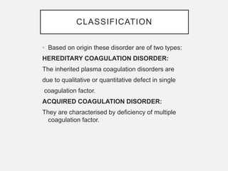
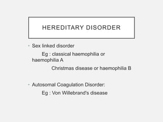
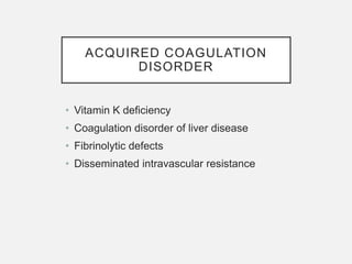
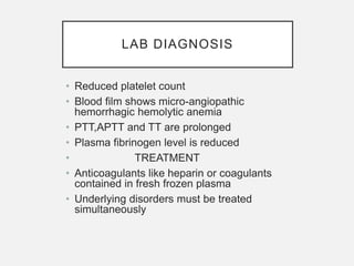
CLOTTING
DISORDER/COAGULATION
DISORDER
Dr. R.RAMYA
1st Year Post Graduate Student
Department of Periodontics and
Implantology
DEFINITION
• They are group of disease caused
due to deficiency of clotting factor
and leads to defect in normal clot
formation process.
CLASSIFICATION
• Based on origin these disorder are of two types:
HEREDITARY COAGULATION DISORDER:
The inherited plasma coagulation disorders are
due to qualitative or quantitative defect in single
coagulation factor.
ACQUIRED COAGULATION DISORDER:
They are characterised by deficiency of multiple
coagulation factor.
HEREDITARY DISORDER
• Sex linked disorder
Eg : classical haemophilia or
haemophilia A
Christmas disease or haemophilia B
• Autosomal Coagulation Disorder:
Eg : Von Willebrand's disease
ACQUIRED COAGULATION
DISORDER
• Vitamin K deficiency
• Coagulation disorder of liver disease
• Fibrinolytic defects
• Disseminated intravascular resistance
HEREDITARY COAGULATION DISORDER
CLASSICAL HEMOPHILIA
• It is the second most common cause of
hereditary coagulation disorder
• It is inherited as x linked recessive trait.so
more common in males while females are
carriers.
PATHOGENESIS
• 1. quantitative reduction in factor VIII(In 90%
of cases)
• 2. normal or increased levels of factor VIII(in
10% of cases)
• Factor VIII is synthesized in hepatocytes in
the intrinsic coagulation pathway, factor IXa
complexes with VIIIa, this complex in
presence of platelets, PL and ca2+ activates
factor X to Xa
CLINICAL FEATURES
• Symptoms are elicited when factor VIII
reduced to less than 25%.pt suffers bleeding
from hours to days and severity is based on
plasma level of factor VIII
• Recurrent painful hemarthroses and muscle
hematoma and sometimes hematuria
LAB DIAGNOSIS
• whole blood coagulation is raised only in
severe cases
• Prothrombin time is usually normal
• Partial thromboplastin time (PTT) is usually
prolonged
• Specific assays for factor VIII shows
reduced activity
TREATMENT
• Symptomatic patients with bleeding
episodes are treated with factor 8
replacement therapy consists of factor 8
concentrates or plasma cryoprecipitates.
CHRISTMAS
DISEASE/HEMOPHILIA B
• X linked recessive disease
• Rarer than hemophilia A
• Incidence ratio 1:100000 male birth
• inheritance pattern and clinical features are
similar to classical hemophilia
LAB DIAGNOSIS &
TREATMENT
• Assay of factor IX is lowered and other lab
findings are similar to hemophilia A
• Infusion of fresh frozen plasma or plasma
enriched with factor ix
VON WILLIBRAND’S DISEASE
Most common hereditary coagulation disorder
Inherited as autosomal dominant trait
Incidence ratio 1:1000 of either sex
Occurs due to qualitative or quantitative defect in
VWF
FACTOR VIII- Von Willebrand complex
consist of 99% Vwf of 1% factor VIII , they circulate
together as unit and perform important function in
clotting and facilitate platelet adhesion to
subendothelial collagen.
CLINICAL FEATURES
• Spontaneous bleeding from mucous
membrane
• excessive bleeding from nose and
menorrhagia
TYPES
• Type1 : it is the most common and
characterized by mild to moderate decrease
in plasma Vwd factor. synthesis is normal
but its release of its multimeter is inhibited
• TYPE 2 : It is much less common and
characterized by normal or near levels of
Vwd factor which was functionally defective
• TYPE 3: It is extremely rare and severe form
of disease. These patients have no
detectable Vwd activity and may have
sufficiently low quantity of factor VIII
LAB DIAGNOSIS
• Prolonged bleeding time
• Normal platelet count
• Reduced plasma Vwd concentration
• Defective platelet aggregation with
rostocetin, an antibiotic
• Reduced factor VIII activity
• TREATMENT
• Cryoprecipitates or factor VIII concentrates
ACQUIRED COAGULATION DISORDER
VITAMIN K DEFICIENCY
• Vitamin k serves as a cofactor in formation of
6 prothrombin complex proteins(vitamin k
dependent coagulation factor) synthesized in
liver. factor II VII IX and x, protein C and
protein S
• CAUSES OF VIT K DEFICIENCY
• Obstructive jaundice
• Chronic diarrhea
• Liver disease
• Hemorrhagic states in infants
LAB DIGNOSIS
• Prolonged PTT and APTT
• TREATMENT
• prolonged administration of vitamin k causes
complete recovery in 48 hours
COAGULATION DISORDER IN
LIVER DISEASE
• Liver is the site of synthesis and metabolism
of coagulation factors.
• Liver disease leads to hypercoagulability
and predispose DIC and systemic
fibrinolysis.
• Major causes of bleeding in liver disease are
• Morphological lesions
• portal hypertension , gastritis and peptic
ulceration
• Hepatic dysfunction
• impaired hepatic synthesis of coagulation
factors and inhibitors , Impaired metabolism
and absorption of vit k and failure to clear
activated coagulation factor
• Complication of therapy
• Massive transfusion leading to platelet and
clotting factor dilution , following heparin
therapy.
LAB DIAGNOSIS
• Prolonged PTT and APTT
• Mild thrombocytopenia
• Normal thrombocytopenia and decreased
hepatic stores of vitamin k
DISSEMINATED INTRAVASCULAR
COAGULATION
• Also called defibrinate syndrome is a
complex thrombo-hemorrhagic disease
occurring as secondary complication of
some systemic disease.
• MAJOR DISORDERS ASSOCIATED WITH
DIC
• Obstetrics complications: abruption
placentae, retained dead fetus, amniotic
fluid embolism.
• Infections: gram negative sepsis
,meningococcemia, rocky mounted spotted
fever and malaria
• Neoplasms: carcinoma of prostate,
pancreas, lung and stomach
• Massive tissue injury: trauma ,burns and
extensive surgery
• Miscellaneous: acute intravascular
hemolysis, snake bite, giant hemangioma ,
shock, heat stroke.
PATHOGENESIS
• Although in each case, a distinct triggering
mechanism has been identified, the
sequence of events, in general, can be
summarised as under,
1.Activation of coagulation
2. Thrombotic phase
3. consumption phase
4. Secondary fibrinolysis
1.ACTIVATION OF COAGULATION:
The etiologic factors listed above initiated widespread activation
of coagulation pathway by release of tissue factor.
2. THROMBOTIC PHASE:
Endothelial damage from the various thrombogenic stimuli
causes generalised platelet aggregation and adhesion with resultant
deposition of small thrombi and emboli throughout the microvasculature.
.3.CONSUMPTION PHASE:
The early thrombotic phase is followed by a phase of
consumption of coagulation factors and platelets.
4. SECONDARY FIBRINOLYSIS:
As a protective mechanism, fibrinolysis system is secondarily
activated at the site of intravascular coagulation. Secondary fibrinolysis
causes breakdown of fibrin resulting in formation of FDPs in the
circulation.
CLINICAL FEATURES
• Widespread fibrin deposition within
microcirculation leading to ischemia of
organs like brain and kidney.
• BLEEDING DIATHESIS: ensues as platelets
and clotting factors are consumed and there
are secondary release of plasminogen
activator but also digest factor v and VIII
thereby reducing their concentration further.
LAB DIAGNOSIS
• Reduced platelet count
• Blood film shows micro-angiopathic
hemorrhagic hemolytic anemia
• PTT,APTT and TT are prolonged
• Plasma fibrinogen level is reduced
• TREATMENT
• Anticoagulants like heparin or coagulants
contained in fresh frozen plasma
• Underlying disorders must be treated
simultaneously
THANK YOU

Clotting Disorders.pptx

  • 1.
    CLOTTING DISORDER/COAGULATION DISORDER Dr. R.RAMYA 1st YearPost Graduate Student Department of Periodontics and Implantology
  • 2.
    DEFINITION • They aregroup of disease caused due to deficiency of clotting factor and leads to defect in normal clot formation process.
  • 3.
    CLASSIFICATION • Based onorigin these disorder are of two types: HEREDITARY COAGULATION DISORDER: The inherited plasma coagulation disorders are due to qualitative or quantitative defect in single coagulation factor. ACQUIRED COAGULATION DISORDER: They are characterised by deficiency of multiple coagulation factor.
  • 4.
    HEREDITARY DISORDER • Sexlinked disorder Eg : classical haemophilia or haemophilia A Christmas disease or haemophilia B • Autosomal Coagulation Disorder: Eg : Von Willebrand's disease
  • 5.
    ACQUIRED COAGULATION DISORDER • VitaminK deficiency • Coagulation disorder of liver disease • Fibrinolytic defects • Disseminated intravascular resistance
  • 6.
  • 7.
    CLASSICAL HEMOPHILIA • Itis the second most common cause of hereditary coagulation disorder • It is inherited as x linked recessive trait.so more common in males while females are carriers.
  • 8.
    PATHOGENESIS • 1. quantitativereduction in factor VIII(In 90% of cases) • 2. normal or increased levels of factor VIII(in 10% of cases) • Factor VIII is synthesized in hepatocytes in the intrinsic coagulation pathway, factor IXa complexes with VIIIa, this complex in presence of platelets, PL and ca2+ activates factor X to Xa
  • 10.
    CLINICAL FEATURES • Symptomsare elicited when factor VIII reduced to less than 25%.pt suffers bleeding from hours to days and severity is based on plasma level of factor VIII • Recurrent painful hemarthroses and muscle hematoma and sometimes hematuria
  • 11.
    LAB DIAGNOSIS • wholeblood coagulation is raised only in severe cases • Prothrombin time is usually normal • Partial thromboplastin time (PTT) is usually prolonged • Specific assays for factor VIII shows reduced activity
  • 12.
    TREATMENT • Symptomatic patientswith bleeding episodes are treated with factor 8 replacement therapy consists of factor 8 concentrates or plasma cryoprecipitates.
  • 13.
    CHRISTMAS DISEASE/HEMOPHILIA B • Xlinked recessive disease • Rarer than hemophilia A • Incidence ratio 1:100000 male birth • inheritance pattern and clinical features are similar to classical hemophilia
  • 14.
    LAB DIAGNOSIS & TREATMENT •Assay of factor IX is lowered and other lab findings are similar to hemophilia A • Infusion of fresh frozen plasma or plasma enriched with factor ix
  • 15.
    VON WILLIBRAND’S DISEASE Mostcommon hereditary coagulation disorder Inherited as autosomal dominant trait Incidence ratio 1:1000 of either sex Occurs due to qualitative or quantitative defect in VWF FACTOR VIII- Von Willebrand complex consist of 99% Vwf of 1% factor VIII , they circulate together as unit and perform important function in clotting and facilitate platelet adhesion to subendothelial collagen.
  • 16.
    CLINICAL FEATURES • Spontaneousbleeding from mucous membrane • excessive bleeding from nose and menorrhagia
  • 17.
    TYPES • Type1 :it is the most common and characterized by mild to moderate decrease in plasma Vwd factor. synthesis is normal but its release of its multimeter is inhibited • TYPE 2 : It is much less common and characterized by normal or near levels of Vwd factor which was functionally defective • TYPE 3: It is extremely rare and severe form of disease. These patients have no detectable Vwd activity and may have sufficiently low quantity of factor VIII
  • 18.
    LAB DIAGNOSIS • Prolongedbleeding time • Normal platelet count • Reduced plasma Vwd concentration • Defective platelet aggregation with rostocetin, an antibiotic • Reduced factor VIII activity • TREATMENT • Cryoprecipitates or factor VIII concentrates
  • 19.
  • 20.
    VITAMIN K DEFICIENCY •Vitamin k serves as a cofactor in formation of 6 prothrombin complex proteins(vitamin k dependent coagulation factor) synthesized in liver. factor II VII IX and x, protein C and protein S • CAUSES OF VIT K DEFICIENCY • Obstructive jaundice • Chronic diarrhea • Liver disease • Hemorrhagic states in infants
  • 21.
    LAB DIGNOSIS • ProlongedPTT and APTT • TREATMENT • prolonged administration of vitamin k causes complete recovery in 48 hours
  • 22.
    COAGULATION DISORDER IN LIVERDISEASE • Liver is the site of synthesis and metabolism of coagulation factors. • Liver disease leads to hypercoagulability and predispose DIC and systemic fibrinolysis.
  • 23.
    • Major causesof bleeding in liver disease are • Morphological lesions • portal hypertension , gastritis and peptic ulceration • Hepatic dysfunction • impaired hepatic synthesis of coagulation factors and inhibitors , Impaired metabolism and absorption of vit k and failure to clear activated coagulation factor • Complication of therapy • Massive transfusion leading to platelet and clotting factor dilution , following heparin therapy.
  • 24.
    LAB DIAGNOSIS • ProlongedPTT and APTT • Mild thrombocytopenia • Normal thrombocytopenia and decreased hepatic stores of vitamin k
  • 25.
    DISSEMINATED INTRAVASCULAR COAGULATION • Alsocalled defibrinate syndrome is a complex thrombo-hemorrhagic disease occurring as secondary complication of some systemic disease.
  • 26.
    • MAJOR DISORDERSASSOCIATED WITH DIC • Obstetrics complications: abruption placentae, retained dead fetus, amniotic fluid embolism. • Infections: gram negative sepsis ,meningococcemia, rocky mounted spotted fever and malaria • Neoplasms: carcinoma of prostate, pancreas, lung and stomach • Massive tissue injury: trauma ,burns and extensive surgery • Miscellaneous: acute intravascular hemolysis, snake bite, giant hemangioma , shock, heat stroke.
  • 27.
    PATHOGENESIS • Although ineach case, a distinct triggering mechanism has been identified, the sequence of events, in general, can be summarised as under, 1.Activation of coagulation 2. Thrombotic phase 3. consumption phase 4. Secondary fibrinolysis
  • 28.
    1.ACTIVATION OF COAGULATION: Theetiologic factors listed above initiated widespread activation of coagulation pathway by release of tissue factor. 2. THROMBOTIC PHASE: Endothelial damage from the various thrombogenic stimuli causes generalised platelet aggregation and adhesion with resultant deposition of small thrombi and emboli throughout the microvasculature. .3.CONSUMPTION PHASE: The early thrombotic phase is followed by a phase of consumption of coagulation factors and platelets. 4. SECONDARY FIBRINOLYSIS: As a protective mechanism, fibrinolysis system is secondarily activated at the site of intravascular coagulation. Secondary fibrinolysis causes breakdown of fibrin resulting in formation of FDPs in the circulation.
  • 30.
    CLINICAL FEATURES • Widespreadfibrin deposition within microcirculation leading to ischemia of organs like brain and kidney. • BLEEDING DIATHESIS: ensues as platelets and clotting factors are consumed and there are secondary release of plasminogen activator but also digest factor v and VIII thereby reducing their concentration further.
  • 31.
    LAB DIAGNOSIS • Reducedplatelet count • Blood film shows micro-angiopathic hemorrhagic hemolytic anemia • PTT,APTT and TT are prolonged • Plasma fibrinogen level is reduced • TREATMENT • Anticoagulants like heparin or coagulants contained in fresh frozen plasma • Underlying disorders must be treated simultaneously
  • 32.