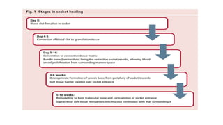
Platelet-rich fibrin to preserve alveolar bone sockets following
tooth extraction: A randomized controlled trial
Kanokporn Areewong DDS| Montri Chantaramungkorn DDS |
Pathawee Khongkhunthian DDS, DMD
PRESENTED BY;
SHUNMUGA PRASANTH D
1ST YEAR,POST GRADUATE
GUIDED BY: Dr.DEEPA
Clinical Implant Dentistry and Related Research
IMPACT FACTOR: 3.22
• Abstract
• Background: Platelet-rich fibrin (PRF) can be used in the alveolar socket
preservation(ARP). However, the hard tissue-regeneration property of PRF in
alveolar socket preservation is still unclear.
• Purpose: To compare the new bone formation ratio between using PRF as a
socket preservation material and normal wound healing, by means of
histomorphometric analysis.
• Materials and methods: Thirty-three healthy volunteers were recruited and
randomized into PRF and control group. Minimally traumatic extractions
were performed.Eighteen patients were treated with ARP using PRF, while
the rest were left to heal naturally. Bone specimens were harvested using
trephine bur 2 months after the extraction process. Histomorphometric
analysis of new bone formation area compared with total socket area was
performed using the software Fiji Is Just Image J (version 2, GNU General
Public License).
• Results: Thirty-three volunteers were participated. Twenty-eight bone
specimens were collected. The new bone formation ratio was higher
in PRF group than in control group (31.33 ± 18% and 26.33 ± 19.63%,
respectively). However, there was no statistically significant difference
in the ratio between the PRF and control groups (P = .431).
• Conclusions: It may be concluded that the use of PRF in ARP does not
statistically significant enhance new bone formation after tooth
extraction compared to normal wound healing (P > .05).
INTRODUCTION
• Protein molecules that play important roles in wound healing are often secreted from
cells surrounding the wound.
• Once tissue damage occurs, platelets, protein-molecule secretory units, are the first
units approaching the wound and activating the clot-formation cascade.
• Platelet granules release several growth factors, which are involved in every step of
wound healing.
• These growth factors help the healing to proceed smoothly; hence, the concentration of
those molecules are influential to the wound healing capacity.
• The higher concentration,the faster rate of healing. Autogenous fibrin glue is the first
generation of platelet concentrates and is a derivative product of platelet-rich plasma
(PRP).
• The fibrin glue has a platelet concentration over 300 times higher than in platelets in the
blood stream.
•Clinically,platelet-concentrate applications are available for many treatment procedures,
such as hemostasis, thrombocytopenia treatment chemotherapy, and so on. Moreover, the
fibrin glue is used as a postoperative bleeding control agent.
•Platelet-rich fibrin (PRF) was introduced by Choukroun.
•It is a three-dimensional network of various important platelet growth factors, the network
gradually releases growth factors for a longer time than with PRP.
•Various studies have suggested that PRF has efficacy in hard and soft tissue regeneration.
Marx and co-workers observed the efficiency of platelet-concentrate as a bone graft
supplement.
•They concluded that platelet concentrate gives significantly increased new bone
regeneration when compared to normal wound healing.
•In addition, they claimed that growth factors in platelet concentrate increase the rates of
bone formation and bone mineralization in first 6 months.
• Recently, Kim and co-workers cultured osteoblasts with PRF, and measured the activity of
the cells.
• The results showed progression of the rates of osteoblast DNA synthesis and alkaline
phosphatase activity.
• They concluded that the use of PRF improves bone regenerative capacity.
• Currently, one obstacle in implant treatment is insufficient bone for optimal dental implant
placement.
• Alveolar bone is formed to support tooth structures.
• Following tooth extraction, the alveolar bone is remodeled.
• According to a systematic review by Tan and co-workers in 2012,the ridge resorbs to a
greater extent horizontally than vertically.
• Bone resorption in the anterior alveolar ridge is a serious problem.
• Consequently, there have been many attempts to solve the issue of insufficient bone;
hence, alveolar ridge preservation (ARP) was introduced.
• ARP simplifies dental implant treatment by reduction of the amount of dimensional
change in the alveolar ridge, and promotes new bone formation.
• Various studies have observed the roles of PRF in ARP.
• A systematic review and meta-analysis of Del Fabbro and co-workers reported only
one study that evaluated the new bone formation percentage, after using platelet
concentrate following mandibular molar extraction, by means of histomorphometric
analysis;after 12 weeks of extraction, significantly more new bone formation was
found in a platelet concentrate group than in a control group that did not receive
platelet concentrate.
• A systematic review by Castro and co-workers found four studies involving the use of
PRF in ARP.
• Of those, two radiologic assessment studies reported higher bone density in PRF groups
than in controls, whereas the other two studies reported no difference in bone healing
capacity between PRF and control groups.
• Recently, Du Toit and co-workers conducted a randomized human histomorphometric
analysis study.
• They investigated the bone healing capacity of PRF with histological analysis 90 days after
nonmolar tooth extraction.
• Their results showed no significant difference in the amount of new bone formation when
PRF (the autogenous graft material) was compared with natural wound healing.
• To date, the capacity of PRF to stimulate bone formation is controversial; few randomized
controlled studies are available.
• Besides, histomorphometric analysis with bone staining is another way which may show
the effectiveness of PRF using in ARP.
• Thus, the purpose of this study was to compare new bone formation using PRF as a socket
preservation material with normal woundhealing, by means of histomorphometric
analysis.
GENERAL REVIEW
ARP
• Alveolar ridge preservation (ARP) is a procedure to arrest or minimise alveolar
ridge resorption following tooth extraction for future prosthodontic treatment
including placement of dental implants.
• ARP was frst described as ‘bone maintenance’ in 1982 and is synonymous with
socket preservation, ridge preservation, socket grafing and socket augmentation.
ARP commits to delaying implant placement by at least three to six months after
extraction with potentially longer treatment times compared with immediate
and early (up to four months) implant placement.
• The aim of ARP is to maintain horizontal and vertical alveolar ridge form using
bone grafs (autografs, allografs, xenografs or alloplastic materials); soft tissue
grafts; guided bone regeneration (GBR) (with resorbable or non-resorbable
barriers), biologically-active materials (growth factors) or combinations to reduce
the loss of coronal alveolar bone height and width.
INDICATION
• The following indications are based on an Osteology Workshop’s recommendations
for ARP. ARP is used where tooth extraction is required and delayed dental implant
placement in that site is likely at some point:
• Implant placement is planned at a time point later than tooth extraction, such as:
1. When immediate or early implantation is not recommended
2. When patients are not available for immediate or early implant placement
3. When primary stability of an implant cannot be obtained
4. In adolescent people
• Contouring of the ridge for conventional prosthetic treatment; for example, pontic
site development
• The cost/benefit ratio is positive. Consideration may be given to the cost of ARP at
the time of tooth extraction compared with the likelihood of GBR at a later date and
its associated cost.
• Reducing the need for elevation of the sinus foor.
PRF
Historical background
• Regenerative potential of platelets had been considered for use in wound healing since
the 1970s and by 1980s their clinical applications had begun with the following timeline:
• 1970- Fibrin Glue by Matras
• 1997- Platelet Rich Plasma by Whitman et al
• 1999- Plasma Rich In Growth Factor (PRGF) by Anitua et al
• 2006- Platelet Rich Fibrin by Choukroun et al
• 2006- Concentrated Growth Factor (CGF) by Sacci
• In the initial years, the use of fibrin adhesives clinically was limited due to the complex
preparation procedure and the cross-infection risks that it carries.
• PRP is prepared by collecting patient’s own blood in a test tube in which anticoagulants
are added. After the first spin, three different layers are formed.
Platelets and Platelet Rich Fibrin:
Platelets are tiny, irregularly-shaped cells,devoid of nucleus which are derived
from larger cells called megakaryocytes. They have a life span of 7 – 10 days.
During any injury, when the platelets come in contact with an exposed
endothelium, they release certain growth factors to facilitate wound healing.
Various growth factors and their functions
1. Platelet-derived growth factors (PDGFs) – Cell proliferation, cellular
migration, Collagen production to repair wound.
2. Transforming growth Factor (TGF) –Angiogenesis, Re-epithelialisation,
Regeneration of Connective tissues.
3. Vascular endothelial growth factor (VEGF) – Angiogenesis, Tissue
Remodelling.
4. Endothelial growth factor – Proliferation and multiplication of endothelial
and mesenchymal cells leading to epithelialisation
• The platelet concentrates can be classified depending on their leukocyte
content and fibrin architecture as follows
First Generation:
1. Platelet Rich Plasma (PRP)
2. Pure Platelet Rich Plasma (P-PRP) / Anitua’s Plasma Rich Growth Factor
(PRGF)
3. Leucocyte Platelet Rich Plasma (L-PRP) / L-PRP Gel)
Second Generation:
1. Platelet Rich Fibrin (PRF)
2. Pure Platelet Rich Fibrin (P-PRF)
3. Leucocyte Platelet Rich Fibrin (L-PRF)
4. Concentrated Growth Factor (CGF) (Solid fibrin material with leucocyte)
5. Advanced Platelet Rich Fibrin (A-PRF)
6. lnjectable Platelet Rich Fibrin (I-PRF): (used to prepare sticky bone)
7. Titanium Platelet Rich Fibrin (T-PRF)
Clinical implications of PRF:
1. In sinus floor elevation procedures, PRF can be combined with bone grafts
to accelerate healing.
2. In ridge augmentation procedures – to stabilize and protect graft materials.
3. To preserve the sockets following extraction.
4. In root coverage procedures for single and multiple tooth recession.
5. In 3 – walled osseous defects for regeneration.
6. Treatment of furcation defects.
7. For cystic cavity filling.
8. In Implantology, PRF is used for a better osseointegration on the surface of
implant.
9. In the Guided tissue regeneration of periodontal ligament, alveolar bone
and cementum.
Advantages of using PRF over PRP:
(1) The preparation technique is simple and cost effective with a single step
centrifugation.
(2) It is obtained from an autologous blood sample that requires minimal
manipulation.
(3) An external addition of Bovine thrombin is not required, thus averting
immunological reactions.
(4) It allows a favourable healing due to a low and completely natural
polymerization process.
(5) It can be used solely or in combination with bone grafts to accelerate the
healing of the grafted bone.
(6) In case a fibrin membrane is used, a surgery at the donor site becomes
unnecessary, thus reducing patient discomfort.
Disadvantages:
1. Since an autologous blood is used, the final amount obtained is low.
2. It is highly technique sensitive and the PRF protocol has to be strictly
followed.
3. The clot polymerization necessitates use of a glass-coated tube.
4. An experienced clinician is required for PRF manipulation.
ROLE OF PRF IN ENHANCING BONE FORMATION
• Socket preservation procedures include the principles of guided tissue regeneration,
minimally traumatic tooth removal, and the protection of the bloodclot.
• Current methods include the use of autografts, allografts, alloplasts, or xenografts and
naturally derived synthetic membranes that are either bioresorbable or nonresorbable.
• However, the useof grafting materials in the fresh extraction socket has been questioned
as they appeared to interfere with the normal healing process.
• Platelet-rich fibrin membranes (PRF) are a newly developed platelet concentrate
allowing a simplified preparation without biochemical blood handling. The slow
polymerization of the fibrin during PRF preparation creates a fibrin network very similar
to the natural one with incorporation of the platelets in the fibrin meshes, which leads
to efficient cell migration and proliferation.
• Platelets are also activated and this results in their massive degranulation with an
important cytokine release(PDGF, TGF-beta, VEGF, TSP-1,EGF) during thefibrin matrix
remodeling.
• PRF may remain exposed in the oral cavity and creates an accelerated
tissue cicatrization due to its function inimmune control, circulating
stem cell trapping,angiogenesis, and wound-covering
epithelialization.
• PRF was used as a source of growth factors that enabled the acceleration of
the initial steps of healing, thus shortening the rehabilitation time required of
an edentulous site.
• In addition, the fibrin plays the role of a biomaterial with a positive effect on
early vascularization where fibrin, fibronectin, and thrombospondin act as a
provisory extracellular matrix, which may enhance bone formation in the
socket.
REVIEW OF LITERATURE
Journal of Oral and Maxillofacial Surgery
Nilima Kumar , MDS,
Dr., Kavitha prasad ,
MDS, Dr., R.M. Lalitha- ,
MDS, Dr., K.
Ranganath , MDS, Dr.,
Jayashree Dexith , MDS,
Dr., Abhishek Chauhan ,
MDS, Dr.
MATERIALS AND METHODS
Ethical approval
• This prospective randomized clinical study was conducted at the
Center of Excellence for Dental Implantology, Faculty of Dentistry,
Chiang
• Mai University (Chiang Mai, Thailand) during August 2017 to
December 2018.
• The clinical study was approved by the Human Experimentation
Committee, Office of Research Ethics, Faculty of Dentistry, Chiang Mai
University (No. 40/2017) and with Thai clinical trials registry ID
number TCTR20180112002
POPULATION
• Sample size calculation using G*Power software program (G*Power
version 3.1.9.4, Heinrich-Heine Universität Düsseldorf, Düsseldorf,
Germany)was based on an alpha error of 5%, a power of 80% and effect
side from pilot study was 0.97.
• The sample size was 18 in each group. Thirty-six extraction sockets were
divided into two groups:
1. PRF group, tooth extraction following by socket preservation using PRF.
2. Control group, the alveolar sockets were left for spontaneous healing.
• Foremost, a blinded third party took responsibility for the random
arrangement of each study group.
• Sealed envelopes containing the group numbers (1 for the PRF group and 2
for the control group) were used for the randomizing.
PRF PREPARATION
• Following the suggestion of Choukroun,blood was
collected via common superficial veins within the cubital
fossa, including the median cubital, basilica, cephalic, and
antebrachial veins along with their attendant tributaries.
• Ten milliliters of blood were drawn and put into a glass
tube.
• The centrifuge device (IntraSpin, Intra-Lock, Nice,France)
speed was 2700 rpm for at least 12 minutes.
• A fibrin clot was formed at the middle of the tube
between the platelet-poor plasma layer and the red
blood cell layer.
• The clot was removed from the glass tube using
sterilized forceps and the lower, red, portion was cut.
• An Xpression box (Intra-Lock, Nice, France) was used to
form the fibrin clot into a fibrin plug (Figure 2).
TOOTH EXTRACTION PROCEDURE
• Local anesthetics containing 4% articaine hydrochloride (Septanest SP 1:100000;
Septodont Inc., Cedex, France) were used at the extraction site.
• A minimally traumatic extraction technique was performed.
• The periodontal ligaments were gently cut with a Piezotome (Implant Center 2,
Acteon, Nice, France).
• The tooth was carefully mobilized using forceps without flap reflection.
• The granulation tissue was removed from alveolar bone socket followed by normal
saline solution irrigation. The alveolar socket was treated differently in each group
• Control group: Blood clots were left naturally within extraction sockets. The sockets
were sutured with nylon using the figure-of- eight method.
• PRF group: Alveolar bone sockets were filled with PRF plugs. The sockets were
sutured using the same technique as in the control group.
• Analgesic and antibiotic drugs were prescribed after the treatment
procedure.
• Acetaminophen (500 mg.) was administered for pain relief.
Amoxicillin (500 mg.) or clindamycin (300 mg.), for patients who had
penicillin allergy, was suggested three times per day or two times per
day, respectively, for infection control.
• The healing events were followed up at the first, second, and eighth
weeks after tooth extraction.
• Any complications found were recorded.
BONE BIOPSY AND HISTOMORPHOMETRIC PREPARATION
• After 2 months of healing, new bone formation in the alveolar
ridge following tooth extraction was investigated using
histomorphometric analysis.
• The alveolar ridge was scanned using cone-bean computed
technology before dental implant placement.
• The localization of the reference planes (sagittal, axial, and
coronal) was determined using a surgical template.
• Local anesthesia was administered at the site. Full
mucoperiosteal buccal and lingual flaps were reflected.
• To ensure that the bone biopsies will be at the center part of
the former extracted sockets, a surgical trephine bur (Komet
Dental, Lemgo, Germany) with a 2-mm inner diameter and 6-
mm length together with the surgical template, was used as a
pilot drill at the implant site which was correspondence to the
center of the former extracted alveolar bone sockets
• The bony core trapped within the trephine bur was harvested and prepared for placement on a
histological slide.
• The alveolar bone was prepared for the appropriate size of dental implant,10 mm in minimum length.
Primary stability of the implant was assured using resonance frequency analysis (RFA).
• Using the method of Donath and Breune,28 the specimens were fixed with 10% formalin and the fixed
bone was dehydrated with serial alcohol concentrations (60%, 80%, 96%, and 100%).
• The specimens were rinsed and embedded in methylmetacrylate blocks.
• The polymerization blocks were cut along the longitudinal axis, using a 150-μm diamond disc in a
grinding machine (EXAKT 400CS, Serve Science, Bangkok, Thailand). The Final thickness of the
specimen was~10 to 15 μm.
• The prepared specimens were stained with toluidine blue followed by basic fuchsin dye.
• Twenty-eight slides were examined under a stereo microscope (Stemi 350, Zeiss, Jena, Germany) with
×4 magnification for each specimen.
• The area of new bone formation in each specimen and the total area of the specimen were calculated
using the software Fiji Is Bone sample harvesting with trephine bur Just Image J, version 2, GNU
General Public License developed by stallman.
• The ratio of new bone to total area was calculated using the following
equation:
• Percentage of new bone area = new bone area/total area × 100
• The data were determined by two independent examiners, with an
intraclass correlation coefficient (ICC) calculated at 0.98.
STATISTICAL ANALYSIS
• SPSS software (SPSS version 17, SPSS Inc., Chicago,
Illinois) was used to perform the statistical analysis. Data
were tested for normality using the Kolmogorov-
Smirnov Test.
• If the data were normally distributed,the independent
t-test was applied to compare the differences in ratio
between the two groups.
• Otherwise, the Mann-Whitney U test was used. The
level of statistical significance was set at a P < .05.
RESULTS
• Thirty-three volunteers enrolled for the study. Three
volunteers had two extracted alveolar sockets.
Therefore, total 36 alveolar bone sockets were included
in the study. The alveolar sockets were randomized
divided into the PRF group and control group equally (n
= 18)
• There were two smokers (<5 cigarettes per day), one in the PRF
group and one in the control group who were recruited in the
study.
• Demographic data of the PRF and control groups are shown in Table
2.
• The volunteers mean age in the PRF group was 48.17 years while in
the control group it was 52.94 years (overall minimum to maximum:
22-73, total mean age: 50.67).
• All alveolar sockets healed uneventfully.
• The new bone forming area was calculated by comparing with the
total bony area (Figure5).
• The results are reported as a percentage ratio. New bone
formation was found in 28 former sockets, while the remaining
eight alveolar sockets were found with no new bone formation at
the center of the alveolar sockets (three cases in the PRF group, five
cases in the control group).
• The data distribution was analyzed using the Kolmogorov-Smirnov
test, and the data were normally distributed in both groups.
• The mean new bone formation ratios
were 31.33 ± 18% in the PRF group and
26.33 ± 19.63% in the control group,
respectively (Figure 6). The results of
the data distribution were normal in
both the PRF group (P = .2 in the male
group, and 0.13 in the female group)
and in the control group (P = .2 in both
groups).
• The mean new bone formation ratios
between males and females in each
group were calculated. New bone
formation in the PRF group was 28.84 ±
20% in males and 33.82 ± 16.05% in
females (Figure 7).
• Simultaneously, the control group
showed that new bone formation in
males was 28.71 ± 23.88% and 25.14 ±
18.21% in females (Figure 8).
• All of the data were calculated with
independent sample t-tests, and the
results demonstrated no statistically
significant difference between the PRF
group and control group (P = .431;
Table 3).
• Concerning gender, the outcome
showed no statistical difference in
newbone formation ratios in either
the PRF group (P = .573) or the control
group (P = .72)
DISCUSSION
• Bone regeneration was regularly observed in the first few weeks of the wound healing process.
According to the study of Cardaropoli and co-workers, observations were made of new bone
regeneration events in dogs at different time intervals, and the authors stated that bone
healing events represented the series of tissue deposition.
• The series began with coagulum filling in the socket on the first day.
• Two weeks after tooth extraction, the provisional connective tissue matrix was formed,
followed by the formation of new bone at the eighth week.
• Finally, new bone was mineralized and turned into lamellar bone at 3 months after the
extraction procedure.
• It can be concluded that woven bone, the mineralized newly formed bone, was clearly noticed
at 8 weeks after tooth extraction.
• Moreover, referring to the study of Choukroun and co-workers, they used PRF as a graft
material in the cystic cavity, and within a period of 2 months the defect was totally filled with
the bone. Therefore, to confirm the capacity of new bone regeneration of PRF, the eighth week
seems to be an appropriate time for the outcome observation in our study
• Observation methods of the PRF property vary. A number of studies examined bone
regeneration proportion with the assistance of radiology. Girish and co-workers
observed the bone regeneration
• value of PRF compared with natural wound healing by using serial radiographs (RVG)
at immediate postop, first month, third month, and sixth month. They found higher
mean pixels of new bone formation at each time interval.
• However, there was no statistically significant difference between the two groups.
• Another study by Alzahrani and co-workers monitored the change in alveolar bone
dimension after ARP with PRF using cast model analysis together with radiographic
analysis via computer graphics software.
• The results showed that the change of ridge dimension in the test group was
statistically less than in the control group.
• Moreover, the average radiographic bone fill was significantly greater than in the
control.
• In addition, a systematic review by Strauss and co-workers reported three
histomorphometric studies. Within the limits of the study, they suggested the use of
PRF could enhance the alveolar ridge preservation.
• In contrast, a randomized clinical trial from Farina and coworkers observed the
efficacy of platelet-rich growth factor (PRGF),one of the platelet concentrate
families, to stimulate early bone deposition at the fourth and eighth weeks.
• The mean bone volume from radiographic analysis at 4 weeks in the control group
was 3.1± 3.4 mm3 and in the PRGF group it was 1.4 ± 2 mm3
• At 8 weeks the results showed 4.5 ± 2.7 mm3 in the control group and 3.2 ± 2.9
mm3 in the PRGF group.
• The results showed no statistically significantly difference either at 4 weeks or 8
weeks (P > .05). In addition to their study, histomorphometric analysis was
conducted using the calculation of the mean area of CD68+ cell staining, and the
number of macrophage and giant cells.
• The results indicated that there was a higher prevalence of CD68+ cells in the PRGF
group (97.8 ± 45.7 cells in the 4-week group and 105.4 ± 93.9 cells in the 8-week
group) at both observation times compared to the control group (97.2 ± 159.1 cells
in the 4-week group and 105.6 cells in the 8-week group).
• However,the results showed that PRGF failed to promote bone formation in this
period of time compared to the control group (P > .05)
• Alternatively, Knapen and co-workers36 conducted a study in rabbits to measure
regenerative bone quantity subsequent to filling with different biomaterials, which were L-
PRF, bovine hydroxyapatite(BHA), a combination of BHA and L-PRF, and no material filled
at all used as the control, into the prepared chamber of rabbit skulls at three different
time points which were 1 week, 5 weeks, and 12 weeks.
• Histomorphometric analysis of the results was presented in bone quantity percentage
ratios. They found that bone quantity increased significantly in co-ordination with time
rather than with the use of biomaterials (P < .0001).
• However, bone formation quantity was not statistically significantly different between
groups (P > .05).
• They concluded that platelet fibrin has insufficient potential to stimulate more bone
regeneration in the control group.
• Similar to our study,the null hypothesis assumed that there was no significant difference
in new bone formation in socket preservation with PRF after tooth extraction compared to
conventional wound healing.
• The statistical results showed no significant difference between the two groups.
• Although, there was no statistically significant difference between the PRF and
control groups, the mean bone formation in the PRF groupwas generally greater
than in the control group.
• From our study, there are eight sockets (three in PRF group, five in control group)
with no bone formation found in the histologic preparations.
• These may be the incomplete process of the bone healing since bone formation
of the socket is coming from the lateral wall of the socket.
• The trephine bone drills used in the study have only 2 mm in diameter, therefore
the bone formation in the center of the socket may not found. However, after the
installation of the implants, all of the implants are completely osseointegrated
after 3 months.
• In addition, wound healing is a multifactorial process. Among those factors, tobacco
is one of the most important in retarding the bone healing process.
• The study of Zhao and co-worker in 2018 showed the effects of smoking on human
alveolar bone marrow mesenchymal stem cells (hABMMSCs).
• In six patients with dental implants, the osteogenic gene expression of hABMMSCs
was reduced resulting in poor bone formation.
• Consequently, the success rate of implant treatment was significantly reduced (P<
.05).
• In our study,two smokers, who were randomly assigned to the different
groups,demonstrated no new bone formation in the central part of the extraction
sockets.
• This finding supported the finding that smoking may play an important factor
affecting bone formation. However,more smokers are required for future studies.
• Gender is one of the possible wound healing related factors, which may
affect time consumption in the wound healing cascade.
• According to Engeland and co-workers in 2006,39 a clinical experiment
measured the wound healing condition using photographs from the first
day of wound healing until the 7th day.
• They found that wound healing in women was delayed compared to
males (P = .0008).
• However, our study showed no statistically significant difference in the
bone regeneration rate of PRF compared to the control group in those
males and females at the eighth week period.
• This may need a more appropriate study design with a larger sample
size in the future.
• The increasing use of PRF is commonly found, especially in private
practice, although the benefits of using PRF are still controversial.
• The use of PRF is somehow able to assist the dentist for wound coverage.
• The regenerative effect of PRF or specific PRF still requires more studies to
confirm the results.
CONCLUSIONS
• Within the limitations of this study, it may be concluded that the use of
PRF in alveolar socket preservation compared to natural bone healing
shows no statistically significant differences.
• The use of PRF may not enhance new bone regeneration in this treatment
option.
• More clinical studies are required to prove the use of PRF in other clinical
situations.
JOURNAL CLUB (2).pptx

JOURNAL CLUB (2).pptx

  • 1.
    Platelet-rich fibrin topreserve alveolar bone sockets following tooth extraction: A randomized controlled trial Kanokporn Areewong DDS| Montri Chantaramungkorn DDS | Pathawee Khongkhunthian DDS, DMD PRESENTED BY; SHUNMUGA PRASANTH D 1ST YEAR,POST GRADUATE GUIDED BY: Dr.DEEPA Clinical Implant Dentistry and Related Research IMPACT FACTOR: 3.22
  • 2.
    • Abstract • Background:Platelet-rich fibrin (PRF) can be used in the alveolar socket preservation(ARP). However, the hard tissue-regeneration property of PRF in alveolar socket preservation is still unclear. • Purpose: To compare the new bone formation ratio between using PRF as a socket preservation material and normal wound healing, by means of histomorphometric analysis. • Materials and methods: Thirty-three healthy volunteers were recruited and randomized into PRF and control group. Minimally traumatic extractions were performed.Eighteen patients were treated with ARP using PRF, while the rest were left to heal naturally. Bone specimens were harvested using trephine bur 2 months after the extraction process. Histomorphometric analysis of new bone formation area compared with total socket area was performed using the software Fiji Is Just Image J (version 2, GNU General Public License).
  • 3.
    • Results: Thirty-threevolunteers were participated. Twenty-eight bone specimens were collected. The new bone formation ratio was higher in PRF group than in control group (31.33 ± 18% and 26.33 ± 19.63%, respectively). However, there was no statistically significant difference in the ratio between the PRF and control groups (P = .431). • Conclusions: It may be concluded that the use of PRF in ARP does not statistically significant enhance new bone formation after tooth extraction compared to normal wound healing (P > .05).
  • 4.
    INTRODUCTION • Protein moleculesthat play important roles in wound healing are often secreted from cells surrounding the wound. • Once tissue damage occurs, platelets, protein-molecule secretory units, are the first units approaching the wound and activating the clot-formation cascade. • Platelet granules release several growth factors, which are involved in every step of wound healing. • These growth factors help the healing to proceed smoothly; hence, the concentration of those molecules are influential to the wound healing capacity. • The higher concentration,the faster rate of healing. Autogenous fibrin glue is the first generation of platelet concentrates and is a derivative product of platelet-rich plasma (PRP). • The fibrin glue has a platelet concentration over 300 times higher than in platelets in the blood stream.
  • 5.
    •Clinically,platelet-concentrate applications areavailable for many treatment procedures, such as hemostasis, thrombocytopenia treatment chemotherapy, and so on. Moreover, the fibrin glue is used as a postoperative bleeding control agent. •Platelet-rich fibrin (PRF) was introduced by Choukroun. •It is a three-dimensional network of various important platelet growth factors, the network gradually releases growth factors for a longer time than with PRP. •Various studies have suggested that PRF has efficacy in hard and soft tissue regeneration. Marx and co-workers observed the efficiency of platelet-concentrate as a bone graft supplement. •They concluded that platelet concentrate gives significantly increased new bone regeneration when compared to normal wound healing. •In addition, they claimed that growth factors in platelet concentrate increase the rates of bone formation and bone mineralization in first 6 months.
  • 6.
    • Recently, Kimand co-workers cultured osteoblasts with PRF, and measured the activity of the cells. • The results showed progression of the rates of osteoblast DNA synthesis and alkaline phosphatase activity. • They concluded that the use of PRF improves bone regenerative capacity. • Currently, one obstacle in implant treatment is insufficient bone for optimal dental implant placement. • Alveolar bone is formed to support tooth structures. • Following tooth extraction, the alveolar bone is remodeled. • According to a systematic review by Tan and co-workers in 2012,the ridge resorbs to a greater extent horizontally than vertically. • Bone resorption in the anterior alveolar ridge is a serious problem.
  • 7.
    • Consequently, therehave been many attempts to solve the issue of insufficient bone; hence, alveolar ridge preservation (ARP) was introduced. • ARP simplifies dental implant treatment by reduction of the amount of dimensional change in the alveolar ridge, and promotes new bone formation. • Various studies have observed the roles of PRF in ARP. • A systematic review and meta-analysis of Del Fabbro and co-workers reported only one study that evaluated the new bone formation percentage, after using platelet concentrate following mandibular molar extraction, by means of histomorphometric analysis;after 12 weeks of extraction, significantly more new bone formation was found in a platelet concentrate group than in a control group that did not receive platelet concentrate. • A systematic review by Castro and co-workers found four studies involving the use of PRF in ARP.
  • 8.
    • Of those,two radiologic assessment studies reported higher bone density in PRF groups than in controls, whereas the other two studies reported no difference in bone healing capacity between PRF and control groups. • Recently, Du Toit and co-workers conducted a randomized human histomorphometric analysis study. • They investigated the bone healing capacity of PRF with histological analysis 90 days after nonmolar tooth extraction. • Their results showed no significant difference in the amount of new bone formation when PRF (the autogenous graft material) was compared with natural wound healing. • To date, the capacity of PRF to stimulate bone formation is controversial; few randomized controlled studies are available. • Besides, histomorphometric analysis with bone staining is another way which may show the effectiveness of PRF using in ARP. • Thus, the purpose of this study was to compare new bone formation using PRF as a socket preservation material with normal woundhealing, by means of histomorphometric analysis.
  • 9.
  • 10.
    ARP • Alveolar ridgepreservation (ARP) is a procedure to arrest or minimise alveolar ridge resorption following tooth extraction for future prosthodontic treatment including placement of dental implants. • ARP was frst described as ‘bone maintenance’ in 1982 and is synonymous with socket preservation, ridge preservation, socket grafing and socket augmentation. ARP commits to delaying implant placement by at least three to six months after extraction with potentially longer treatment times compared with immediate and early (up to four months) implant placement. • The aim of ARP is to maintain horizontal and vertical alveolar ridge form using bone grafs (autografs, allografs, xenografs or alloplastic materials); soft tissue grafts; guided bone regeneration (GBR) (with resorbable or non-resorbable barriers), biologically-active materials (growth factors) or combinations to reduce the loss of coronal alveolar bone height and width.
  • 13.
    INDICATION • The followingindications are based on an Osteology Workshop’s recommendations for ARP. ARP is used where tooth extraction is required and delayed dental implant placement in that site is likely at some point: • Implant placement is planned at a time point later than tooth extraction, such as: 1. When immediate or early implantation is not recommended 2. When patients are not available for immediate or early implant placement 3. When primary stability of an implant cannot be obtained 4. In adolescent people • Contouring of the ridge for conventional prosthetic treatment; for example, pontic site development • The cost/benefit ratio is positive. Consideration may be given to the cost of ARP at the time of tooth extraction compared with the likelihood of GBR at a later date and its associated cost. • Reducing the need for elevation of the sinus foor.
  • 15.
    PRF Historical background • Regenerativepotential of platelets had been considered for use in wound healing since the 1970s and by 1980s their clinical applications had begun with the following timeline: • 1970- Fibrin Glue by Matras • 1997- Platelet Rich Plasma by Whitman et al • 1999- Plasma Rich In Growth Factor (PRGF) by Anitua et al • 2006- Platelet Rich Fibrin by Choukroun et al • 2006- Concentrated Growth Factor (CGF) by Sacci • In the initial years, the use of fibrin adhesives clinically was limited due to the complex preparation procedure and the cross-infection risks that it carries. • PRP is prepared by collecting patient’s own blood in a test tube in which anticoagulants are added. After the first spin, three different layers are formed.
  • 16.
    Platelets and PlateletRich Fibrin: Platelets are tiny, irregularly-shaped cells,devoid of nucleus which are derived from larger cells called megakaryocytes. They have a life span of 7 – 10 days. During any injury, when the platelets come in contact with an exposed endothelium, they release certain growth factors to facilitate wound healing. Various growth factors and their functions 1. Platelet-derived growth factors (PDGFs) – Cell proliferation, cellular migration, Collagen production to repair wound. 2. Transforming growth Factor (TGF) –Angiogenesis, Re-epithelialisation, Regeneration of Connective tissues. 3. Vascular endothelial growth factor (VEGF) – Angiogenesis, Tissue Remodelling. 4. Endothelial growth factor – Proliferation and multiplication of endothelial and mesenchymal cells leading to epithelialisation
  • 17.
    • The plateletconcentrates can be classified depending on their leukocyte content and fibrin architecture as follows First Generation: 1. Platelet Rich Plasma (PRP) 2. Pure Platelet Rich Plasma (P-PRP) / Anitua’s Plasma Rich Growth Factor (PRGF) 3. Leucocyte Platelet Rich Plasma (L-PRP) / L-PRP Gel) Second Generation: 1. Platelet Rich Fibrin (PRF) 2. Pure Platelet Rich Fibrin (P-PRF) 3. Leucocyte Platelet Rich Fibrin (L-PRF) 4. Concentrated Growth Factor (CGF) (Solid fibrin material with leucocyte) 5. Advanced Platelet Rich Fibrin (A-PRF) 6. lnjectable Platelet Rich Fibrin (I-PRF): (used to prepare sticky bone) 7. Titanium Platelet Rich Fibrin (T-PRF)
  • 18.
    Clinical implications ofPRF: 1. In sinus floor elevation procedures, PRF can be combined with bone grafts to accelerate healing. 2. In ridge augmentation procedures – to stabilize and protect graft materials. 3. To preserve the sockets following extraction. 4. In root coverage procedures for single and multiple tooth recession. 5. In 3 – walled osseous defects for regeneration. 6. Treatment of furcation defects. 7. For cystic cavity filling. 8. In Implantology, PRF is used for a better osseointegration on the surface of implant. 9. In the Guided tissue regeneration of periodontal ligament, alveolar bone and cementum.
  • 19.
    Advantages of usingPRF over PRP: (1) The preparation technique is simple and cost effective with a single step centrifugation. (2) It is obtained from an autologous blood sample that requires minimal manipulation. (3) An external addition of Bovine thrombin is not required, thus averting immunological reactions. (4) It allows a favourable healing due to a low and completely natural polymerization process. (5) It can be used solely or in combination with bone grafts to accelerate the healing of the grafted bone. (6) In case a fibrin membrane is used, a surgery at the donor site becomes unnecessary, thus reducing patient discomfort.
  • 20.
    Disadvantages: 1. Since anautologous blood is used, the final amount obtained is low. 2. It is highly technique sensitive and the PRF protocol has to be strictly followed. 3. The clot polymerization necessitates use of a glass-coated tube. 4. An experienced clinician is required for PRF manipulation.
  • 21.
    ROLE OF PRFIN ENHANCING BONE FORMATION • Socket preservation procedures include the principles of guided tissue regeneration, minimally traumatic tooth removal, and the protection of the bloodclot. • Current methods include the use of autografts, allografts, alloplasts, or xenografts and naturally derived synthetic membranes that are either bioresorbable or nonresorbable. • However, the useof grafting materials in the fresh extraction socket has been questioned as they appeared to interfere with the normal healing process. • Platelet-rich fibrin membranes (PRF) are a newly developed platelet concentrate allowing a simplified preparation without biochemical blood handling. The slow polymerization of the fibrin during PRF preparation creates a fibrin network very similar to the natural one with incorporation of the platelets in the fibrin meshes, which leads to efficient cell migration and proliferation. • Platelets are also activated and this results in their massive degranulation with an important cytokine release(PDGF, TGF-beta, VEGF, TSP-1,EGF) during thefibrin matrix remodeling.
  • 22.
    • PRF mayremain exposed in the oral cavity and creates an accelerated tissue cicatrization due to its function inimmune control, circulating stem cell trapping,angiogenesis, and wound-covering epithelialization. • PRF was used as a source of growth factors that enabled the acceleration of the initial steps of healing, thus shortening the rehabilitation time required of an edentulous site. • In addition, the fibrin plays the role of a biomaterial with a positive effect on early vascularization where fibrin, fibronectin, and thrombospondin act as a provisory extracellular matrix, which may enhance bone formation in the socket.
  • 23.
  • 25.
    Journal of Oraland Maxillofacial Surgery Nilima Kumar , MDS, Dr., Kavitha prasad , MDS, Dr., R.M. Lalitha- , MDS, Dr., K. Ranganath , MDS, Dr., Jayashree Dexith , MDS, Dr., Abhishek Chauhan , MDS, Dr.
  • 27.
    MATERIALS AND METHODS Ethicalapproval • This prospective randomized clinical study was conducted at the Center of Excellence for Dental Implantology, Faculty of Dentistry, Chiang • Mai University (Chiang Mai, Thailand) during August 2017 to December 2018. • The clinical study was approved by the Human Experimentation Committee, Office of Research Ethics, Faculty of Dentistry, Chiang Mai University (No. 40/2017) and with Thai clinical trials registry ID number TCTR20180112002
  • 28.
    POPULATION • Sample sizecalculation using G*Power software program (G*Power version 3.1.9.4, Heinrich-Heine Universität Düsseldorf, Düsseldorf, Germany)was based on an alpha error of 5%, a power of 80% and effect side from pilot study was 0.97. • The sample size was 18 in each group. Thirty-six extraction sockets were divided into two groups: 1. PRF group, tooth extraction following by socket preservation using PRF. 2. Control group, the alveolar sockets were left for spontaneous healing. • Foremost, a blinded third party took responsibility for the random arrangement of each study group. • Sealed envelopes containing the group numbers (1 for the PRF group and 2 for the control group) were used for the randomizing.
  • 30.
    PRF PREPARATION • Followingthe suggestion of Choukroun,blood was collected via common superficial veins within the cubital fossa, including the median cubital, basilica, cephalic, and antebrachial veins along with their attendant tributaries. • Ten milliliters of blood were drawn and put into a glass tube. • The centrifuge device (IntraSpin, Intra-Lock, Nice,France) speed was 2700 rpm for at least 12 minutes. • A fibrin clot was formed at the middle of the tube between the platelet-poor plasma layer and the red blood cell layer. • The clot was removed from the glass tube using sterilized forceps and the lower, red, portion was cut. • An Xpression box (Intra-Lock, Nice, France) was used to form the fibrin clot into a fibrin plug (Figure 2).
  • 31.
    TOOTH EXTRACTION PROCEDURE •Local anesthetics containing 4% articaine hydrochloride (Septanest SP 1:100000; Septodont Inc., Cedex, France) were used at the extraction site. • A minimally traumatic extraction technique was performed. • The periodontal ligaments were gently cut with a Piezotome (Implant Center 2, Acteon, Nice, France). • The tooth was carefully mobilized using forceps without flap reflection. • The granulation tissue was removed from alveolar bone socket followed by normal saline solution irrigation. The alveolar socket was treated differently in each group • Control group: Blood clots were left naturally within extraction sockets. The sockets were sutured with nylon using the figure-of- eight method. • PRF group: Alveolar bone sockets were filled with PRF plugs. The sockets were sutured using the same technique as in the control group.
  • 32.
    • Analgesic andantibiotic drugs were prescribed after the treatment procedure. • Acetaminophen (500 mg.) was administered for pain relief. Amoxicillin (500 mg.) or clindamycin (300 mg.), for patients who had penicillin allergy, was suggested three times per day or two times per day, respectively, for infection control. • The healing events were followed up at the first, second, and eighth weeks after tooth extraction. • Any complications found were recorded.
  • 33.
    BONE BIOPSY ANDHISTOMORPHOMETRIC PREPARATION • After 2 months of healing, new bone formation in the alveolar ridge following tooth extraction was investigated using histomorphometric analysis. • The alveolar ridge was scanned using cone-bean computed technology before dental implant placement. • The localization of the reference planes (sagittal, axial, and coronal) was determined using a surgical template. • Local anesthesia was administered at the site. Full mucoperiosteal buccal and lingual flaps were reflected. • To ensure that the bone biopsies will be at the center part of the former extracted sockets, a surgical trephine bur (Komet Dental, Lemgo, Germany) with a 2-mm inner diameter and 6- mm length together with the surgical template, was used as a pilot drill at the implant site which was correspondence to the center of the former extracted alveolar bone sockets
  • 34.
    • The bonycore trapped within the trephine bur was harvested and prepared for placement on a histological slide. • The alveolar bone was prepared for the appropriate size of dental implant,10 mm in minimum length. Primary stability of the implant was assured using resonance frequency analysis (RFA). • Using the method of Donath and Breune,28 the specimens were fixed with 10% formalin and the fixed bone was dehydrated with serial alcohol concentrations (60%, 80%, 96%, and 100%). • The specimens were rinsed and embedded in methylmetacrylate blocks. • The polymerization blocks were cut along the longitudinal axis, using a 150-μm diamond disc in a grinding machine (EXAKT 400CS, Serve Science, Bangkok, Thailand). The Final thickness of the specimen was~10 to 15 μm. • The prepared specimens were stained with toluidine blue followed by basic fuchsin dye. • Twenty-eight slides were examined under a stereo microscope (Stemi 350, Zeiss, Jena, Germany) with ×4 magnification for each specimen. • The area of new bone formation in each specimen and the total area of the specimen were calculated using the software Fiji Is Bone sample harvesting with trephine bur Just Image J, version 2, GNU General Public License developed by stallman.
  • 35.
    • The ratioof new bone to total area was calculated using the following equation: • Percentage of new bone area = new bone area/total area × 100 • The data were determined by two independent examiners, with an intraclass correlation coefficient (ICC) calculated at 0.98.
  • 36.
    STATISTICAL ANALYSIS • SPSSsoftware (SPSS version 17, SPSS Inc., Chicago, Illinois) was used to perform the statistical analysis. Data were tested for normality using the Kolmogorov- Smirnov Test. • If the data were normally distributed,the independent t-test was applied to compare the differences in ratio between the two groups. • Otherwise, the Mann-Whitney U test was used. The level of statistical significance was set at a P < .05. RESULTS • Thirty-three volunteers enrolled for the study. Three volunteers had two extracted alveolar sockets. Therefore, total 36 alveolar bone sockets were included in the study. The alveolar sockets were randomized divided into the PRF group and control group equally (n = 18)
  • 37.
    • There weretwo smokers (<5 cigarettes per day), one in the PRF group and one in the control group who were recruited in the study. • Demographic data of the PRF and control groups are shown in Table 2. • The volunteers mean age in the PRF group was 48.17 years while in the control group it was 52.94 years (overall minimum to maximum: 22-73, total mean age: 50.67). • All alveolar sockets healed uneventfully. • The new bone forming area was calculated by comparing with the total bony area (Figure5). • The results are reported as a percentage ratio. New bone formation was found in 28 former sockets, while the remaining eight alveolar sockets were found with no new bone formation at the center of the alveolar sockets (three cases in the PRF group, five cases in the control group). • The data distribution was analyzed using the Kolmogorov-Smirnov test, and the data were normally distributed in both groups.
  • 38.
    • The meannew bone formation ratios were 31.33 ± 18% in the PRF group and 26.33 ± 19.63% in the control group, respectively (Figure 6). The results of the data distribution were normal in both the PRF group (P = .2 in the male group, and 0.13 in the female group) and in the control group (P = .2 in both groups). • The mean new bone formation ratios between males and females in each group were calculated. New bone formation in the PRF group was 28.84 ± 20% in males and 33.82 ± 16.05% in females (Figure 7).
  • 39.
    • Simultaneously, thecontrol group showed that new bone formation in males was 28.71 ± 23.88% and 25.14 ± 18.21% in females (Figure 8). • All of the data were calculated with independent sample t-tests, and the results demonstrated no statistically significant difference between the PRF group and control group (P = .431; Table 3). • Concerning gender, the outcome showed no statistical difference in newbone formation ratios in either the PRF group (P = .573) or the control group (P = .72)
  • 40.
    DISCUSSION • Bone regenerationwas regularly observed in the first few weeks of the wound healing process. According to the study of Cardaropoli and co-workers, observations were made of new bone regeneration events in dogs at different time intervals, and the authors stated that bone healing events represented the series of tissue deposition. • The series began with coagulum filling in the socket on the first day. • Two weeks after tooth extraction, the provisional connective tissue matrix was formed, followed by the formation of new bone at the eighth week. • Finally, new bone was mineralized and turned into lamellar bone at 3 months after the extraction procedure. • It can be concluded that woven bone, the mineralized newly formed bone, was clearly noticed at 8 weeks after tooth extraction. • Moreover, referring to the study of Choukroun and co-workers, they used PRF as a graft material in the cystic cavity, and within a period of 2 months the defect was totally filled with the bone. Therefore, to confirm the capacity of new bone regeneration of PRF, the eighth week seems to be an appropriate time for the outcome observation in our study
  • 41.
    • Observation methodsof the PRF property vary. A number of studies examined bone regeneration proportion with the assistance of radiology. Girish and co-workers observed the bone regeneration • value of PRF compared with natural wound healing by using serial radiographs (RVG) at immediate postop, first month, third month, and sixth month. They found higher mean pixels of new bone formation at each time interval. • However, there was no statistically significant difference between the two groups. • Another study by Alzahrani and co-workers monitored the change in alveolar bone dimension after ARP with PRF using cast model analysis together with radiographic analysis via computer graphics software. • The results showed that the change of ridge dimension in the test group was statistically less than in the control group. • Moreover, the average radiographic bone fill was significantly greater than in the control. • In addition, a systematic review by Strauss and co-workers reported three histomorphometric studies. Within the limits of the study, they suggested the use of PRF could enhance the alveolar ridge preservation.
  • 42.
    • In contrast,a randomized clinical trial from Farina and coworkers observed the efficacy of platelet-rich growth factor (PRGF),one of the platelet concentrate families, to stimulate early bone deposition at the fourth and eighth weeks. • The mean bone volume from radiographic analysis at 4 weeks in the control group was 3.1± 3.4 mm3 and in the PRGF group it was 1.4 ± 2 mm3 • At 8 weeks the results showed 4.5 ± 2.7 mm3 in the control group and 3.2 ± 2.9 mm3 in the PRGF group. • The results showed no statistically significantly difference either at 4 weeks or 8 weeks (P > .05). In addition to their study, histomorphometric analysis was conducted using the calculation of the mean area of CD68+ cell staining, and the number of macrophage and giant cells. • The results indicated that there was a higher prevalence of CD68+ cells in the PRGF group (97.8 ± 45.7 cells in the 4-week group and 105.4 ± 93.9 cells in the 8-week group) at both observation times compared to the control group (97.2 ± 159.1 cells in the 4-week group and 105.6 cells in the 8-week group). • However,the results showed that PRGF failed to promote bone formation in this period of time compared to the control group (P > .05)
  • 43.
    • Alternatively, Knapenand co-workers36 conducted a study in rabbits to measure regenerative bone quantity subsequent to filling with different biomaterials, which were L- PRF, bovine hydroxyapatite(BHA), a combination of BHA and L-PRF, and no material filled at all used as the control, into the prepared chamber of rabbit skulls at three different time points which were 1 week, 5 weeks, and 12 weeks. • Histomorphometric analysis of the results was presented in bone quantity percentage ratios. They found that bone quantity increased significantly in co-ordination with time rather than with the use of biomaterials (P < .0001). • However, bone formation quantity was not statistically significantly different between groups (P > .05). • They concluded that platelet fibrin has insufficient potential to stimulate more bone regeneration in the control group. • Similar to our study,the null hypothesis assumed that there was no significant difference in new bone formation in socket preservation with PRF after tooth extraction compared to conventional wound healing.
  • 44.
    • The statisticalresults showed no significant difference between the two groups. • Although, there was no statistically significant difference between the PRF and control groups, the mean bone formation in the PRF groupwas generally greater than in the control group. • From our study, there are eight sockets (three in PRF group, five in control group) with no bone formation found in the histologic preparations. • These may be the incomplete process of the bone healing since bone formation of the socket is coming from the lateral wall of the socket. • The trephine bone drills used in the study have only 2 mm in diameter, therefore the bone formation in the center of the socket may not found. However, after the installation of the implants, all of the implants are completely osseointegrated after 3 months.
  • 45.
    • In addition,wound healing is a multifactorial process. Among those factors, tobacco is one of the most important in retarding the bone healing process. • The study of Zhao and co-worker in 2018 showed the effects of smoking on human alveolar bone marrow mesenchymal stem cells (hABMMSCs). • In six patients with dental implants, the osteogenic gene expression of hABMMSCs was reduced resulting in poor bone formation. • Consequently, the success rate of implant treatment was significantly reduced (P< .05). • In our study,two smokers, who were randomly assigned to the different groups,demonstrated no new bone formation in the central part of the extraction sockets. • This finding supported the finding that smoking may play an important factor affecting bone formation. However,more smokers are required for future studies.
  • 46.
    • Gender isone of the possible wound healing related factors, which may affect time consumption in the wound healing cascade. • According to Engeland and co-workers in 2006,39 a clinical experiment measured the wound healing condition using photographs from the first day of wound healing until the 7th day. • They found that wound healing in women was delayed compared to males (P = .0008). • However, our study showed no statistically significant difference in the bone regeneration rate of PRF compared to the control group in those males and females at the eighth week period. • This may need a more appropriate study design with a larger sample size in the future.
  • 47.
    • The increasinguse of PRF is commonly found, especially in private practice, although the benefits of using PRF are still controversial. • The use of PRF is somehow able to assist the dentist for wound coverage. • The regenerative effect of PRF or specific PRF still requires more studies to confirm the results. CONCLUSIONS • Within the limitations of this study, it may be concluded that the use of PRF in alveolar socket preservation compared to natural bone healing shows no statistically significant differences. • The use of PRF may not enhance new bone regeneration in this treatment option. • More clinical studies are required to prove the use of PRF in other clinical situations.