SlideShare a Scribd company logo
PLANOVALGUS FOOT (PES PLANUS , FLAT FOOT)
CLINICAL EXAMINATION
HUDA ALFATAFTA, MASTERS BY RESEARCH
UNIVERSITY OF SALFORD, UK
LECTURER AT UNIVERSITY OF JORDAN/ ORTHOTICS AND PROSTHETICS DEPARTMENT
FLAT FOOT
EXAMINATION
2018
Introduction Flat Foot
Risk
factors
Clinical
symptoms
Gait
patterns
Clinical
tests
Gait analysis
Treatments
PT P&O surgery The
end
Huda Alfatafta
Foot print tests
FOOT
▪ The foot is a complex anatomical structure.
▪ Functions:
 It acts to transmit force between the lower limb and the ground, allowing stable
ambulation and stance.
 During gait the foot functions as a flexible shock-absorber, deforming to uneven
surfaces before undergoing a series of biomechanical changes which allow it to act
as a rigid lever to exert force.
Huda Alfatafta
ARCHES OF FOOT
▪ Three arches : medial and lateral longitudinal arches, and transverse
arche.
▪ The medial longitudinal arch consists of the calcaneum, talus, navicular,
medial, intermediate and lateral cuneiforms and the first three
metatarsals.
▪ The talus sits at the apex of the arch (key stone) and confers stability by
acting as a wedge between the calcaneum and navicular
▪ Half the body-weight passes through the apex of the arch whilst
standing.
▪ The ends of the ach are unable to move apart due to the tight plantar
fascia which connects them . Hence arch height is maintained
“notrmaly”
Huda Alfatafta
THE MEDIAL
LONGITUDINAL
ARCH
Stabilizing factors
Stabilizing models
Huda Alfatafta
STABILIZING FACTORS (STATIC AND DYNAMIC)
Static factors
▪ The most important primary stabilizer
is the plantar fascia,
▪ followed by the long and short plantar
ligaments and then the spring
ligament.
▪ Then, bony congruency
Huda Alfatafta
Dynamic factors
▪ tibialis posterior and other long flexors
are important dynamic stabilizers.
▪ releasing these structures without
releasing any static stabilizers has only
a modest effect on arch height.
▪ One study found the result of releasing
posterior tibial tension is associate
with 0.5 mm reduction in arch height
Huda Alfatafta
STABILIZING MODELS
THE BEAM MODEL
▪ in the beam model, a load is applied to
the apex of the arch generating
compressive forces on the dorsal
surface and tensile forces on the
plantar surface.
▪ Stability in this model results from
bony congruency and ligamentous
attachments
THE TRUSS MODEL.
▪ In the truss model, there is a triangular
arrangement of structures.
▪ The bones of the arch are able to pivot
about their apex whilst the tough
plantar fascia forms the third side.
▪ This is firmly attached to the medial
and lateral calcaneal tuberosities
proximally and its slips insert into the
plantar plate and the fibrous flexor
sheathes distally
Huda Alfatafta
Huda Alfatafta
▪ the beam model ▪ the truss model.
WHAT IS FLAT FOOT
▪ collapse or disappearance of the medial
longitudinal foot arch and is associated with several
three-dimensional foot deformities associated with
a rotational hindfoot abnormality and heel valgus
▪ All infants are born with flatfoot, and the
longitudinal arch of the foot develops during the
first decade, but the prevalence of flatfoot is 37%–
59.7% in children 2–6 years of age and 4%–19.1% in
those 8–13 years of age .
Huda Alfatafta
RISK FACTORS
GROUP A
▪ BMI, OBESITY
▪ Age
▪ Pregnancy
▪ heredity
GROUP B
▪ tibialis posterior dysfunction.
▪ Ligament laxity
▪ rotational abnormalities of the lower
limbs
Huda Alfatafta
CLINICAL SYMPTOMS
▪ Pain on the sole side of the forefoot
▪ plantar pressure under the talar head
▪ Pain between the tip of the fibula and
the calcaneus.
pain
Low
activity
level
significant
change in
foot shape
Huda Alfatafta
GAIT PATTERNS
▪ Eversion of the foot at the
talonavicular and talocalcaneal joints
▪ Valgus foot from posterior  the
medial border of the foot approaching
the ground
Huda Alfatafta
CLINICAL FOOT CLASSIFICATION MEASURES
FLAT FOOT
STEPS OF EXAMINATION
▪ Look at standing foot posture, swelling, hindfoot alignment, shoe and assess gait.
▪ Feel for localized tenderness and the nature of any swelling.
▪ Move each joint systematically, checking for pain, crepitus or stiffness.
▪ Special tests
▪ Radiographs
Huda Alfatafta
REARFOOT ANGLE (RFA)
▪ Briefly, four locations were palpated and marked using a skin
marker pen (Fig. 1A). These were:
▪ (1) the base of the calcaneus; (2) the Achilles tendon attachment;
(3) the centre of the Achilles tendon at the height of the medial
malleoli; (4.) the centre of the posterior aspect of the shank 15
cm above marker three.
▪ The RFA was measured using a goniometer. The arms of the
goniometer were aligned with the line connecting marker one
and two (line 1) and the other arm with the lines connecting
marker three and four (line 2).
▪ The RFA was measured as the acute angle between the projection
of line one and line two.
▪ RFA ≥ 5° valgus represented a pronated foot type, 4° valgus to 4°
varus a neutral foot and ≥ 5° varus a supinated foot
Huda Alfatafta
MEDIAL
LONGITUDINAL ARCH
ANGLE (MLAA)
▪ the midpoint of the medial malleolus, the most prominent aspect
of the navicular tuberosity and the most medial prominence of
the first metatarsal head were palpated and marked using a skin
marker pen.
▪ The MLAA was measured using a goniometer with the centre of
the goniometer aligned with the navicular mark and the arms
aligned to connect the navicular mark with the medial malleolus
and first metatarsal head markings. The obtuse angle was
recorded as the MLAA.
MLAA < 130° represented a pronated
foot type, 130° to 150° a neutral foot
type and > 150° a supinated foot type
Huda Alfatafta
NAVICULAR DROP
TEST (NDT).
▪ Navicular Drop Test (NDT). Initially the most prominent aspect of
the navicular tuberosity was palpated and marked with a skin
marker pen. A piece of card was placed next to the medial aspect
of the foot and the height of the navicular in a relaxed standing
position marked on the card. The foot was then manipulated into
subtalar joint neutral as determined by congruence of the talar
head, and the process outlined above repeated.
▪ ND was recorded as the difference in navicular height between
STJN and relaxed standing.
The normal foot group included
subjects with an NDT of 5–9mm
ND > 9 mm represented a pronated foot
type, 5 to 9 mm a neutral foot and < 5
mm a supinated foot
Huda Alfatafta
FOOT POSTURE
INDEX (FPI-6)
▪ The FPI is a six-item clinical assessment tool used to evaluate foot
posture. It has acceptable validity and good intra-rater reliability
(ICC = 0.893–0.958).
The FPI evaluates the multi-segmental nature of
foot posture in all three planes;
It does not require the use of specialised
equipment.
Each item of the FPI is scored between 2 and +2.
Foot type was classified according to normative
values with scores of ≥ 8 representing a pronated
foot type, 0 to 5 a neutral foot and ≤ -1 a
supinated foot
Huda Alfatafta
Huda Alfatafta
THE LEVEL OF AGREEMENT BETWEEN COMMON
CLINICAL FOOT CLASSIFICATION MEASURES.
▪ Foot classification across the two test occasions was almost perfect for MLAA (Kw =
0.92) and FPI-6 (Kw =0 .92), moderate for RFA (Kw = 0.60) and fair for ND (Kw =0 .40)
for comparison within the measures.
▪ Overall agreement between the measures for foot classification was moderate (Kf =
0.58).
▪ Conclusion: The findings reported in this study highlight discrepancies between the
chosen foot classification measures. The FPI-6 was a reliable multi-planar measure
whereas navicular drop emerged as an unreliable measure with only fair agreement
across test sessions. The use of this measure for foot assessment is discouraged.
Huda Alfatafta
FOOT PRINT
▪ Arch index (AI)
▪ calculated as the ratio of the area of the middle third
of the footprint to the entire footprint area. The foot
was defined as high-arched if the AI was lower than
21% or flat if the AI was higher than 28%.
▪ AI= B/ (A+B+C)* 100%
Huda Alfatafta
FOOT PRINT
▪ Russian author method
▪ If the foot print occupies three out of five fields, it is the first degree; four out of five
means the second degree, while five out of five means the third degree of the
suspended foot.
Huda Alfatafta
RADIOGRAPHIC MEASURES
▪ Simple standing anteroposterior and lateral radiographs of weight-bearing feet were
obtained in a digitalized manner from each subject. We measured the four angles
commonly used to assess flatfoot on the lateral radiograph, including 1) lateral talo-
first metatarsal angle (Meary’s angle), 2) talo-calcaneal angle, 3) metatarsal angle,
and 4) calcaneal pitch
Huda Alfatafta
Normal Supination (high
arch )
Pronation (flat
foot)
lateral talo-first
metatarsal angle
(Meary’s angle)
Zero- 4 upward
or downward
4 or more
upward
4 or more
downward.
mild <15º
moderate: 15-
30º
severe: 30º
talo-calcaneal
angle
25-45 degrees Less than 25
degrees
over 45 degrees
metatarsal angle Not common to be used
calcaneal pitch 18 to 20° More than 20 Less than 18
Huda Alfatafta
The red highlighted
tests are widely used
GAIT ANALYSIS AND PRESSURE MEASUREMENT
▪ Platform with force-sensitive sensors
Huda Alfatafta
FLAT FOOT STAGES
Huda Alfatafta
Stage
Stage I by symptoms of pain and swelling along the posterior tibial tendon without
visible changes of foot alignment
Stage II asymmetry of alignment with hindfoot valgus and forefoot abduction
exaggerated on the symptomatic foot. have weakness with inversion and
difficulty performing a single foot heel raise.
Stage III flatfoot represents a progression of all the clinical signs but not rigid
Stage VI Rigid ankle valgus deformity
FLEXIBLE OR RIGID ?
 Bony structure on not ?
Huda Alfatafta
Jack’s test Tip toe standing
REHABILITATION TREATMENT OPTIONS
FLAT FOOT
TREATMENTS
PHYSIOTHERAPIST
▪ foot exercises
ORTHOTIST
▪ Arch support
▪ Wedge
▪ SMO
Huda Alfatafta
vs. SURGEON
Calcaneal osteotomy
Subtalar arthrodesis
 Triple arthrodesis
vs.
PT
EXERCISES OPTIONS
Huda Alfatafta
P&O
ORTHOTICS INTERVENTIONS
Huda Alfatafta
SURGERY
REFERENCES
▪ Jankowicz-Szymańska, A., Wódka, K., Kołpa, M., & Mikołajczyk, E. (2018). Foot longitudinal arches in obese, overweight
and normal weight females who differ in age. HOMO, 69(1-2), 37-42.
▪ Pardo, F. J. V., del Amo, A. L., Rios, M. P., Gijon-Nogueron, G., & Yuste, C. C. (2018). Changes in foot posture during
pregnancy and their relation with musculoskeletal pain: A longitudinal cohort study. Women and Birth, 31(2), e84-e88.
▪ Kangas, J., Dankaerts, W., & Staes, F. (2011). New approach to the diagnosis and classification of chronic foot and ankle
disorders: Identifying motor control and movement impairments. Manual therapy, 16(6), 522-530.
▪ Lee, J. S., Kim, K. B., Jeong, J. O., Kwon, N. Y., & Jeong, S. M. (2015). Correlation of foot posture index with plantar
pressure and radiographic measurements in pediatric flatfoot. Annals of rehabilitation medicine, 39(1), 10-17.
▪ Young, C. C., Niedfeldt, M. W., Morris, G. A., & Eerkes, K. J. (2005). Clinical examination of the foot and ankle. Primary
Care: Clinics in Office Practice, 32(1), 105-132.
▪ Cebulski-Delebarre, A., Boutry, N., Szymanski, C., Maynou, C., Lefebvre, G., Amzallag-Bellenger, E., & Cotten, A. (2016).
Correlation between primary flat foot and lower extremity rotational misalignment in adults. Diagnostic and
interventional imaging, 97(11), 1151-1157.
▪ Lever, C. J., & Hennessy, M. S. (2016). Adult flat foot deformity. Orthopaedics and Trauma, 30(1), 41-50.
Huda Alfatafta
CONT.
▪ Roche, A., Hunter, L., Pocock, N., & Brown, D. (2009). Physical examination of the foot and ankle by
orthopaedic and accident and emergency clinicians. Injury, 40(2), 136-138.
▪ Langley, B., Cramp, M., & Morrison, S. C. (2016). Clinical measures of static foot posture do not
agree. Journal of foot and ankle research, 9(1), 45.
▪ DELAND, J. T. (2004). ANATOMY AND BIOMECHANICS OF THE FOOT AND ANKLE. Foot and Ankle, 1.
▪ Rao, S., Riskowski, J. L., & Hannan, M. T. (2012). Musculoskeletal conditions of the foot and ankle:
assessments and treatment options. Best practice & research Clinical rheumatology, 26(3), 345-
368.
▪ Brockett, C. L., & Chapman, G. J. (2016). Biomechanics of the ankle. Orthopaedics and
trauma, 30(3), 232-238.
▪ Haendlmayer, K. T., & Harris, N. J. (2009). (ii) Flatfoot deformity: an overview. Orthopaedics and
Trauma, 23(6), 395-403.
▪ Pauk, J., & Ezerskiy, V. (2011). The effect of foot orthotics on arch height: prediction of arch height
correction in flat-foot children. Biocybernetics and Biomedical Engineering, 31(1), 51-62.
Huda Alfatafta

More Related Content

What's hot

Tests for shoulder joint
Tests for shoulder jointTests for shoulder joint
Tests for shoulder jointAarti Sareen
 
Genu varum, Genu valgum, Genu recurvatum
Genu varum, Genu valgum, Genu recurvatumGenu varum, Genu valgum, Genu recurvatum
Genu varum, Genu valgum, Genu recurvatum
Murugesh M Kurani
 
examination of foot and ankle
examination of foot and ankleexamination of foot and ankle
examination of foot and ankle
manoj das
 
Hand deformity in rheumatoid arthritis
Hand deformity in rheumatoid arthritisHand deformity in rheumatoid arthritis
Hand deformity in rheumatoid arthritis
sushilonlines
 
Shoulder examination
Shoulder examination Shoulder examination
Shoulder examination
Dhananjaya Sabat
 
Anterior cruciate ligament-Injury & management
Anterior cruciate ligament-Injury & managementAnterior cruciate ligament-Injury & management
Anterior cruciate ligament-Injury & managementAnand Rao
 
Flat foot and Cavus foot
 Flat foot and Cavus foot Flat foot and Cavus foot
Flat foot and Cavus foot
Dr Thouseef Abdul Majeed
 
Wrist and hand examination
Wrist and hand examinationWrist and hand examination
Wrist and hand examination
zahramp
 
Shoulder impingement syndrome
Shoulder impingement syndromeShoulder impingement syndrome
Shoulder impingement syndrome
Ratan Khuman
 
Thomas Test.pdf
Thomas Test.pdfThomas Test.pdf
Thomas Test.pdf
mhmad farooq
 
Genu valgus
Genu valgusGenu valgus
Genu valgus
Santosh Batajoo
 
Floor reaction orthosis
Floor reaction orthosisFloor reaction orthosis
Floor reaction orthosis
Indra Singh
 
Carrying Angle
Carrying AngleCarrying Angle
Carrying Angle
mrinal joshi
 
Acl reconstruction
Acl reconstructionAcl reconstruction
Acl reconstruction
Morteza Dehnookhalaji
 
Prosthetic gait deviation
Prosthetic gait deviationProsthetic gait deviation
Prosthetic gait deviation
ayan ghosal
 
Coxa vara
Coxa varaCoxa vara
Coxa vara
manoj das
 
Iliotibial Band Syndrome (Itbs)
Iliotibial Band Syndrome (Itbs)Iliotibial Band Syndrome (Itbs)
Iliotibial Band Syndrome (Itbs)
colinmasterson
 
Pes planus / Flat Foot
Pes planus / Flat Foot Pes planus / Flat Foot
Pes planus / Flat Foot
Saloni Patil
 

What's hot (20)

Tests for shoulder joint
Tests for shoulder jointTests for shoulder joint
Tests for shoulder joint
 
Genu varum, Genu valgum, Genu recurvatum
Genu varum, Genu valgum, Genu recurvatumGenu varum, Genu valgum, Genu recurvatum
Genu varum, Genu valgum, Genu recurvatum
 
examination of foot and ankle
examination of foot and ankleexamination of foot and ankle
examination of foot and ankle
 
Hand deformity in rheumatoid arthritis
Hand deformity in rheumatoid arthritisHand deformity in rheumatoid arthritis
Hand deformity in rheumatoid arthritis
 
Shoulder examination
Shoulder examination Shoulder examination
Shoulder examination
 
Anterior cruciate ligament-Injury & management
Anterior cruciate ligament-Injury & managementAnterior cruciate ligament-Injury & management
Anterior cruciate ligament-Injury & management
 
Flat foot and Cavus foot
 Flat foot and Cavus foot Flat foot and Cavus foot
Flat foot and Cavus foot
 
Wrist and hand examination
Wrist and hand examinationWrist and hand examination
Wrist and hand examination
 
Shoulder impingement syndrome
Shoulder impingement syndromeShoulder impingement syndrome
Shoulder impingement syndrome
 
Thomas Test.pdf
Thomas Test.pdfThomas Test.pdf
Thomas Test.pdf
 
Genu valgus
Genu valgusGenu valgus
Genu valgus
 
Floor reaction orthosis
Floor reaction orthosisFloor reaction orthosis
Floor reaction orthosis
 
Carrying Angle
Carrying AngleCarrying Angle
Carrying Angle
 
Pcl pp
Pcl ppPcl pp
Pcl pp
 
Common peroneal nerve lesions
Common peroneal nerve lesionsCommon peroneal nerve lesions
Common peroneal nerve lesions
 
Acl reconstruction
Acl reconstructionAcl reconstruction
Acl reconstruction
 
Prosthetic gait deviation
Prosthetic gait deviationProsthetic gait deviation
Prosthetic gait deviation
 
Coxa vara
Coxa varaCoxa vara
Coxa vara
 
Iliotibial Band Syndrome (Itbs)
Iliotibial Band Syndrome (Itbs)Iliotibial Band Syndrome (Itbs)
Iliotibial Band Syndrome (Itbs)
 
Pes planus / Flat Foot
Pes planus / Flat Foot Pes planus / Flat Foot
Pes planus / Flat Foot
 

Similar to clinical examination of flat foot

Flat foot By Dr.Mahbub
Flat foot By Dr.MahbubFlat foot By Dr.Mahbub
Flat foot By Dr.Mahbub
dr_mhb21
 
Dr tarek ankle pain1
Dr tarek ankle pain1Dr tarek ankle pain1
Dr tarek ankle pain1
al azhar universty
 
Physical examination signs
Physical examination signsPhysical examination signs
Physical examination signscoolboy101pk
 
Flatfoot in Indian population.docx
Flatfoot in Indian population.docxFlatfoot in Indian population.docx
Flatfoot in Indian population.docx
AngelMaurya1
 
Flatfoot in Indian population.docx
Flatfoot in Indian population.docxFlatfoot in Indian population.docx
Flatfoot in Indian population.docx
HarshitMishar
 
examination of the hip joint
examination of the hip jointexamination of the hip joint
examination of the hip joint
Pallav Agrawal
 
Special Tests for Lower Leg, Ankle, and Foot
 Special Tests for Lower Leg, Ankle, and Foot Special Tests for Lower Leg, Ankle, and Foot
Special Tests for Lower Leg, Ankle, and Foot
Julie Jane
 
Knee clinical examination for orthopaedic residents.dr mohamed ashraf TD medi...
Knee clinical examination for orthopaedic residents.dr mohamed ashraf TD medi...Knee clinical examination for orthopaedic residents.dr mohamed ashraf TD medi...
Knee clinical examination for orthopaedic residents.dr mohamed ashraf TD medi...
drashraf369
 
Examination of knee joint
Examination of knee jointExamination of knee joint
Examination of knee joint
Fadzlina Zabri
 
Ctev
CtevCtev
flat foot.ppt [pes planus ] #physio.# rehabilitation
flat foot.ppt [pes planus ] #physio.# rehabilitationflat foot.ppt [pes planus ] #physio.# rehabilitation
flat foot.ppt [pes planus ] #physio.# rehabilitation
Priyankaranawat4
 
TalipesEquinoVarus.pptx
TalipesEquinoVarus.pptxTalipesEquinoVarus.pptx
TalipesEquinoVarus.pptx
Owen342285
 
Biomechanics of Gait, Posture and ADL
Biomechanics of Gait, Posture and ADLBiomechanics of Gait, Posture and ADL
Biomechanics of Gait, Posture and ADL
Soniya Lohana
 
Adult acquired flat foot deformity
Adult acquired flat foot deformityAdult acquired flat foot deformity
Adult acquired flat foot deformity
Ponnilavan Ponz
 
Club foot
Club foot Club foot
Club foot
Sufindc
 
praetip ankle fracture
praetip ankle fracturepraetip ankle fracture
praetip ankle fracture
Nattapon Panupinthu
 
FRO.ppt
FRO.pptFRO.ppt
CTEV
CTEVCTEV
AJM Sheet: pes cavus
AJM Sheet: pes cavusAJM Sheet: pes cavus
AJM Sheet: pes cavus
Podiatry Town
 

Similar to clinical examination of flat foot (20)

Flat foot By Dr.Mahbub
Flat foot By Dr.MahbubFlat foot By Dr.Mahbub
Flat foot By Dr.Mahbub
 
Dr tarek ankle pain1
Dr tarek ankle pain1Dr tarek ankle pain1
Dr tarek ankle pain1
 
Physical examination signs
Physical examination signsPhysical examination signs
Physical examination signs
 
Flatfoot in Indian population.docx
Flatfoot in Indian population.docxFlatfoot in Indian population.docx
Flatfoot in Indian population.docx
 
Flatfoot in Indian population.docx
Flatfoot in Indian population.docxFlatfoot in Indian population.docx
Flatfoot in Indian population.docx
 
examination of the hip joint
examination of the hip jointexamination of the hip joint
examination of the hip joint
 
Special Tests for Lower Leg, Ankle, and Foot
 Special Tests for Lower Leg, Ankle, and Foot Special Tests for Lower Leg, Ankle, and Foot
Special Tests for Lower Leg, Ankle, and Foot
 
Knee clinical examination for orthopaedic residents.dr mohamed ashraf TD medi...
Knee clinical examination for orthopaedic residents.dr mohamed ashraf TD medi...Knee clinical examination for orthopaedic residents.dr mohamed ashraf TD medi...
Knee clinical examination for orthopaedic residents.dr mohamed ashraf TD medi...
 
Examination of knee joint
Examination of knee jointExamination of knee joint
Examination of knee joint
 
Ctev
CtevCtev
Ctev
 
flat foot.ppt [pes planus ] #physio.# rehabilitation
flat foot.ppt [pes planus ] #physio.# rehabilitationflat foot.ppt [pes planus ] #physio.# rehabilitation
flat foot.ppt [pes planus ] #physio.# rehabilitation
 
TalipesEquinoVarus.pptx
TalipesEquinoVarus.pptxTalipesEquinoVarus.pptx
TalipesEquinoVarus.pptx
 
Biomechanics of Gait, Posture and ADL
Biomechanics of Gait, Posture and ADLBiomechanics of Gait, Posture and ADL
Biomechanics of Gait, Posture and ADL
 
Adult acquired flat foot deformity
Adult acquired flat foot deformityAdult acquired flat foot deformity
Adult acquired flat foot deformity
 
Club foot
Club foot Club foot
Club foot
 
praetip ankle fracture
praetip ankle fracturepraetip ankle fracture
praetip ankle fracture
 
FRO.ppt
FRO.pptFRO.ppt
FRO.ppt
 
CTEV
CTEVCTEV
CTEV
 
Assessing the motor
Assessing the motor Assessing the motor
Assessing the motor
 
AJM Sheet: pes cavus
AJM Sheet: pes cavusAJM Sheet: pes cavus
AJM Sheet: pes cavus
 

More from huda alfatafta

activity level evaluation_conference paper
activity level evaluation_conference paper  activity level evaluation_conference paper
activity level evaluation_conference paper
huda alfatafta
 
Knee joint-kinematics-and-kinetics-during-the-force-plates-attachable-interla...
Knee joint-kinematics-and-kinetics-during-the-force-plates-attachable-interla...Knee joint-kinematics-and-kinetics-during-the-force-plates-attachable-interla...
Knee joint-kinematics-and-kinetics-during-the-force-plates-attachable-interla...
huda alfatafta
 
Effect of a_knee_ankle_foot_orthosis_on_knee.10
Effect of a_knee_ankle_foot_orthosis_on_knee.10Effect of a_knee_ankle_foot_orthosis_on_knee.10
Effect of a_knee_ankle_foot_orthosis_on_knee.10
huda alfatafta
 
K level presenation
K level presenationK level presenation
K level presenation
huda alfatafta
 
Activ pal technologies
Activ pal technologies Activ pal technologies
Activ pal technologies
huda alfatafta
 
Wrist joint overuse and sport injuries
Wrist joint overuse and sport injuriesWrist joint overuse and sport injuries
Wrist joint overuse and sport injuries
huda alfatafta
 

More from huda alfatafta (6)

activity level evaluation_conference paper
activity level evaluation_conference paper  activity level evaluation_conference paper
activity level evaluation_conference paper
 
Knee joint-kinematics-and-kinetics-during-the-force-plates-attachable-interla...
Knee joint-kinematics-and-kinetics-during-the-force-plates-attachable-interla...Knee joint-kinematics-and-kinetics-during-the-force-plates-attachable-interla...
Knee joint-kinematics-and-kinetics-during-the-force-plates-attachable-interla...
 
Effect of a_knee_ankle_foot_orthosis_on_knee.10
Effect of a_knee_ankle_foot_orthosis_on_knee.10Effect of a_knee_ankle_foot_orthosis_on_knee.10
Effect of a_knee_ankle_foot_orthosis_on_knee.10
 
K level presenation
K level presenationK level presenation
K level presenation
 
Activ pal technologies
Activ pal technologies Activ pal technologies
Activ pal technologies
 
Wrist joint overuse and sport injuries
Wrist joint overuse and sport injuriesWrist joint overuse and sport injuries
Wrist joint overuse and sport injuries
 

Recently uploaded

HEAT WAVE presented by priya bhojwani..pptx
HEAT WAVE presented by priya bhojwani..pptxHEAT WAVE presented by priya bhojwani..pptx
HEAT WAVE presented by priya bhojwani..pptx
priyabhojwani1200
 
ABDOMINAL COMPARTMENT SYSNDROME
ABDOMINAL COMPARTMENT SYSNDROMEABDOMINAL COMPARTMENT SYSNDROME
ABDOMINAL COMPARTMENT SYSNDROME
Rommel Luis III Israel
 
Deep Leg Vein Thrombosis (DVT): Meaning, Causes, Symptoms, Treatment, and Mor...
Deep Leg Vein Thrombosis (DVT): Meaning, Causes, Symptoms, Treatment, and Mor...Deep Leg Vein Thrombosis (DVT): Meaning, Causes, Symptoms, Treatment, and Mor...
Deep Leg Vein Thrombosis (DVT): Meaning, Causes, Symptoms, Treatment, and Mor...
The Lifesciences Magazine
 
CHAPTER 1 SEMESTER V - ROLE OF PEADIATRIC NURSE.pdf
CHAPTER 1 SEMESTER V - ROLE OF PEADIATRIC NURSE.pdfCHAPTER 1 SEMESTER V - ROLE OF PEADIATRIC NURSE.pdf
CHAPTER 1 SEMESTER V - ROLE OF PEADIATRIC NURSE.pdf
Sachin Sharma
 
Empowering ACOs: Leveraging Quality Management Tools for MIPS and Beyond
Empowering ACOs: Leveraging Quality Management Tools for MIPS and BeyondEmpowering ACOs: Leveraging Quality Management Tools for MIPS and Beyond
Empowering ACOs: Leveraging Quality Management Tools for MIPS and Beyond
Health Catalyst
 
Navigating Challenges: Mental Health, Legislation, and the Prison System in B...
Navigating Challenges: Mental Health, Legislation, and the Prison System in B...Navigating Challenges: Mental Health, Legislation, and the Prison System in B...
Navigating Challenges: Mental Health, Legislation, and the Prison System in B...
Guillermo Rivera
 
CONSTRUCTION OF TEST IN MANAGEMENT .docx
CONSTRUCTION OF TEST IN MANAGEMENT .docxCONSTRUCTION OF TEST IN MANAGEMENT .docx
CONSTRUCTION OF TEST IN MANAGEMENT .docx
PGIMS Rohtak
 
.Metabolic.disordersYYSSSFFSSSSSSSSSSDDD
.Metabolic.disordersYYSSSFFSSSSSSSSSSDDD.Metabolic.disordersYYSSSFFSSSSSSSSSSDDD
.Metabolic.disordersYYSSSFFSSSSSSSSSSDDD
samahesh1
 
ICH Guidelines for Pharmacovigilance.pdf
ICH Guidelines for Pharmacovigilance.pdfICH Guidelines for Pharmacovigilance.pdf
ICH Guidelines for Pharmacovigilance.pdf
NEHA GUPTA
 
The Docs PPG - 30.05.2024.pptx..........
The Docs PPG - 30.05.2024.pptx..........The Docs PPG - 30.05.2024.pptx..........
The Docs PPG - 30.05.2024.pptx..........
TheDocs
 
VVIP Dehradun Girls 9719300533 Heat-bake { Dehradun } Genteel ℂall Serviℂe By...
VVIP Dehradun Girls 9719300533 Heat-bake { Dehradun } Genteel ℂall Serviℂe By...VVIP Dehradun Girls 9719300533 Heat-bake { Dehradun } Genteel ℂall Serviℂe By...
VVIP Dehradun Girls 9719300533 Heat-bake { Dehradun } Genteel ℂall Serviℂe By...
rajkumar669520
 
10 Ideas for Enhancing Your Meeting Experience
10 Ideas for Enhancing Your Meeting Experience10 Ideas for Enhancing Your Meeting Experience
10 Ideas for Enhancing Your Meeting Experience
ranishasharma67
 
Neuro Saphirex Cranial Brochure
Neuro Saphirex Cranial BrochureNeuro Saphirex Cranial Brochure
Neuro Saphirex Cranial Brochure
RXOOM Healthcare Pvt. Ltd. ​
 
GLOBAL WARMING BY PRIYA BHOJWANI @..pptx
GLOBAL WARMING BY PRIYA BHOJWANI @..pptxGLOBAL WARMING BY PRIYA BHOJWANI @..pptx
GLOBAL WARMING BY PRIYA BHOJWANI @..pptx
priyabhojwani1200
 
the IUA Administrative Board and General Assembly meeting
the IUA Administrative Board and General Assembly meetingthe IUA Administrative Board and General Assembly meeting
the IUA Administrative Board and General Assembly meeting
ssuser787e5c1
 
POLYCYSTIC OVARIAN SYNDROME (PCOS)......
POLYCYSTIC OVARIAN SYNDROME (PCOS)......POLYCYSTIC OVARIAN SYNDROME (PCOS)......
POLYCYSTIC OVARIAN SYNDROME (PCOS)......
Ameena Kadar
 
ventilator, child on ventilator, newborn
ventilator, child on ventilator, newbornventilator, child on ventilator, newborn
ventilator, child on ventilator, newborn
Pooja Rani
 
Leading the Way in Nephrology: Dr. David Greene's Work with Stem Cells for Ki...
Leading the Way in Nephrology: Dr. David Greene's Work with Stem Cells for Ki...Leading the Way in Nephrology: Dr. David Greene's Work with Stem Cells for Ki...
Leading the Way in Nephrology: Dr. David Greene's Work with Stem Cells for Ki...
Dr. David Greene Arizona
 
💘Ludhiana ℂall Girls 📞]][89011★83002][[ 📱 ❤ESCORTS service in Ludhiana💃💦Ludhi...
💘Ludhiana ℂall Girls 📞]][89011★83002][[ 📱 ❤ESCORTS service in Ludhiana💃💦Ludhi...💘Ludhiana ℂall Girls 📞]][89011★83002][[ 📱 ❤ESCORTS service in Ludhiana💃💦Ludhi...
💘Ludhiana ℂall Girls 📞]][89011★83002][[ 📱 ❤ESCORTS service in Ludhiana💃💦Ludhi...
ranishasharma67
 
The Importance of Community Nursing Care.pdf
The Importance of Community Nursing Care.pdfThe Importance of Community Nursing Care.pdf
The Importance of Community Nursing Care.pdf
AD Healthcare
 

Recently uploaded (20)

HEAT WAVE presented by priya bhojwani..pptx
HEAT WAVE presented by priya bhojwani..pptxHEAT WAVE presented by priya bhojwani..pptx
HEAT WAVE presented by priya bhojwani..pptx
 
ABDOMINAL COMPARTMENT SYSNDROME
ABDOMINAL COMPARTMENT SYSNDROMEABDOMINAL COMPARTMENT SYSNDROME
ABDOMINAL COMPARTMENT SYSNDROME
 
Deep Leg Vein Thrombosis (DVT): Meaning, Causes, Symptoms, Treatment, and Mor...
Deep Leg Vein Thrombosis (DVT): Meaning, Causes, Symptoms, Treatment, and Mor...Deep Leg Vein Thrombosis (DVT): Meaning, Causes, Symptoms, Treatment, and Mor...
Deep Leg Vein Thrombosis (DVT): Meaning, Causes, Symptoms, Treatment, and Mor...
 
CHAPTER 1 SEMESTER V - ROLE OF PEADIATRIC NURSE.pdf
CHAPTER 1 SEMESTER V - ROLE OF PEADIATRIC NURSE.pdfCHAPTER 1 SEMESTER V - ROLE OF PEADIATRIC NURSE.pdf
CHAPTER 1 SEMESTER V - ROLE OF PEADIATRIC NURSE.pdf
 
Empowering ACOs: Leveraging Quality Management Tools for MIPS and Beyond
Empowering ACOs: Leveraging Quality Management Tools for MIPS and BeyondEmpowering ACOs: Leveraging Quality Management Tools for MIPS and Beyond
Empowering ACOs: Leveraging Quality Management Tools for MIPS and Beyond
 
Navigating Challenges: Mental Health, Legislation, and the Prison System in B...
Navigating Challenges: Mental Health, Legislation, and the Prison System in B...Navigating Challenges: Mental Health, Legislation, and the Prison System in B...
Navigating Challenges: Mental Health, Legislation, and the Prison System in B...
 
CONSTRUCTION OF TEST IN MANAGEMENT .docx
CONSTRUCTION OF TEST IN MANAGEMENT .docxCONSTRUCTION OF TEST IN MANAGEMENT .docx
CONSTRUCTION OF TEST IN MANAGEMENT .docx
 
.Metabolic.disordersYYSSSFFSSSSSSSSSSDDD
.Metabolic.disordersYYSSSFFSSSSSSSSSSDDD.Metabolic.disordersYYSSSFFSSSSSSSSSSDDD
.Metabolic.disordersYYSSSFFSSSSSSSSSSDDD
 
ICH Guidelines for Pharmacovigilance.pdf
ICH Guidelines for Pharmacovigilance.pdfICH Guidelines for Pharmacovigilance.pdf
ICH Guidelines for Pharmacovigilance.pdf
 
The Docs PPG - 30.05.2024.pptx..........
The Docs PPG - 30.05.2024.pptx..........The Docs PPG - 30.05.2024.pptx..........
The Docs PPG - 30.05.2024.pptx..........
 
VVIP Dehradun Girls 9719300533 Heat-bake { Dehradun } Genteel ℂall Serviℂe By...
VVIP Dehradun Girls 9719300533 Heat-bake { Dehradun } Genteel ℂall Serviℂe By...VVIP Dehradun Girls 9719300533 Heat-bake { Dehradun } Genteel ℂall Serviℂe By...
VVIP Dehradun Girls 9719300533 Heat-bake { Dehradun } Genteel ℂall Serviℂe By...
 
10 Ideas for Enhancing Your Meeting Experience
10 Ideas for Enhancing Your Meeting Experience10 Ideas for Enhancing Your Meeting Experience
10 Ideas for Enhancing Your Meeting Experience
 
Neuro Saphirex Cranial Brochure
Neuro Saphirex Cranial BrochureNeuro Saphirex Cranial Brochure
Neuro Saphirex Cranial Brochure
 
GLOBAL WARMING BY PRIYA BHOJWANI @..pptx
GLOBAL WARMING BY PRIYA BHOJWANI @..pptxGLOBAL WARMING BY PRIYA BHOJWANI @..pptx
GLOBAL WARMING BY PRIYA BHOJWANI @..pptx
 
the IUA Administrative Board and General Assembly meeting
the IUA Administrative Board and General Assembly meetingthe IUA Administrative Board and General Assembly meeting
the IUA Administrative Board and General Assembly meeting
 
POLYCYSTIC OVARIAN SYNDROME (PCOS)......
POLYCYSTIC OVARIAN SYNDROME (PCOS)......POLYCYSTIC OVARIAN SYNDROME (PCOS)......
POLYCYSTIC OVARIAN SYNDROME (PCOS)......
 
ventilator, child on ventilator, newborn
ventilator, child on ventilator, newbornventilator, child on ventilator, newborn
ventilator, child on ventilator, newborn
 
Leading the Way in Nephrology: Dr. David Greene's Work with Stem Cells for Ki...
Leading the Way in Nephrology: Dr. David Greene's Work with Stem Cells for Ki...Leading the Way in Nephrology: Dr. David Greene's Work with Stem Cells for Ki...
Leading the Way in Nephrology: Dr. David Greene's Work with Stem Cells for Ki...
 
💘Ludhiana ℂall Girls 📞]][89011★83002][[ 📱 ❤ESCORTS service in Ludhiana💃💦Ludhi...
💘Ludhiana ℂall Girls 📞]][89011★83002][[ 📱 ❤ESCORTS service in Ludhiana💃💦Ludhi...💘Ludhiana ℂall Girls 📞]][89011★83002][[ 📱 ❤ESCORTS service in Ludhiana💃💦Ludhi...
💘Ludhiana ℂall Girls 📞]][89011★83002][[ 📱 ❤ESCORTS service in Ludhiana💃💦Ludhi...
 
The Importance of Community Nursing Care.pdf
The Importance of Community Nursing Care.pdfThe Importance of Community Nursing Care.pdf
The Importance of Community Nursing Care.pdf
 

clinical examination of flat foot

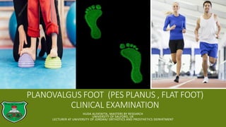
  • 1. PLANOVALGUS FOOT (PES PLANUS , FLAT FOOT) CLINICAL EXAMINATION HUDA ALFATAFTA, MASTERS BY RESEARCH UNIVERSITY OF SALFORD, UK LECTURER AT UNIVERSITY OF JORDAN/ ORTHOTICS AND PROSTHETICS DEPARTMENT
  • 2. FLAT FOOT EXAMINATION 2018 Introduction Flat Foot Risk factors Clinical symptoms Gait patterns Clinical tests Gait analysis Treatments PT P&O surgery The end Huda Alfatafta Foot print tests
  • 3. FOOT ▪ The foot is a complex anatomical structure. ▪ Functions:  It acts to transmit force between the lower limb and the ground, allowing stable ambulation and stance.  During gait the foot functions as a flexible shock-absorber, deforming to uneven surfaces before undergoing a series of biomechanical changes which allow it to act as a rigid lever to exert force. Huda Alfatafta
  • 4. ARCHES OF FOOT ▪ Three arches : medial and lateral longitudinal arches, and transverse arche. ▪ The medial longitudinal arch consists of the calcaneum, talus, navicular, medial, intermediate and lateral cuneiforms and the first three metatarsals. ▪ The talus sits at the apex of the arch (key stone) and confers stability by acting as a wedge between the calcaneum and navicular ▪ Half the body-weight passes through the apex of the arch whilst standing. ▪ The ends of the ach are unable to move apart due to the tight plantar fascia which connects them . Hence arch height is maintained “notrmaly” Huda Alfatafta
  • 6. STABILIZING FACTORS (STATIC AND DYNAMIC) Static factors ▪ The most important primary stabilizer is the plantar fascia, ▪ followed by the long and short plantar ligaments and then the spring ligament. ▪ Then, bony congruency Huda Alfatafta Dynamic factors ▪ tibialis posterior and other long flexors are important dynamic stabilizers. ▪ releasing these structures without releasing any static stabilizers has only a modest effect on arch height. ▪ One study found the result of releasing posterior tibial tension is associate with 0.5 mm reduction in arch height
  • 8. STABILIZING MODELS THE BEAM MODEL ▪ in the beam model, a load is applied to the apex of the arch generating compressive forces on the dorsal surface and tensile forces on the plantar surface. ▪ Stability in this model results from bony congruency and ligamentous attachments THE TRUSS MODEL. ▪ In the truss model, there is a triangular arrangement of structures. ▪ The bones of the arch are able to pivot about their apex whilst the tough plantar fascia forms the third side. ▪ This is firmly attached to the medial and lateral calcaneal tuberosities proximally and its slips insert into the plantar plate and the fibrous flexor sheathes distally Huda Alfatafta
  • 9. Huda Alfatafta ▪ the beam model ▪ the truss model.
  • 10. WHAT IS FLAT FOOT ▪ collapse or disappearance of the medial longitudinal foot arch and is associated with several three-dimensional foot deformities associated with a rotational hindfoot abnormality and heel valgus ▪ All infants are born with flatfoot, and the longitudinal arch of the foot develops during the first decade, but the prevalence of flatfoot is 37%– 59.7% in children 2–6 years of age and 4%–19.1% in those 8–13 years of age . Huda Alfatafta
  • 11. RISK FACTORS GROUP A ▪ BMI, OBESITY ▪ Age ▪ Pregnancy ▪ heredity GROUP B ▪ tibialis posterior dysfunction. ▪ Ligament laxity ▪ rotational abnormalities of the lower limbs Huda Alfatafta
  • 12. CLINICAL SYMPTOMS ▪ Pain on the sole side of the forefoot ▪ plantar pressure under the talar head ▪ Pain between the tip of the fibula and the calcaneus. pain Low activity level significant change in foot shape Huda Alfatafta
  • 13. GAIT PATTERNS ▪ Eversion of the foot at the talonavicular and talocalcaneal joints ▪ Valgus foot from posterior  the medial border of the foot approaching the ground Huda Alfatafta
  • 14. CLINICAL FOOT CLASSIFICATION MEASURES FLAT FOOT
  • 15. STEPS OF EXAMINATION ▪ Look at standing foot posture, swelling, hindfoot alignment, shoe and assess gait. ▪ Feel for localized tenderness and the nature of any swelling. ▪ Move each joint systematically, checking for pain, crepitus or stiffness. ▪ Special tests ▪ Radiographs Huda Alfatafta
  • 16. REARFOOT ANGLE (RFA) ▪ Briefly, four locations were palpated and marked using a skin marker pen (Fig. 1A). These were: ▪ (1) the base of the calcaneus; (2) the Achilles tendon attachment; (3) the centre of the Achilles tendon at the height of the medial malleoli; (4.) the centre of the posterior aspect of the shank 15 cm above marker three. ▪ The RFA was measured using a goniometer. The arms of the goniometer were aligned with the line connecting marker one and two (line 1) and the other arm with the lines connecting marker three and four (line 2). ▪ The RFA was measured as the acute angle between the projection of line one and line two. ▪ RFA ≥ 5° valgus represented a pronated foot type, 4° valgus to 4° varus a neutral foot and ≥ 5° varus a supinated foot Huda Alfatafta
  • 17. MEDIAL LONGITUDINAL ARCH ANGLE (MLAA) ▪ the midpoint of the medial malleolus, the most prominent aspect of the navicular tuberosity and the most medial prominence of the first metatarsal head were palpated and marked using a skin marker pen. ▪ The MLAA was measured using a goniometer with the centre of the goniometer aligned with the navicular mark and the arms aligned to connect the navicular mark with the medial malleolus and first metatarsal head markings. The obtuse angle was recorded as the MLAA. MLAA < 130° represented a pronated foot type, 130° to 150° a neutral foot type and > 150° a supinated foot type Huda Alfatafta
  • 18. NAVICULAR DROP TEST (NDT). ▪ Navicular Drop Test (NDT). Initially the most prominent aspect of the navicular tuberosity was palpated and marked with a skin marker pen. A piece of card was placed next to the medial aspect of the foot and the height of the navicular in a relaxed standing position marked on the card. The foot was then manipulated into subtalar joint neutral as determined by congruence of the talar head, and the process outlined above repeated. ▪ ND was recorded as the difference in navicular height between STJN and relaxed standing. The normal foot group included subjects with an NDT of 5–9mm ND > 9 mm represented a pronated foot type, 5 to 9 mm a neutral foot and < 5 mm a supinated foot Huda Alfatafta
  • 19. FOOT POSTURE INDEX (FPI-6) ▪ The FPI is a six-item clinical assessment tool used to evaluate foot posture. It has acceptable validity and good intra-rater reliability (ICC = 0.893–0.958). The FPI evaluates the multi-segmental nature of foot posture in all three planes; It does not require the use of specialised equipment. Each item of the FPI is scored between 2 and +2. Foot type was classified according to normative values with scores of ≥ 8 representing a pronated foot type, 0 to 5 a neutral foot and ≤ -1 a supinated foot Huda Alfatafta
  • 21. THE LEVEL OF AGREEMENT BETWEEN COMMON CLINICAL FOOT CLASSIFICATION MEASURES. ▪ Foot classification across the two test occasions was almost perfect for MLAA (Kw = 0.92) and FPI-6 (Kw =0 .92), moderate for RFA (Kw = 0.60) and fair for ND (Kw =0 .40) for comparison within the measures. ▪ Overall agreement between the measures for foot classification was moderate (Kf = 0.58). ▪ Conclusion: The findings reported in this study highlight discrepancies between the chosen foot classification measures. The FPI-6 was a reliable multi-planar measure whereas navicular drop emerged as an unreliable measure with only fair agreement across test sessions. The use of this measure for foot assessment is discouraged. Huda Alfatafta
  • 22. FOOT PRINT ▪ Arch index (AI) ▪ calculated as the ratio of the area of the middle third of the footprint to the entire footprint area. The foot was defined as high-arched if the AI was lower than 21% or flat if the AI was higher than 28%. ▪ AI= B/ (A+B+C)* 100% Huda Alfatafta
  • 23. FOOT PRINT ▪ Russian author method ▪ If the foot print occupies three out of five fields, it is the first degree; four out of five means the second degree, while five out of five means the third degree of the suspended foot. Huda Alfatafta
  • 24. RADIOGRAPHIC MEASURES ▪ Simple standing anteroposterior and lateral radiographs of weight-bearing feet were obtained in a digitalized manner from each subject. We measured the four angles commonly used to assess flatfoot on the lateral radiograph, including 1) lateral talo- first metatarsal angle (Meary’s angle), 2) talo-calcaneal angle, 3) metatarsal angle, and 4) calcaneal pitch Huda Alfatafta
  • 25. Normal Supination (high arch ) Pronation (flat foot) lateral talo-first metatarsal angle (Meary’s angle) Zero- 4 upward or downward 4 or more upward 4 or more downward. mild <15º moderate: 15- 30º severe: 30º talo-calcaneal angle 25-45 degrees Less than 25 degrees over 45 degrees metatarsal angle Not common to be used calcaneal pitch 18 to 20° More than 20 Less than 18 Huda Alfatafta The red highlighted tests are widely used
  • 26. GAIT ANALYSIS AND PRESSURE MEASUREMENT ▪ Platform with force-sensitive sensors Huda Alfatafta
  • 27. FLAT FOOT STAGES Huda Alfatafta Stage Stage I by symptoms of pain and swelling along the posterior tibial tendon without visible changes of foot alignment Stage II asymmetry of alignment with hindfoot valgus and forefoot abduction exaggerated on the symptomatic foot. have weakness with inversion and difficulty performing a single foot heel raise. Stage III flatfoot represents a progression of all the clinical signs but not rigid Stage VI Rigid ankle valgus deformity
  • 28. FLEXIBLE OR RIGID ?  Bony structure on not ? Huda Alfatafta Jack’s test Tip toe standing
  • 30. TREATMENTS PHYSIOTHERAPIST ▪ foot exercises ORTHOTIST ▪ Arch support ▪ Wedge ▪ SMO Huda Alfatafta vs. SURGEON Calcaneal osteotomy Subtalar arthrodesis  Triple arthrodesis vs.
  • 34. REFERENCES ▪ Jankowicz-Szymańska, A., Wódka, K., Kołpa, M., & Mikołajczyk, E. (2018). Foot longitudinal arches in obese, overweight and normal weight females who differ in age. HOMO, 69(1-2), 37-42. ▪ Pardo, F. J. V., del Amo, A. L., Rios, M. P., Gijon-Nogueron, G., & Yuste, C. C. (2018). Changes in foot posture during pregnancy and their relation with musculoskeletal pain: A longitudinal cohort study. Women and Birth, 31(2), e84-e88. ▪ Kangas, J., Dankaerts, W., & Staes, F. (2011). New approach to the diagnosis and classification of chronic foot and ankle disorders: Identifying motor control and movement impairments. Manual therapy, 16(6), 522-530. ▪ Lee, J. S., Kim, K. B., Jeong, J. O., Kwon, N. Y., & Jeong, S. M. (2015). Correlation of foot posture index with plantar pressure and radiographic measurements in pediatric flatfoot. Annals of rehabilitation medicine, 39(1), 10-17. ▪ Young, C. C., Niedfeldt, M. W., Morris, G. A., & Eerkes, K. J. (2005). Clinical examination of the foot and ankle. Primary Care: Clinics in Office Practice, 32(1), 105-132. ▪ Cebulski-Delebarre, A., Boutry, N., Szymanski, C., Maynou, C., Lefebvre, G., Amzallag-Bellenger, E., & Cotten, A. (2016). Correlation between primary flat foot and lower extremity rotational misalignment in adults. Diagnostic and interventional imaging, 97(11), 1151-1157. ▪ Lever, C. J., & Hennessy, M. S. (2016). Adult flat foot deformity. Orthopaedics and Trauma, 30(1), 41-50. Huda Alfatafta
  • 35. CONT. ▪ Roche, A., Hunter, L., Pocock, N., & Brown, D. (2009). Physical examination of the foot and ankle by orthopaedic and accident and emergency clinicians. Injury, 40(2), 136-138. ▪ Langley, B., Cramp, M., & Morrison, S. C. (2016). Clinical measures of static foot posture do not agree. Journal of foot and ankle research, 9(1), 45. ▪ DELAND, J. T. (2004). ANATOMY AND BIOMECHANICS OF THE FOOT AND ANKLE. Foot and Ankle, 1. ▪ Rao, S., Riskowski, J. L., & Hannan, M. T. (2012). Musculoskeletal conditions of the foot and ankle: assessments and treatment options. Best practice & research Clinical rheumatology, 26(3), 345- 368. ▪ Brockett, C. L., & Chapman, G. J. (2016). Biomechanics of the ankle. Orthopaedics and trauma, 30(3), 232-238. ▪ Haendlmayer, K. T., & Harris, N. J. (2009). (ii) Flatfoot deformity: an overview. Orthopaedics and Trauma, 23(6), 395-403. ▪ Pauk, J., & Ezerskiy, V. (2011). The effect of foot orthotics on arch height: prediction of arch height correction in flat-foot children. Biocybernetics and Biomedical Engineering, 31(1), 51-62. Huda Alfatafta