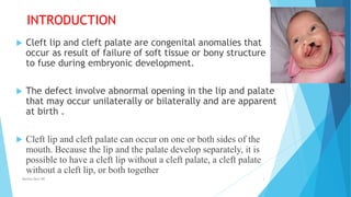
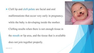
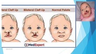
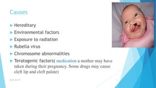
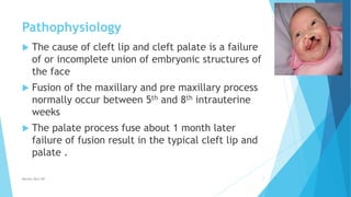
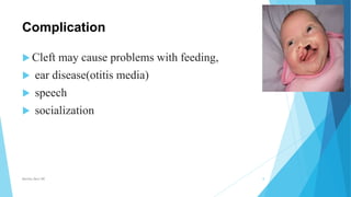
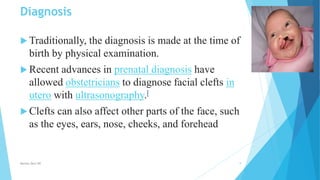
Cleft lip
and
cleft palate
Prepared by
Monika Devi NR
M. Sc. Nursing
GMCH Jammu
INTRODUCTION
 Cleft lip and cleft palate are congenital anomalies that
occur as result of failure of soft tissue or bony structure
to fuse during embryonic development.
 The defect involve abnormal opening in the lip and palate
that may occur unilaterally or bilaterally and are apparent
at birth .
 Cleft lip and cleft palate can occur on one or both sides of the
mouth. Because the lip and the palate develop separately, it is
possible to have a cleft lip without a cleft palate, a cleft palate
without a cleft lip, or both together
Monika Devi NR 3
 Cleft lip and cleft palate are facial and oral
malformations that occur very early in pregnancy,
while the baby is developing inside the mother.
Clefting results when there is not enough tissue in
the mouth or lip area, and the tissue that is available
does not join together properly.
Monika Devi NR 4
Monika Devi NR 5
Causes
 Hereditary
 Environmental factors
 Exposure to radiation
 Rubella virus
 Chromosome abnormalities
 Teratogenic factors( medication a mother may have
taken during their pregnancy. Some drugs may cause
cleft lip and cleft palate)
Monika Devi NR 6
Pathophysiology
 The cause of cleft lip and cleft palate is a failure
of or incomplete union of embryonic structures of
the face
 Fusion of the maxillary and pre maxillary process
normally occur between 5th and 8th intrauterine
weeks
 The palate process fuse about 1 month later
failure of fusion result in the typical cleft lip and
palate .
Monika Devi NR 7
Complication
 Cleft may cause problems with feeding,
 ear disease(otitis media)
 speech
 socialization
Monika Devi NR 8
Diagnosis
 Traditionally, the diagnosis is made at the time of
birth by physical examination.
 Recent advances in prenatal diagnosis have
allowed obstetricians to diagnose facial clefts in
utero with ultrasonography.[
 Clefts can also affect other parts of the face, such
as the eyes, ears, nose, cheeks, and forehead
Monika Devi NR 9
Intervention
 Assess the ability to suck, swallow, handle normal
secretion, and breath without distress.
 Assess fluid and calorie intake daily.
 Monitor daily weight
 Modify feeding technique plan to use specialized
feeding techniques ,
 Hold the infant in an upright position and direct the
formula to side and back of mouth to prevent
aspiration .
Monika Devi NR 10
Surgical management
 A cleft lip may require one or two surgeries depending on
the severity of defect .the indicial surgery is usually
performed at age of 3 month. Common procedures for
repair of cleft lip are Tennison Randall and triangular flap
(z type )cheiloplasty millards rotation
 Clef palate repair after requires the first surgical repair
usually occur when the baby is between 6-12,24 month the
chance of fluid entering the middle ear palatoplasty
Monika Devi NR 11
Monika Devi NR 12
Post operative management
 CLEFT LIP
 Provide lip protection a metal appliance or adhesive strips may be taped
securely to the cheek to prevent trauma to the suture line
 Avoid positioning the infant on the side of repair or in prone position
because these position can cause rubbing of surgical site on the matters
 Keep surgical site clean and dry after feeding gently clean the suture line of
formula
 Apply antibiotic ointment to the sit as prescribed
 Elbow restraint should be used to prevent the infant from injuring or
traumatizing the surgical site
Monika Devi NR 13
Cleft palate
 Oral packing may be secured to the palate (usually removed
in 2 to3day)
 Do not allow the child to brush his or her teeth.
 Instruct the parents to avoid offering hard food items to the
child such as toast .
 Soft elbow and jacket restraints at list every 2 hours (per
agency procedure)
 Avoid use of oral suction ,tongue depressor, thermometer
straws spoons, fork or pacifiers
Monika Devi NR 14
care
 Provide analgesics for pain as prescribed
 Instruct the parents to monitor for sign of infection at
the surgical site such as redness ,swelling, or drainage.
Monika Devi NR 15
Thank you
Monika Devi NR 16

CLIFT LIP AND CLEFT PALATE

  • 1.
    Cleft lip and cleft palate Preparedby Monika Devi NR M. Sc. Nursing GMCH Jammu
  • 3.
    INTRODUCTION  Cleft lipand cleft palate are congenital anomalies that occur as result of failure of soft tissue or bony structure to fuse during embryonic development.  The defect involve abnormal opening in the lip and palate that may occur unilaterally or bilaterally and are apparent at birth .  Cleft lip and cleft palate can occur on one or both sides of the mouth. Because the lip and the palate develop separately, it is possible to have a cleft lip without a cleft palate, a cleft palate without a cleft lip, or both together Monika Devi NR 3
  • 4.
     Cleft lipand cleft palate are facial and oral malformations that occur very early in pregnancy, while the baby is developing inside the mother. Clefting results when there is not enough tissue in the mouth or lip area, and the tissue that is available does not join together properly. Monika Devi NR 4
  • 5.
  • 6.
    Causes  Hereditary  Environmentalfactors  Exposure to radiation  Rubella virus  Chromosome abnormalities  Teratogenic factors( medication a mother may have taken during their pregnancy. Some drugs may cause cleft lip and cleft palate) Monika Devi NR 6
  • 7.
    Pathophysiology  The causeof cleft lip and cleft palate is a failure of or incomplete union of embryonic structures of the face  Fusion of the maxillary and pre maxillary process normally occur between 5th and 8th intrauterine weeks  The palate process fuse about 1 month later failure of fusion result in the typical cleft lip and palate . Monika Devi NR 7
  • 8.
    Complication  Cleft maycause problems with feeding,  ear disease(otitis media)  speech  socialization Monika Devi NR 8
  • 9.
    Diagnosis  Traditionally, thediagnosis is made at the time of birth by physical examination.  Recent advances in prenatal diagnosis have allowed obstetricians to diagnose facial clefts in utero with ultrasonography.[  Clefts can also affect other parts of the face, such as the eyes, ears, nose, cheeks, and forehead Monika Devi NR 9
  • 10.
    Intervention  Assess theability to suck, swallow, handle normal secretion, and breath without distress.  Assess fluid and calorie intake daily.  Monitor daily weight  Modify feeding technique plan to use specialized feeding techniques ,  Hold the infant in an upright position and direct the formula to side and back of mouth to prevent aspiration . Monika Devi NR 10
  • 11.
    Surgical management  Acleft lip may require one or two surgeries depending on the severity of defect .the indicial surgery is usually performed at age of 3 month. Common procedures for repair of cleft lip are Tennison Randall and triangular flap (z type )cheiloplasty millards rotation  Clef palate repair after requires the first surgical repair usually occur when the baby is between 6-12,24 month the chance of fluid entering the middle ear palatoplasty Monika Devi NR 11
  • 12.
  • 13.
    Post operative management CLEFT LIP  Provide lip protection a metal appliance or adhesive strips may be taped securely to the cheek to prevent trauma to the suture line  Avoid positioning the infant on the side of repair or in prone position because these position can cause rubbing of surgical site on the matters  Keep surgical site clean and dry after feeding gently clean the suture line of formula  Apply antibiotic ointment to the sit as prescribed  Elbow restraint should be used to prevent the infant from injuring or traumatizing the surgical site Monika Devi NR 13
  • 14.
    Cleft palate  Oralpacking may be secured to the palate (usually removed in 2 to3day)  Do not allow the child to brush his or her teeth.  Instruct the parents to avoid offering hard food items to the child such as toast .  Soft elbow and jacket restraints at list every 2 hours (per agency procedure)  Avoid use of oral suction ,tongue depressor, thermometer straws spoons, fork or pacifiers Monika Devi NR 14
  • 15.
    care  Provide analgesicsfor pain as prescribed  Instruct the parents to monitor for sign of infection at the surgical site such as redness ,swelling, or drainage. Monika Devi NR 15
  • 16.