z
CLEFT LIP &
CLEFT PALATE
Mr. Pradeep Abothu, M.Sc (N), PhD Scholar,
Associate Professor, Dept. of Child Health(N)
ASRAM College Of Nursing
z
CLEFT LIP
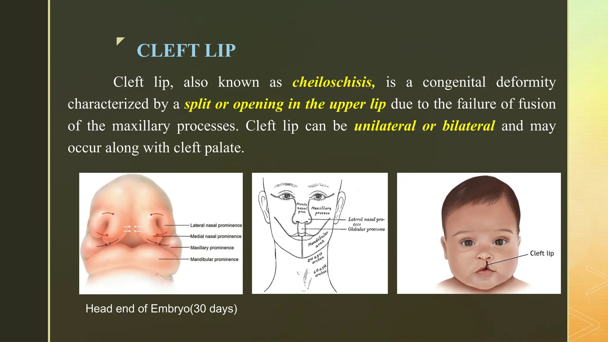
Cleft lip, also known as cheiloschisis, is a congenital deformity
characterized by a split or opening in the upper lip due to the failure of fusion
of the maxillary processes. Cleft lip can be unilateral or bilateral and may
occur along with cleft palate.
Head end of Embryo(30 days)
z
CLEFT PALATE
Cleft palate, also known as palatoschisis, is a congenital condition
characterized by an opening in the roof of the mouth caused by the failure of
fusion of the palatine processes. This condition can involve the hard palate,
soft palate, or both.
z INCIDENCE
 Cleft Lip: The incidence of cleft lip, with or without cleft palate, is
approximately 1 in 1,000 live births. It is more common in males than
females.
 Cleft Palate: The incidence of cleft palate alone is about 1 in 2,500 live
births. This condition is more common in females than males.
z
ETIOLOGY
Genetic Factors:
 Family history of clefts can increase the risk.
 Mutations in genes linked to facial development.
 Chromosomal abnormalities associated with conditions like trisomy 13
and Van der Woude syndrome.
The exact cause of cleft lip and cleft palate is unknown; however,
they are believed due to a combination of genetic and environmental factors.
Environmental Factors:
 Maternal health issues such as diabetes and obesity
contribute.
 Nutritional deficiencies, especially folic acid,
during pregnancy.
 Substance use, including alcohol and tobacco, by
the mother during pregnancy.
 Exposure to X-rays or radiation during pregnancy.
 Overuse of certain medications like anticonvulsants
during pregnancy.
z
PATHOPHYSIOLOGY
Cleft lip and cleft palate result from abnormal fusion of facial structures during
embryonic development.
The lip forms from the merging of the medial nasal processes and maxillary processes,
while the palate develops from the fusion of palatine shelves.
Disruptions in these processes can lead to incomplete formation, resulting in gaps.
These malformations can lead to problems such as difficulty eating, speech delays, ear
infections, and orthodontic issues.
z
COMPLICATIONS
 Feeding Difficulties: Infants may struggle to create a proper seal for breastfeeding or bottle
feeding, leading to inadequate nutrition.
 Speech Delays: Clefts can affect the development of normal speech patterns, requiring
speech therapy.
 Ear Infections: Increased risk of otitis media due to Eustachian tube dysfunction, which can
lead to hearing loss.
 Respiratory Infections: Higher susceptibility to respiratory infections due to compromised
airway function.
 Dental Problems: Misalignment of teeth, missing teeth, and increased risk of cavities.
 Growth and Development Issues: Potential impact on overall growth if feeding difficulties
are significant.
z
DIAGNOSTIC EVALUATION TESTS
 Prenatal Ultrasound: Can identify physical abnormalities, such as cleft
lip/palate, as early as the second trimester.
 Physical Examination: A thorough examination at birth can confirm the
presence of cleft lip/ palate.
z
TREATMENT
 After birth, infants with cleft lip and/or cleft palate require careful
management to ensure their health and prepare them for surgical interventions.
 A multidisciplinary healthcare team is involved, including pediatricians,
speech and feeding specialists, nutritionists, surgeons, and prosthodontists, all
coordinating to monitor overall health and development.
 Once the child is at least 5 kg by 3 months, has an absence of respiratory
infections, and demonstrates adequate nutritional status, the child is
considered healthy enough to proceed with surgery.
z
SURGICAL MANAGEMENT
 Cleft Lip Repair: known as cheiloplasty, is typically performed between 3 to
6 months of age. During the procedure, an incision is made along the natural
contour of the lip, allowing the surgeon to bring the lip edges together and
suture them using techniques such as Z-plasty for improved aesthetics, other
techniques that can be used are Millard repair, Randall-Graham procedure,
and Rose-Thompson procedure.
z
SURGICAL MANAGEMENT
 Cleft Palate Repair: known as palatoplasty, the procedure is usually
conducted between 9 to 18 months of age. An incision is made in the oral
mucosa along the cleft, and tissue flaps from the hard and soft palates are
mobilized and sutured to close the cleft, often utilizing the V-Y pushback
technique.
NURSING
MANAGEMENT
z
Care of the Child immediately after birth:
• Evaluate the extent of the cleft lip and/or palate.
• Monitor for feeding difficulties and signs of aspiration.
• Assess for associated anomalies or syndromes.
• Use specialized feeding bottles and nipples designed for
cleft lip/palate.
• Encourage upright positioning during feeds to reduce the
risk of aspiration.
z
• Give the child enough time to swallow and burp in between feeds.
• Consider using a palatal obturator to help with feeding.
• Monitor for complications, such as otitis media or dental issues, and refer to
specialists as needed.
• Teach parents proper feeding techniques and the use of specialized
bottles/nipples.
• Provide emotional support and encourage bonding with the child.
z
Preoperative Care:
• Conduct a thorough preoperative assessment and ensure all preoperative
investigations are completed.
• Keep the child NPO.
• Educate parents about the surgical procedure, expected outcomes, and
postoperative care.
Postoperative Care:
• Monitor vital signs and surgical site for signs of infection or bleeding.
• Position the child in semi upright position to prevent aspiration.
• Administer pain management as prescribed.
• Use elbow restraints if necessary to prevent the child from touching or
pulling at the surgical site.
z
• Apply a Logan's bow or other protective
devices as directed by the surgeon.
• Start with clear liquids and gradually advance
to a soft diet as tolerated.
• Use a syringe or soft-tipped feeder to avoid
putting pressure on the surgical site.
• Teach parents how to care for the surgical site, including cleaning
and applying any prescribed ointments.
• Instruct parents on signs of infection to watch for and when to
seek medical advice.
z
NURSING DIAGNOSIS
 Impaired Nutrition less than Body Requirements related to feeding
difficulties.
 Risk for Aspiration related to anatomical defects.
 Delayed Growth and Development related to inadequate nutritional intake.
 Risk for Infection related to surgical interventions.
 Acute Pain related to surgical intervention.
 Knowledge Deficit related to lack of information about proper feeding
techniques and postoperative care.
CLEFT LIP AND PALATE: NURSING MANAGEMENT.pptx

CLEFT LIP AND PALATE: NURSING MANAGEMENT.pptx

  • 1.
    z CLEFT LIP & CLEFTPALATE Mr. Pradeep Abothu, M.Sc (N), PhD Scholar, Associate Professor, Dept. of Child Health(N) ASRAM College Of Nursing
  • 2.
    z CLEFT LIP Cleft lip,also known as cheiloschisis, is a congenital deformity characterized by a split or opening in the upper lip due to the failure of fusion of the maxillary processes. Cleft lip can be unilateral or bilateral and may occur along with cleft palate. Head end of Embryo(30 days)
  • 3.
    z CLEFT PALATE Cleft palate,also known as palatoschisis, is a congenital condition characterized by an opening in the roof of the mouth caused by the failure of fusion of the palatine processes. This condition can involve the hard palate, soft palate, or both.
  • 4.
    z INCIDENCE  CleftLip: The incidence of cleft lip, with or without cleft palate, is approximately 1 in 1,000 live births. It is more common in males than females.  Cleft Palate: The incidence of cleft palate alone is about 1 in 2,500 live births. This condition is more common in females than males.
  • 5.
    z ETIOLOGY Genetic Factors:  Familyhistory of clefts can increase the risk.  Mutations in genes linked to facial development.  Chromosomal abnormalities associated with conditions like trisomy 13 and Van der Woude syndrome. The exact cause of cleft lip and cleft palate is unknown; however, they are believed due to a combination of genetic and environmental factors.
  • 6.
    Environmental Factors:  Maternalhealth issues such as diabetes and obesity contribute.  Nutritional deficiencies, especially folic acid, during pregnancy.  Substance use, including alcohol and tobacco, by the mother during pregnancy.  Exposure to X-rays or radiation during pregnancy.  Overuse of certain medications like anticonvulsants during pregnancy.
  • 7.
    z PATHOPHYSIOLOGY Cleft lip andcleft palate result from abnormal fusion of facial structures during embryonic development. The lip forms from the merging of the medial nasal processes and maxillary processes, while the palate develops from the fusion of palatine shelves. Disruptions in these processes can lead to incomplete formation, resulting in gaps. These malformations can lead to problems such as difficulty eating, speech delays, ear infections, and orthodontic issues.
  • 8.
    z COMPLICATIONS  Feeding Difficulties:Infants may struggle to create a proper seal for breastfeeding or bottle feeding, leading to inadequate nutrition.  Speech Delays: Clefts can affect the development of normal speech patterns, requiring speech therapy.  Ear Infections: Increased risk of otitis media due to Eustachian tube dysfunction, which can lead to hearing loss.  Respiratory Infections: Higher susceptibility to respiratory infections due to compromised airway function.  Dental Problems: Misalignment of teeth, missing teeth, and increased risk of cavities.  Growth and Development Issues: Potential impact on overall growth if feeding difficulties are significant.
  • 9.
    z DIAGNOSTIC EVALUATION TESTS Prenatal Ultrasound: Can identify physical abnormalities, such as cleft lip/palate, as early as the second trimester.  Physical Examination: A thorough examination at birth can confirm the presence of cleft lip/ palate.
  • 10.
    z TREATMENT  After birth,infants with cleft lip and/or cleft palate require careful management to ensure their health and prepare them for surgical interventions.  A multidisciplinary healthcare team is involved, including pediatricians, speech and feeding specialists, nutritionists, surgeons, and prosthodontists, all coordinating to monitor overall health and development.  Once the child is at least 5 kg by 3 months, has an absence of respiratory infections, and demonstrates adequate nutritional status, the child is considered healthy enough to proceed with surgery.
  • 11.
    z SURGICAL MANAGEMENT  CleftLip Repair: known as cheiloplasty, is typically performed between 3 to 6 months of age. During the procedure, an incision is made along the natural contour of the lip, allowing the surgeon to bring the lip edges together and suture them using techniques such as Z-plasty for improved aesthetics, other techniques that can be used are Millard repair, Randall-Graham procedure, and Rose-Thompson procedure.
  • 12.
    z SURGICAL MANAGEMENT  CleftPalate Repair: known as palatoplasty, the procedure is usually conducted between 9 to 18 months of age. An incision is made in the oral mucosa along the cleft, and tissue flaps from the hard and soft palates are mobilized and sutured to close the cleft, often utilizing the V-Y pushback technique.
  • 13.
  • 14.
    z Care of theChild immediately after birth: • Evaluate the extent of the cleft lip and/or palate. • Monitor for feeding difficulties and signs of aspiration. • Assess for associated anomalies or syndromes. • Use specialized feeding bottles and nipples designed for cleft lip/palate. • Encourage upright positioning during feeds to reduce the risk of aspiration.
  • 15.
    z • Give thechild enough time to swallow and burp in between feeds. • Consider using a palatal obturator to help with feeding. • Monitor for complications, such as otitis media or dental issues, and refer to specialists as needed. • Teach parents proper feeding techniques and the use of specialized bottles/nipples. • Provide emotional support and encourage bonding with the child.
  • 16.
    z Preoperative Care: • Conducta thorough preoperative assessment and ensure all preoperative investigations are completed. • Keep the child NPO. • Educate parents about the surgical procedure, expected outcomes, and postoperative care. Postoperative Care: • Monitor vital signs and surgical site for signs of infection or bleeding. • Position the child in semi upright position to prevent aspiration. • Administer pain management as prescribed. • Use elbow restraints if necessary to prevent the child from touching or pulling at the surgical site.
  • 17.
    z • Apply aLogan's bow or other protective devices as directed by the surgeon. • Start with clear liquids and gradually advance to a soft diet as tolerated. • Use a syringe or soft-tipped feeder to avoid putting pressure on the surgical site. • Teach parents how to care for the surgical site, including cleaning and applying any prescribed ointments. • Instruct parents on signs of infection to watch for and when to seek medical advice.
  • 18.
    z NURSING DIAGNOSIS  ImpairedNutrition less than Body Requirements related to feeding difficulties.  Risk for Aspiration related to anatomical defects.  Delayed Growth and Development related to inadequate nutritional intake.  Risk for Infection related to surgical interventions.  Acute Pain related to surgical intervention.  Knowledge Deficit related to lack of information about proper feeding techniques and postoperative care.