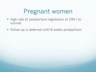
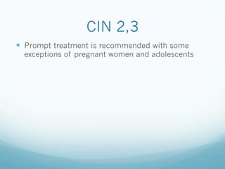
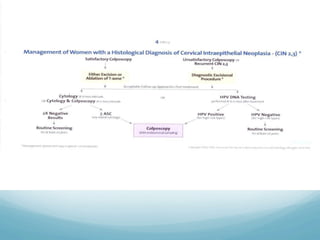
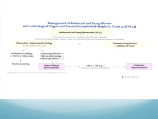
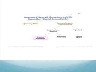
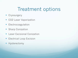
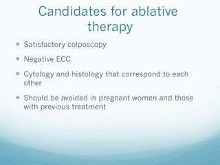
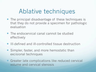
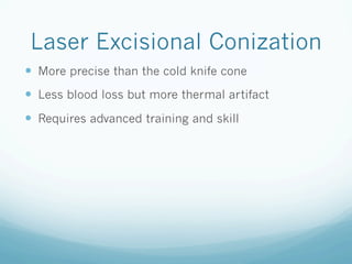
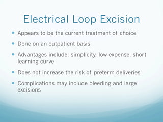
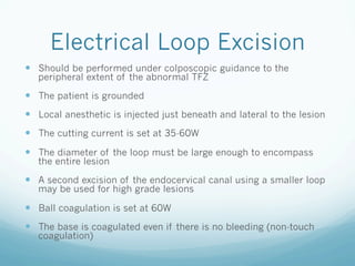
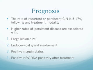
Management of CIN
A. Alobaid, MBBS, FRCS(C), FACOG
Consultant, Gynecologic Oncology
Assistant professor, KSU
Medical Director, Women s Specialized Hospital
King Fahad Medical City
—  Who to treat?
—  When to treat?
—  Treatment options
—  Treatment principles
—  Complications
CIN I
—  CIN I preceded by LGSIL
—  CIN I preceded by HGSIL
CIN I preceded by LGSIL
—  Spontaneous regression is observed in most
women (80%)
—  Expectant follow up is warranted
—  Treatment acceptable if:
1.  CIN I persists for more than 24 months
2.  Relieve patient s anxiety
3.  Patient at very high risk to follow-up
CIN I preceded by HGSIL
—  There is 70% chance of having underlying CIN II, III
or worse
—  Excisional diagnostic procedure Is generally
recommended
—  An alternative approach is expectant management
with intensive monitoring
Adolescent women with
CIN I
—  Undetected high grade disease is uncommon,
invasive cancer is rare, regression to normal is
common
—  Expectant management is preferred
Pregnant women
—  high rate of postpartum regression of CIN I to
normal
—  Follow up is deferred until 6 weeks postpartum
CIN 2,3
—  Prompt treatment is recommended with some
exceptions of pregnant women and adolescents
Treatment options
—  Cryosurgery
—  CO2 Laser Vaporization
—  Electrocoagulation
—  Sharp Conization
—  Laser Excisional Conization
—  Electrical Loop Excision
—  Hysterectomy
Candidates for ablative
therapy
—  Satisfactory colposcopy
—  Negative ECC
—  Cytology and histology that correspond to each
other
—  Should be avoided in pregnant women and those
with previous treatment
Ablative techniques
—  The principal disadvantage of these techniques is
that they do not provide a specimen for pathologic
evaluation
—  The endocervical canal cannot be studied
effectively
—  Ill-defined and ill-controlled tissue destruction
—  Simpler, faster, and more hemostatic than
excisional techniques
—  Greater late complications like reduced cervical
volume and cervical stenosis
Cryotherapy
—  Office procedure using local anesthesia and
NSAID s
—  Using refrigerant gas (CO2 or N2O)
—  The ectocervix must be cooled to -20 C to cause
crystallization of intracellular water and destroy the
lesion
—  It is achieved by forming an ice ball in the cervical
tissue that is at least 5 mm from the tip of the
probe
Cryosurgery
—  A thin layer of water-soluble lubricant is applied
over the tip of the probe to allow more uniform and
rapid freeze of the cervix
—  The probe should cover the entire lesion, and a 4-5
mm ice ball around the probe is required for an
adequate freeze
—  Repeat freeze thaw cycles will produce greater
tissue volume destruction than single freeze cycles
for the same amount of time provided
Cryosurgery
—  There is usually a watery discharge for 10-14 days
—  The patient is re-evaluated after 4 months of the
treatment
—  If the pap smear remains positive 6 months after
therapy, then cryosurgery is considered a failure
and the patient should be reevaluated and retreated
Criocirugia-YouTube.flv
CO2 laser
—  Laser is directed at the lesion under colposcopic
guidance
—  Water in the tissue absorbs the laser energy which
destroys the tissue by vaporization
—  The lesion is ablated to a depth of 5 mm on the
ectocervix and 8-9 mm around the endocervix
Excisional therapy
—  Cold knife conization
—  Laser conization
—  LEEP (LLETZ)
Treatment specifications
—  Perform colposcopy during treatment phase to
obtain accurate view of the entire TFZ
—  3 mm margin is obtained around the abnormal TFZ
to allow for glandular involvement
—  Obtain 1 cm endocervical margin for LGSIL
—  Obtain 1.5 cm endocervical margin for HGSIL
Indications for excisional
therapy
—  Unsatisfactory colposcopy
—  Lesions extending into the endocervical canal
—  +ve ECC
—  Discrepancy between the cytology and biopsy results
—  Suspected microinvasion
—  Suspected AIS
—  Invasive disease suspected
—  Recurrence after previous treatment
Sharp Conization
—  One of the oldest techniques
—  Recommended for women with suspected
microinvasion and AIS
—  The margins of the cone are plotted colposcopically
using acetic acid or Lugol s solution
—  The configuration of the specimen is based on the
extent of disease
—  Complications include: bleeding (immediate or
delayed), cervical stenosis, cervical incompetence
Laser Excisional Conization
—  More precise than the cold knife cone
—  Less blood loss but more thermal artifact
—  Requires advanced training and skill
Colposcop?aCono-l?ser-YouTube.flv
Electrical Loop Excision
—  Appears to be the current treatment of choice
—  Done on an outpatient basis
—  Advantages include: simplicity, low expense, short
learning curve
—  Does not increase the risk of preterm deliveries
—  Complications may include bleeding and large
excisions
Electrical Loop Excision
—  Should be performed under colposcopic guidance to the
peripheral extent of the abnormal TFZ
—  The patient is grounded
—  Local anesthetic is injected just beneath and lateral to the lesion
—  The cutting current is set at 35-60W
—  The diameter of the loop must be large enough to encompass
the entire lesion
—  A second excision of the endocervical canal using a smaller loop
may be used for high grade lesions
—  Ball coagulation is set at 60W
—  The base is coagulated even if there is no bleeding (non-touch
coagulation)
Colposcop?a-ConodeCÈrvix-YouTube.flv
Conizacion cervical v final - YouTube.flv
Complications
—  Intraoperative bleeding
—  Postoperative bleeding: early or delayed
—  Infection
—  Cervical stenosis
Hysterectomy
—  If there is coexistent gynecologic conditions
requiring hysterectomy
—  Patient request and persistent or recurrent CIN 2,3
Reproductive outcome
—  Cold knife conization is the only treatment modality
that increases the risk of perinatal mortality and
preterm delivery
—  During pregnancy, surveillance with serial TV
ultrasound for cervical length measurement is
recommended for patients who had CKC
Prognosis
—  The rate of recurrent or persistent CIN is 5-17%
following any treatment modality
—  Higher rates of persistent disease are associated
with:
1.  Large lesion size
2.  Endocervical gland involvement
3.  Positive margin status
4.  Positive HPV DNA positivity after treatment
Thank you

Cin managment

  • 1.
    Management of CIN A.Alobaid, MBBS, FRCS(C), FACOG Consultant, Gynecologic Oncology Assistant professor, KSU Medical Director, Women s Specialized Hospital King Fahad Medical City
  • 2.
    —  Who totreat? —  When to treat? —  Treatment options —  Treatment principles —  Complications
  • 3.
    CIN I —  CINI preceded by LGSIL —  CIN I preceded by HGSIL
  • 4.
    CIN I precededby LGSIL —  Spontaneous regression is observed in most women (80%) —  Expectant follow up is warranted —  Treatment acceptable if: 1.  CIN I persists for more than 24 months 2.  Relieve patient s anxiety 3.  Patient at very high risk to follow-up
  • 6.
    CIN I precededby HGSIL —  There is 70% chance of having underlying CIN II, III or worse —  Excisional diagnostic procedure Is generally recommended —  An alternative approach is expectant management with intensive monitoring
  • 8.
    Adolescent women with CINI —  Undetected high grade disease is uncommon, invasive cancer is rare, regression to normal is common —  Expectant management is preferred
  • 10.
    Pregnant women —  highrate of postpartum regression of CIN I to normal —  Follow up is deferred until 6 weeks postpartum
  • 11.
    CIN 2,3 —  Prompttreatment is recommended with some exceptions of pregnant women and adolescents
  • 15.
    Treatment options —  Cryosurgery — CO2 Laser Vaporization —  Electrocoagulation —  Sharp Conization —  Laser Excisional Conization —  Electrical Loop Excision —  Hysterectomy
  • 16.
    Candidates for ablative therapy — Satisfactory colposcopy —  Negative ECC —  Cytology and histology that correspond to each other —  Should be avoided in pregnant women and those with previous treatment
  • 17.
    Ablative techniques —  Theprincipal disadvantage of these techniques is that they do not provide a specimen for pathologic evaluation —  The endocervical canal cannot be studied effectively —  Ill-defined and ill-controlled tissue destruction —  Simpler, faster, and more hemostatic than excisional techniques —  Greater late complications like reduced cervical volume and cervical stenosis
  • 18.
    Cryotherapy —  Office procedureusing local anesthesia and NSAID s —  Using refrigerant gas (CO2 or N2O) —  The ectocervix must be cooled to -20 C to cause crystallization of intracellular water and destroy the lesion —  It is achieved by forming an ice ball in the cervical tissue that is at least 5 mm from the tip of the probe
  • 19.
    Cryosurgery —  A thinlayer of water-soluble lubricant is applied over the tip of the probe to allow more uniform and rapid freeze of the cervix —  The probe should cover the entire lesion, and a 4-5 mm ice ball around the probe is required for an adequate freeze —  Repeat freeze thaw cycles will produce greater tissue volume destruction than single freeze cycles for the same amount of time provided
  • 20.
    Cryosurgery —  There isusually a watery discharge for 10-14 days —  The patient is re-evaluated after 4 months of the treatment —  If the pap smear remains positive 6 months after therapy, then cryosurgery is considered a failure and the patient should be reevaluated and retreated
  • 21.
  • 22.
    CO2 laser —  Laseris directed at the lesion under colposcopic guidance —  Water in the tissue absorbs the laser energy which destroys the tissue by vaporization —  The lesion is ablated to a depth of 5 mm on the ectocervix and 8-9 mm around the endocervix
  • 23.
    Excisional therapy —  Coldknife conization —  Laser conization —  LEEP (LLETZ)
  • 24.
    Treatment specifications —  Performcolposcopy during treatment phase to obtain accurate view of the entire TFZ —  3 mm margin is obtained around the abnormal TFZ to allow for glandular involvement —  Obtain 1 cm endocervical margin for LGSIL —  Obtain 1.5 cm endocervical margin for HGSIL
  • 25.
    Indications for excisional therapy — Unsatisfactory colposcopy —  Lesions extending into the endocervical canal —  +ve ECC —  Discrepancy between the cytology and biopsy results —  Suspected microinvasion —  Suspected AIS —  Invasive disease suspected —  Recurrence after previous treatment
  • 26.
    Sharp Conization —  Oneof the oldest techniques —  Recommended for women with suspected microinvasion and AIS —  The margins of the cone are plotted colposcopically using acetic acid or Lugol s solution —  The configuration of the specimen is based on the extent of disease —  Complications include: bleeding (immediate or delayed), cervical stenosis, cervical incompetence
  • 33.
    Laser Excisional Conization — More precise than the cold knife cone —  Less blood loss but more thermal artifact —  Requires advanced training and skill
  • 41.
  • 42.
    Electrical Loop Excision — Appears to be the current treatment of choice —  Done on an outpatient basis —  Advantages include: simplicity, low expense, short learning curve —  Does not increase the risk of preterm deliveries —  Complications may include bleeding and large excisions
  • 43.
    Electrical Loop Excision — Should be performed under colposcopic guidance to the peripheral extent of the abnormal TFZ —  The patient is grounded —  Local anesthetic is injected just beneath and lateral to the lesion —  The cutting current is set at 35-60W —  The diameter of the loop must be large enough to encompass the entire lesion —  A second excision of the endocervical canal using a smaller loop may be used for high grade lesions —  Ball coagulation is set at 60W —  The base is coagulated even if there is no bleeding (non-touch coagulation)
  • 53.
  • 54.
    Conizacion cervical vfinal - YouTube.flv
  • 55.
    Complications —  Intraoperative bleeding — Postoperative bleeding: early or delayed —  Infection —  Cervical stenosis
  • 56.
    Hysterectomy —  If thereis coexistent gynecologic conditions requiring hysterectomy —  Patient request and persistent or recurrent CIN 2,3
  • 57.
    Reproductive outcome —  Coldknife conization is the only treatment modality that increases the risk of perinatal mortality and preterm delivery —  During pregnancy, surveillance with serial TV ultrasound for cervical length measurement is recommended for patients who had CKC
  • 58.
    Prognosis —  The rateof recurrent or persistent CIN is 5-17% following any treatment modality —  Higher rates of persistent disease are associated with: 1.  Large lesion size 2.  Endocervical gland involvement 3.  Positive margin status 4.  Positive HPV DNA positivity after treatment
  • 59.