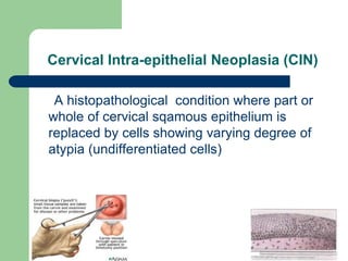
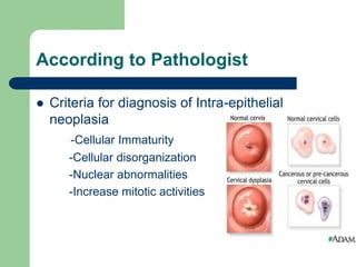
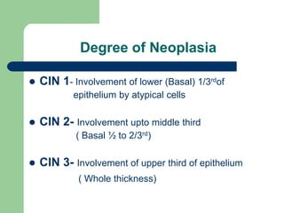
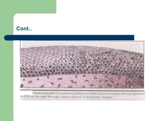
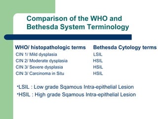
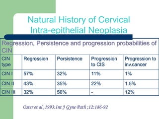
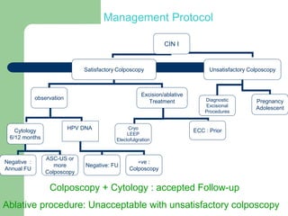
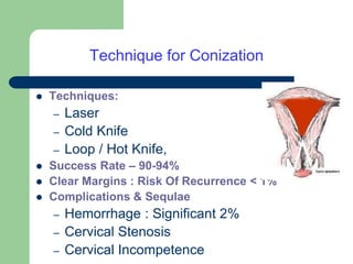
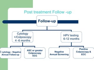
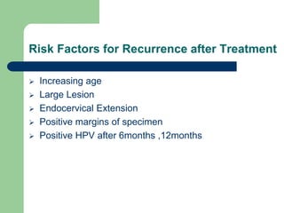
MANAGEMENT OF CERVICAL
INTRA-EPITHELIAL NEOPLASIA
(CIN)
Dr. Parul Sinha
Dept. of Obstetrics &
Gynaecology
Cervical Intra-epithelial Neoplasia (CIN)
A histopathological condition where part or
whole of cervical sqamous epithelium is
replaced by cells showing varying degree of
atypia (undifferentiated cells)
According to Pathologist
 Criteria for diagnosis of Intra-epithelial
neoplasia
-Cellular Immaturity
-Cellular disorganization
-Nuclear abnormalities
-Increase mitotic activities
Degree of Neoplasia
 CIN 1- Involvement of lower (Basal) 1/3rdof
epithelium by atypical cells
 CIN 2- Involvement upto middle third
( Basal ½ to 2/3rd)
 CIN 3- Involvement of upper third of epithelium
( Whole thickness)
Cont..
Comparison of the WHO and
Bethesda System Terminology
WHO/ histopathologic terms Bethesda Cytology terms
CIN 1/ Mild dysplasia LSIL
CIN 2/ Moderate dysplasia HSIL
CIN 3/ Severe dysplasia HSIL
CIN 3/ Carcinoma in Situ HSIL
*LSIL : Low grade Sqamous Intra-epithelial Lesion
*HSIL : High grade Sqamous Intra-epithelial Lesion
Why We Want To Treat CIN?
Regression, Persistence and progression probabilities of
CIN
CIN
type
Regression Persistence Progression
to CIS
Progression to
inv.cancer
CIN I 57% 32% 11% 1%
CIN II 43% 35% 22% 1.5%
CIN III 32% 56% - 12%
Natural History of Cervical
Intra-epithelial Neoplasia
Oster et al ,1993:Int J Gyne Path ;12:186-92
Management of CIN1
 We have 2 option
-Observation: Follow up without treatment
-Treatment : Ablative/ Excisional Procedure
 As spontaneous regression in >50% in 1-2 years
 so both options are accepted for CIN with satisfactory
colposcopy.
Follow up without Treatment
 CIN 1: Satisfactory Colposcopy
Based on : Age,parity,Education of patient & willingness , to come for follow up.
 Options : Any one of the following
-Cytology at 6 month & 1year : If 2 smear –Ve annual FU.
-HPV DNA at 1year : If –Ve annual FU
-Colposcopy + Cytology at 1year :If – Ve annual FU
 If the lesion progress during follow up or persist at 2years
-Treatment should be performed
(Regression after 2years decreases)
Immediate Treatment For CIN 1
Recommended :
-Extensive lesion
-Unsatisfactory Colposcopy
-Persistent lesion
-Not reliable for follow up
Management Protocol
CIN I
Satisfactory Colposcopy Unsatisfactory Colposcopy
observation
Excision/ablative
Treatment Diagnostic
Excisional
Procedures
Pregnancy
Adolescent
Cytology
6/12 months
HPV DNA
Negative :
Annual FU
ASC-US or
more
Colposcopy
Negative: FU
+ve :
Colposcopy
Cryo
LEEP
Electofulgration
ECC : Prior
Colposcopy + Cytology : accepted Follow-up
Ablative procedure: Unacceptable with unsatisfactory colposcopy
Management of CIN 2 or 3 /
Moderate or severe dysplasia
 All cases with CIN II/III should be treated
 Spontaneous regression only 30-40%, progression
22%,progress to cancer 12-15% in 5-15yrs
 Preferred treatment for CIN 2& 3 is excisional
 Persistent & recurrent disease rates are 4%to 10%
 Strict follow-up
 Observation in pregnancy or occasionally in very young
patients.
Treatment Modalities For CIN
* Ablative methods:
– Cryotherapy
– Electro coagulation diathermy
– LASER vaporization
* Excisional methods
– Loop Electrosurgical Excision Procedure (LEEP/LLETZ)
– Conisation : Laser, Cold Knife, LOOP
*Hysterectomy
Selection of Treatment Modality
Depends on :
 Lesion size, grade, placement
 Size & contour of cervix
 Pt’s age & Reproductive history
 Compliance for follow up
 Associated gynecological condition
 Clinician skill & Experience
 Equipment available
Ablative Treatment
 Ablation Suitable for
 CIN grade 1-2
 Small lesions<_2 adjacent quadrants
 Lesion located on the ectocervix & can be seen
entirely
 Negative ECC
 No evidence of micro invasive / invasive on
cytology,colposcopy& ECC
Cryotherapy :Principle
 Destruction of tissue is by freezing
 Consist of a probe Tip of probe is cooled below
freezing point (- 60*C) by rapid expansion of gas which
pass through it
 Freezing Cellular dehydration by crystallization of
Intracellular water Cell death
Cryotherapy : Eligibility Criteria
 Entire lesion visible on ectocervix
 No extension on to vagina/canal
 Lesion should be covered by largest available
Cryotherapy probe.
 CIN confirmed by biopsy/Colposcopy
 No invasive carcinoma.
 Endocervical canal normal.
 No evidence of glandular dysplasia.
Cryotherapy Equipment
• CO2 /N2O : refrigerants
of choice.
• The probe must cover
the lesion and the entire
TZ.
Cryotherapy : Procedure
 OPD procedure done without anaesthesia
 Postmenstrual phase
 Informed Consent
 Clear mucus & debris of cervix with cotton swab
 Choose correct size Probe
 Apply appropriate Probe to the cervix
Cont.
 Freezing activated
 3 mts Freeze-5mts Thaw-3mts Freeze
 When Ice ball 4-5 mm is obtained beyond the edge
of the probe stop Freezing
 The probe thawed and remove
 Probe should not pulled out until it separate
 Depth of cryo-destruction is 5mm
Cervical Appearance after Cryotherapy
Immediately after After 2 weeks After 1 year
Follow-up Instructions
 Watery discharge for 4-6 weeks.
 Avoid intercourse for 1 month
 Should report immediately if bleeding, fever, foul
smelling discharge, severe pain
 Granulation – 2-3 weeks
 Total wound healing : 8-12 weeks
 Follow-up at 4,8,12,18 &24 months
 Cytology/VIA/Colposcopy
Disadvantages
 Ablative method.
 No tissue for confirmation
 Not adequate for larger lesions, lesions involving
Endocervical canal, vagina
 5-10% failure rate.
 Complications : Infection, Bleeding &
Cervical stenosis(1%),
Ablative Procedure- Laser
Laser Vaporization by CO2 Laser
Useful When:
 Large lesion involving >2 quadrants
 Irregular Cervix with deep depth
 When disease extends into vagina
 Satellite lesions in vagina / valva
Advantages - Laser vaporization
 Even in high grade lesion success is upto 90-95%
( recurrence 2-10%)
 Ability to control depth and width of destruction
 Ablate till 7mm depth
 Can combine Laser & Laser Cone
 Rapid healing
Disadvantage- Laser
 Expensive
 Lack of availability of Equipment
 Skill required – Necessity for special training
Excisional Procedure
Should be considered
 Lesions are large involve >2 quadrants
 High grade
 >5mm in cervical os
 Recurrent CIN- Prior ablation performed
Advantages
 Tissue available for H/P
 Diagnosis of unsuspected invasive disease
Loop Electrosurgical Excision Procedure
(LEEP / LLETZ)
 A loop (2-3cm) of very thin (0.5mm) stainless steel,
tungsten wire is used for excision of TZ
 Blended current (cutting & coagulation), low voltage
output is used
 The crater produced by LEEP is fulgurated with a ball
electrode for adequate haemostasis
Cont.
 Simple / Effective procedure
 Can be done under LA/ GA
 Both diagnostic and therapeutic
 Tissue up to a depth of 10mm or more can removed
Eligibility : LEEP
 Biopsy confirmed CIN whenever possible
 Entire TZ must be seen. If the lesion extends in
cervical canal, the upper end of lesion must be seen.
 The depth inside cervical canal must not be more than
1 cm.
 No glandular dysplasia/ Invasive carcinoma.
• Use the loop wider than the lesion
• Depth of the loop should be minimum 5 mm.
• Excision : Single pass
Excision of large Lesions : Multiple passes
LEEP :Complications
 Severe bleeding : <2%
 Secondary Hemorrhage
 Infection
 Cervical stenosis : 1-4%
 Squamocolumnar Junction in EC canal
 Recurrence Rate : 2-4%
Cervix : One year after LEEP
Excisional Procedure
Conization
 Indications
1.Unsatisfactory Colposcopy with HGCIN
2.Endocervical cytology / ECC +ve for CIN 2or3
3.Lack of correlation between cytology, biopsy and
colposcopy
4.Microinvasion suspected on cytology, biopsy or
colposcopy
5.Adenocarcinoma in situ on biopsy or ECC
Technique for Conization
 Techniques:
– Laser
– Cold Knife
– Loop / Hot Knife,
 Success Rate – 90-94%
 Clear Margins : Risk Of Recurrence < 1%
 Complications & Sequlae
– Hemorrhage : Significant 2%
– Cervical Stenosis
– Cervical Incompetence
Excisional Procedures- Results
Cochrane Database Review
( Martin Hirst . 2002 )
 With satisfactory colpo.& proper case selection
 Results with LEEP,Laser & Cold knife conisation
are comparable
 Choice depends on equipment available, skill
and cost factor
Post treatment Follow -up
Follow-up
Cytology
+/Colposcopy
4 -6 months
HPV testing
6-12 months
Negative
Annual Screening
Positive
Colposcopy &
ECC
3 cytology : Negative
Annual Follow-up
ASC or greater
Colposcopy
ECC
Risk Factors for Recurrence after Treatment
 Increasing age
 Large Lesion
 Endocervical Extension
 Positive margins of specimen
 Positive HPV after 6months ,12months
Cone Margin or ECC +VE
Counsel her – She needs very vigilant follow up
 According to ASCCP guide line there is role of follow up
with Colpo + ECC +HPV at 6 months interval- Preffered
 If Pt. insist-Repeat diagnostic procedure- Acceptable
 If above not possible – Hysterectomy –Acceptable
On Follow up ,Recurrent / Persistent CIN 2,3
Hysterectomy - Acceptable
Role of Hysterectomy-CIN
To be considered if :
 Microinvasion
 Coexistent Gynecological aliment e.g. Fibroid Uterus
 Poor compliance with follow-up.
 HGCIN in postmenopausal woman
 Adenocarcinoma in situ/ Microinvasion
 Histologically confirmed recurrent high grade CIN
Conclusion
 Sqamous metaplasia - a physiological change needs
no Tt.
 All HGSIL should have colpo.+ ECC
 Individualize patient care
 Spontaneous regression observed in most of CIN1
 so expectant management is ‘preferred’ in reliable pt.
with satisfactory colposcopy.
Cont.
 All HGCIN must be treated except in adolescent &
pregnancy
 Satisfactory colpo – Ablation/ Excision -Acceptable
 Unsatisfactory colpo – Diagnostic Excisional procedure
 Recurrent CIN 2,3 – Diagnostic Excisional procedure /
Hysterectomy
 All treated case should follow up – Cyto + colpo or by HPV
6 monthly. If any +ve – Biopsy +ECC.
CIN.ppt

CIN.ppt

  • 1.
    MANAGEMENT OF CERVICAL INTRA-EPITHELIALNEOPLASIA (CIN) Dr. Parul Sinha Dept. of Obstetrics & Gynaecology
  • 2.
    Cervical Intra-epithelial Neoplasia(CIN) A histopathological condition where part or whole of cervical sqamous epithelium is replaced by cells showing varying degree of atypia (undifferentiated cells)
  • 3.
    According to Pathologist Criteria for diagnosis of Intra-epithelial neoplasia -Cellular Immaturity -Cellular disorganization -Nuclear abnormalities -Increase mitotic activities
  • 4.
    Degree of Neoplasia CIN 1- Involvement of lower (Basal) 1/3rdof epithelium by atypical cells  CIN 2- Involvement upto middle third ( Basal ½ to 2/3rd)  CIN 3- Involvement of upper third of epithelium ( Whole thickness)
  • 5.
  • 6.
    Comparison of theWHO and Bethesda System Terminology WHO/ histopathologic terms Bethesda Cytology terms CIN 1/ Mild dysplasia LSIL CIN 2/ Moderate dysplasia HSIL CIN 3/ Severe dysplasia HSIL CIN 3/ Carcinoma in Situ HSIL *LSIL : Low grade Sqamous Intra-epithelial Lesion *HSIL : High grade Sqamous Intra-epithelial Lesion
  • 7.
    Why We WantTo Treat CIN?
  • 8.
    Regression, Persistence andprogression probabilities of CIN CIN type Regression Persistence Progression to CIS Progression to inv.cancer CIN I 57% 32% 11% 1% CIN II 43% 35% 22% 1.5% CIN III 32% 56% - 12% Natural History of Cervical Intra-epithelial Neoplasia Oster et al ,1993:Int J Gyne Path ;12:186-92
  • 9.
    Management of CIN1 We have 2 option -Observation: Follow up without treatment -Treatment : Ablative/ Excisional Procedure  As spontaneous regression in >50% in 1-2 years  so both options are accepted for CIN with satisfactory colposcopy.
  • 10.
    Follow up withoutTreatment  CIN 1: Satisfactory Colposcopy Based on : Age,parity,Education of patient & willingness , to come for follow up.  Options : Any one of the following -Cytology at 6 month & 1year : If 2 smear –Ve annual FU. -HPV DNA at 1year : If –Ve annual FU -Colposcopy + Cytology at 1year :If – Ve annual FU  If the lesion progress during follow up or persist at 2years -Treatment should be performed (Regression after 2years decreases)
  • 11.
    Immediate Treatment ForCIN 1 Recommended : -Extensive lesion -Unsatisfactory Colposcopy -Persistent lesion -Not reliable for follow up
  • 12.
    Management Protocol CIN I SatisfactoryColposcopy Unsatisfactory Colposcopy observation Excision/ablative Treatment Diagnostic Excisional Procedures Pregnancy Adolescent Cytology 6/12 months HPV DNA Negative : Annual FU ASC-US or more Colposcopy Negative: FU +ve : Colposcopy Cryo LEEP Electofulgration ECC : Prior Colposcopy + Cytology : accepted Follow-up Ablative procedure: Unacceptable with unsatisfactory colposcopy
  • 13.
    Management of CIN2 or 3 / Moderate or severe dysplasia  All cases with CIN II/III should be treated  Spontaneous regression only 30-40%, progression 22%,progress to cancer 12-15% in 5-15yrs  Preferred treatment for CIN 2& 3 is excisional  Persistent & recurrent disease rates are 4%to 10%  Strict follow-up  Observation in pregnancy or occasionally in very young patients.
  • 14.
    Treatment Modalities ForCIN * Ablative methods: – Cryotherapy – Electro coagulation diathermy – LASER vaporization * Excisional methods – Loop Electrosurgical Excision Procedure (LEEP/LLETZ) – Conisation : Laser, Cold Knife, LOOP *Hysterectomy
  • 15.
    Selection of TreatmentModality Depends on :  Lesion size, grade, placement  Size & contour of cervix  Pt’s age & Reproductive history  Compliance for follow up  Associated gynecological condition  Clinician skill & Experience  Equipment available
  • 16.
    Ablative Treatment  AblationSuitable for  CIN grade 1-2  Small lesions<_2 adjacent quadrants  Lesion located on the ectocervix & can be seen entirely  Negative ECC  No evidence of micro invasive / invasive on cytology,colposcopy& ECC
  • 17.
    Cryotherapy :Principle  Destructionof tissue is by freezing  Consist of a probe Tip of probe is cooled below freezing point (- 60*C) by rapid expansion of gas which pass through it  Freezing Cellular dehydration by crystallization of Intracellular water Cell death
  • 18.
    Cryotherapy : EligibilityCriteria  Entire lesion visible on ectocervix  No extension on to vagina/canal  Lesion should be covered by largest available Cryotherapy probe.  CIN confirmed by biopsy/Colposcopy  No invasive carcinoma.  Endocervical canal normal.  No evidence of glandular dysplasia.
  • 19.
    Cryotherapy Equipment • CO2/N2O : refrigerants of choice. • The probe must cover the lesion and the entire TZ.
  • 20.
    Cryotherapy : Procedure OPD procedure done without anaesthesia  Postmenstrual phase  Informed Consent  Clear mucus & debris of cervix with cotton swab  Choose correct size Probe  Apply appropriate Probe to the cervix
  • 21.
    Cont.  Freezing activated 3 mts Freeze-5mts Thaw-3mts Freeze  When Ice ball 4-5 mm is obtained beyond the edge of the probe stop Freezing  The probe thawed and remove  Probe should not pulled out until it separate  Depth of cryo-destruction is 5mm
  • 22.
    Cervical Appearance afterCryotherapy Immediately after After 2 weeks After 1 year
  • 23.
    Follow-up Instructions  Waterydischarge for 4-6 weeks.  Avoid intercourse for 1 month  Should report immediately if bleeding, fever, foul smelling discharge, severe pain  Granulation – 2-3 weeks  Total wound healing : 8-12 weeks  Follow-up at 4,8,12,18 &24 months  Cytology/VIA/Colposcopy
  • 24.
    Disadvantages  Ablative method. No tissue for confirmation  Not adequate for larger lesions, lesions involving Endocervical canal, vagina  5-10% failure rate.  Complications : Infection, Bleeding & Cervical stenosis(1%),
  • 25.
    Ablative Procedure- Laser LaserVaporization by CO2 Laser Useful When:  Large lesion involving >2 quadrants  Irregular Cervix with deep depth  When disease extends into vagina  Satellite lesions in vagina / valva
  • 26.
    Advantages - Laservaporization  Even in high grade lesion success is upto 90-95% ( recurrence 2-10%)  Ability to control depth and width of destruction  Ablate till 7mm depth  Can combine Laser & Laser Cone  Rapid healing
  • 27.
    Disadvantage- Laser  Expensive Lack of availability of Equipment  Skill required – Necessity for special training
  • 28.
    Excisional Procedure Should beconsidered  Lesions are large involve >2 quadrants  High grade  >5mm in cervical os  Recurrent CIN- Prior ablation performed Advantages  Tissue available for H/P  Diagnosis of unsuspected invasive disease
  • 29.
    Loop Electrosurgical ExcisionProcedure (LEEP / LLETZ)  A loop (2-3cm) of very thin (0.5mm) stainless steel, tungsten wire is used for excision of TZ  Blended current (cutting & coagulation), low voltage output is used  The crater produced by LEEP is fulgurated with a ball electrode for adequate haemostasis
  • 30.
    Cont.  Simple /Effective procedure  Can be done under LA/ GA  Both diagnostic and therapeutic  Tissue up to a depth of 10mm or more can removed
  • 31.
    Eligibility : LEEP Biopsy confirmed CIN whenever possible  Entire TZ must be seen. If the lesion extends in cervical canal, the upper end of lesion must be seen.  The depth inside cervical canal must not be more than 1 cm.  No glandular dysplasia/ Invasive carcinoma.
  • 32.
    • Use theloop wider than the lesion • Depth of the loop should be minimum 5 mm. • Excision : Single pass
  • 33.
    Excision of largeLesions : Multiple passes
  • 34.
    LEEP :Complications  Severebleeding : <2%  Secondary Hemorrhage  Infection  Cervical stenosis : 1-4%  Squamocolumnar Junction in EC canal  Recurrence Rate : 2-4%
  • 35.
    Cervix : Oneyear after LEEP
  • 36.
    Excisional Procedure Conization  Indications 1.UnsatisfactoryColposcopy with HGCIN 2.Endocervical cytology / ECC +ve for CIN 2or3 3.Lack of correlation between cytology, biopsy and colposcopy 4.Microinvasion suspected on cytology, biopsy or colposcopy 5.Adenocarcinoma in situ on biopsy or ECC
  • 37.
    Technique for Conization Techniques: – Laser – Cold Knife – Loop / Hot Knife,  Success Rate – 90-94%  Clear Margins : Risk Of Recurrence < 1%  Complications & Sequlae – Hemorrhage : Significant 2% – Cervical Stenosis – Cervical Incompetence
  • 38.
    Excisional Procedures- Results CochraneDatabase Review ( Martin Hirst . 2002 )  With satisfactory colpo.& proper case selection  Results with LEEP,Laser & Cold knife conisation are comparable  Choice depends on equipment available, skill and cost factor
  • 39.
    Post treatment Follow-up Follow-up Cytology +/Colposcopy 4 -6 months HPV testing 6-12 months Negative Annual Screening Positive Colposcopy & ECC 3 cytology : Negative Annual Follow-up ASC or greater Colposcopy ECC
  • 40.
    Risk Factors forRecurrence after Treatment  Increasing age  Large Lesion  Endocervical Extension  Positive margins of specimen  Positive HPV after 6months ,12months
  • 41.
    Cone Margin orECC +VE Counsel her – She needs very vigilant follow up  According to ASCCP guide line there is role of follow up with Colpo + ECC +HPV at 6 months interval- Preffered  If Pt. insist-Repeat diagnostic procedure- Acceptable  If above not possible – Hysterectomy –Acceptable On Follow up ,Recurrent / Persistent CIN 2,3 Hysterectomy - Acceptable
  • 42.
    Role of Hysterectomy-CIN Tobe considered if :  Microinvasion  Coexistent Gynecological aliment e.g. Fibroid Uterus  Poor compliance with follow-up.  HGCIN in postmenopausal woman  Adenocarcinoma in situ/ Microinvasion  Histologically confirmed recurrent high grade CIN
  • 43.
    Conclusion  Sqamous metaplasia- a physiological change needs no Tt.  All HGSIL should have colpo.+ ECC  Individualize patient care  Spontaneous regression observed in most of CIN1  so expectant management is ‘preferred’ in reliable pt. with satisfactory colposcopy.
  • 44.
    Cont.  All HGCINmust be treated except in adolescent & pregnancy  Satisfactory colpo – Ablation/ Excision -Acceptable  Unsatisfactory colpo – Diagnostic Excisional procedure  Recurrent CIN 2,3 – Diagnostic Excisional procedure / Hysterectomy  All treated case should follow up – Cyto + colpo or by HPV 6 monthly. If any +ve – Biopsy +ECC.