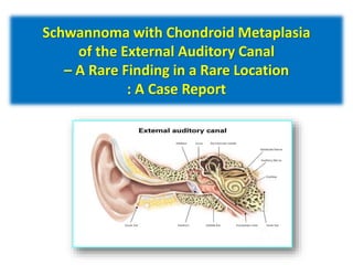
In epidemiology, case reports are defined as singular reports on one individual patient. In our presentation, we explain about a case report titled Schwannoma with Chondroid Metaplasia of the External Auditory Canal.
This document discusses brain biopsies, including their history, indications, and procedure. Brain biopsies are used to determine the cause of diffuse or multifocal brain diseases when the specific diagnosis cannot be established by other means. Key indications include infections, vasculitides, pediatric neurodegenerative diseases, and atypical dementias. The procedure involves making a burr hole in the skull under general anesthesia and removing a small sample of brain tissue for analysis. Potential complications include hemorrhage, swelling, neurological deficits, seizures, and infections. A multidisciplinary team approach is recommended for planning and evaluating brain biopsies.
Salivary gland imaging and sialochemistry (radiological and biochemistry)Coco Mathew
A through guide in understanding salivary gland disorders, it radiographic interpretation and components of saliva, its function along with treatment aspects.
This document discusses principles and techniques of biopsy. It outlines the steps involved in evaluating a patient with an oral lesion including taking a health history, examining the lesion history, performing a clinical and radiographic exam, and potentially obtaining laboratory tests. It describes different types of biopsies including incisional, excisional, and intraosseous biopsies. Principles of biopsy such as anesthesia, hemostasis, handling specimens, and closure techniques are also outlined. The goal is to obtain sufficient tissue for accurate histopathologic evaluation.
This document provides an overview of sialendoscopy procedures. It begins with the anatomy of the parotid and submandibular salivary glands. It then discusses sialolithiasis (salivary gland stones), the indications for sialendoscopy including removal of stones and treatment of strictures, and the technique of sialendoscopy including identification of ducts, instrumentation, and methods for removing stones such as grasping, lithotripsy, and extracorporeal shockwave lithotripsy. Complications are also briefly mentioned.
Sialendoscopy is a minimally invasive endoscopic technique used to both diagnose and treat salivary gland disorders. Some key developments in sialendoscopy include Katz introducing a flexible endoscope in 1991 to diagnose and treat salivary gland stones. Sialendoscopy uses thin endoscopes and mini instruments to visualize and treat obstructive pathologies like sialoliths, strictures, ductal polyps and mucous plugs in the major salivary glands. It allows for direct visualization of the ductal system and less invasive interventional procedures compared to open surgery. Complications can include local pain, lingual nerve changes, infection and ductal strictures.
Sialendoscopy is a minimally invasive endoscopic technique used to both diagnose and treat obstructive pathologies of the salivary glands. It involves inserting a sialendoscope into the ducts of the parotid and submandibular glands under local anesthesia. This allows direct visualization of the ductal system to identify structures causing obstruction like sialoliths, strictures, or polyps. Sialendoscopy can remove small stones and dilate strictures in a single procedure. It has advantages over surgery as it is less invasive, avoids gland removal, and maintains gland function post-procedure. Potential complications include ductal perforation, lingual nerve paresthesia, and stenosis.
This case report describes the use of cone beam computed tomography (CBCT) sialography to diagnose and treat a patient's salivary gland condition. A 60-year-old female presented with chronic pain in her left parotid gland. Previous investigations including plain films and ultrasound were non-diagnostic. CBCT sialography identified two sialoliths, one in the deep lobe and one in the superior lobe. The patient underwent sialoendoscopy where the two sialoliths were successfully retrieved, providing pain relief. CBCT sialography is a novel technique that overcomes limitations of other imaging modalities. It allows for precise localization of radiolucent sialoliths to guide minimally
External ear canal cholesteatoma after ventilation tube insertionPrasanna Datta
1. This case report describes an external ear canal cholesteatoma that developed in a 16-year-old boy after he underwent mastoidectomy and ventilation tube insertion surgery for acute purulent mastoiditis.
2. Computed tomography imaging showed a soft tissue mass completely filling the external ear canal with bone erosion. The mass was connected to the margins of the tympanic membrane incision from the ventilation tube placement.
3. The patient underwent revision surgery to remove the cholesteatoma and ventilation tube. Regular follow-ups showed no recurrence of the disease. The report discusses potential mechanisms for how external ear canal cholesteatomas may develop after surgeries involving ventilation tube placement
This document discusses malignant tumors of the paranasal sinuses and skull base. It covers:
1) Epidemiology, risk factors, histology, clinical presentation and staging of these tumors.
2) Preoperative evaluation including imaging, biopsy and the role of PET-CT in accurate staging.
3) Treatment approaches including surgery, radiation therapy and chemotherapy, both as single modalities and in combination.
4) Post-treatment outcomes and factors influencing survival. Management of the neck is also addressed.
This document describes contact endoscopy (CE), a non-invasive optical technique that uses a magnifying endoscope to provide real-time visualization and examination of the cellular architecture and vascular patterns of mucosal tissues. CE allows in vivo assessment of precancerous and cancerous lesions without biopsy. Several contact endoscope models from Karl Storz are described. The document outlines CE's applications in examining various head and neck tissues and its ability to detect abnormalities. The benefits of CE include its non-invasive nature, ability to examine large areas quickly, and provision of immediate results.
1. A 31-year-old male presented with a swelling in the right parotid region for 1 year. On examination, a 2cmx3cm firm, non-tender swelling was found in the right parotid gland.
2. A provisional diagnosis of pleomorphic adenoma of the right parotid gland was made.
3. The anatomy and clinical features of the parotid gland were discussed, along with differential diagnoses and management of parotid tumors.
Cholesteatoma is an abnormal skin growth in the middle ear space or mastoid bone that is non-cancerous. It can be present at birth or develop later due to repeated ear infections which cause negative pressure and inward pulling of the eardrum. Left untreated, cholesteatomas can grow large, cause hearing loss and facial paralysis, and potentially spread to the brain. Diagnosis involves examination of the ear and sometimes CT scans or audiology tests. Treatment is primarily surgical removal of the cholesteatoma and repair of any ear damage to preserve hearing.
This document discusses vestibular schwannoma (VS), also known as acoustic neuroma. It provides details on:
1. The anatomy, pathology, genetics, histology, symptoms, diagnosis and treatment options for VS. Common symptoms include unilateral hearing loss and tinnitus. Diagnosis involves audiometry, imaging like MRI and evaluation of cranial nerve function.
2. Surgical removal and stereotactic radiotherapy are common treatment options. Observation may be appropriate for older patients or small, slow-growing tumors not affecting hearing or brain function.
3. Comprehensive details are given on the staging, imaging, and growth patterns of VS to aid in predicting hearing loss, nerve damage and
This document defines and classifies odontogenic cysts, which are epithelium-lined sacs that arise from odontogenic epithelium. It discusses the most common types, including periapical (radicular) cysts, dentigerous cysts, odontogenic keratocysts, glandular odontogenic cysts, and calcifying odontogenic cysts. For each cyst type, it describes characteristics such as prevalence, location, radiographic appearance, histopathology, treatment involving enucleation or marsupialization, and prognosis. It also discusses the basal cell nevus syndrome that can be associated with odontogenic keratocysts.
The document discusses cholesteatoma, including:
1) Its composition of keratin debris, keratinizing squamous epithelium matrix, and subepithelial connective tissue matrix.
2) Its pathogenesis involving retraction pocket theory of retraction of the tympanic membrane leading to proliferation.
3) Its classification including attic, sinus, and pars tensa cholesteatomas depending on location of retraction.
Mr. Manjunath Beth presents on tumors of the ear. He discusses that tumors can occur in the external ear, ear canal, middle ear, or inner ear. The document focuses on three main types of ear tumors: cancers of the ear canal which can be benign or malignant; glomus tumors of the middle ear; and vestibular schwannomas (acoustic neuromas) of the inner ear. For each type of tumor, the document describes their characteristics, symptoms, diagnostic methods such as biopsy and imaging, and treatment options including surgery and radiation therapy. The goal is to educate on properly diagnosing and treating tumors based on their location in the ear.
Presentation1.pptx, radiological imaging of choleteatoma.Abdellah Nazeer
Cholesteatoma is a non-neoplastic keratinizing squamous epithelial lesion that grows in the middle ear. It has an osteolytic potential due to collagenase production. The document discusses the pathogenesis, classification, imaging features, and complications of cholesteatoma. Key findings on imaging include hyperintensity on diffusion weighted MRI sequences indicating restricted diffusion, which is highly specific for cholesteatoma. Complications include ossicular erosion, facial nerve paralysis, and labyrinthine fistula formation.
The document discusses the anatomy and physiology of the pituitary gland and sphenoid sinus. It then covers pituitary adenomas including classification, clinical features, investigations and approaches to transsphenoid hypophysectomy surgery. Specific surgical steps are outlined including opening the sphenoid sinus, removing the bony walls, and dissecting the dura, capsule and gland tissue to remove an adenoma. Potential complications of the surgery are also mentioned.
This document discusses the history and developments in neuroendoscopy. It describes how Walter Dandy is considered the father of neuroendoscopy for using an endoscope to inspect the ventricles in 1922. Significant technological developments include the invention of fiber optic cables in the 1960s and charge-coupled device cameras in 1969, which improved image quality for endoscopy. The document outlines the use of neuroendoscopy for diagnostic and therapeutic applications like biopsy of tumors and relief of hydrocephalus through third ventriculostomy.
This document discusses the anatomy, clinical presentation, management, and treatment of cancers affecting the paranasal sinuses. It begins with an overview of the anatomy of the different paranasal sinuses and their relationships to surrounding structures. It then discusses the clinical presentation of sinus cancers, which can include nasal obstruction, eye symptoms, facial pain or numbness. The document reviews imaging techniques like CT and MRI to evaluate tumor extent. Management options are also summarized, including surgical resection with or without radiation therapy or chemotherapy based on tumor size and spread. Post-operative radiation techniques like IMRT and proton beam therapy are mentioned. Overall survival rates from historical studies on paranasal sinus cancer treatment are provided.
This document discusses various surgical approaches for pituitary adenomas. It begins with a brief history of pituitary surgery dating back to 1893. There are two main categories of approaches - extracranial (transnasal, transmaxillary) and intracranial (subfrontal, subtemporal). The transnasal transsphenoidal approach is the most common due to its advantages. Extended approaches are sometimes needed for large tumors. Complications can include CSF leaks, hormonal issues, and visual changes. Landmarks, techniques, and closure methods are outlined for different procedures. Indications for alternative approaches like transcranial are also summarized.
Tumors of the middle ear can be benign or malignant. Benign tumors include glomus tumors, which are the most common benign neoplasm of the middle ear. They arise from paraganglionic cells and present with slowly progressive conductive hearing loss and pulsatile tinnitus. Surgical removal is the primary treatment. Malignant tumors can be primary, such as squamous cell carcinoma which occurs more often in older smoking males, or secondary from adjacent sites. Squamous cell carcinoma presents as an ulcerating mass and treatment involves radical mastoidectomy and neck dissection. Malignant tumors can also metastasize to distant sites. CT, MRI, and biopsy are used in evaluation and diagnosis of
Glomus tumours, also known as paragangliomas or chemodectomas, are rare benign tumours that arise from parasympathetic nerve tissue. They most commonly occur in the middle ear (glomus tympanicum), jugular bulb (glomus jugulare), or vagus nerve in the neck (glomus vagale). Presenting symptoms depend on location but often include pulsatile tinnitus and conductive hearing loss. Diagnosis involves imaging such as CT or MRI to determine size and involvement of structures. Surgical resection is the main treatment, with preoperative embolization for larger tumours, while radiotherapy may be used for inoperable or residual cases.
This document discusses various cytology techniques including exfoliative cytology, abrasive cytology, and fine needle aspiration cytology (FNAC). It focuses on the use of FNAC and ultrasound guidance for sampling deep seated lesions. FNAC is a useful tool for diagnosing lesions without significant trauma. Ultrasound guidance allows real-time visualization of the needle for precise and safe sampling of deep lesions. Complications are usually minor bleeding or infection when the technique is performed appropriately. Cytology provides valuable diagnostic information with minimal risk to patients.
The document discusses salivary gland disorders. It begins with definitions and classifications of salivary glands. It then discusses the anatomy, functions, and disorders of the parotid, submandibular, and sublingual salivary glands. Diagnostic aids are outlined including clinical history, physical examination, imaging such as CT, MRI, ultrasound and sialography. Cystic conditions of the minor salivary glands such as mucoceles are also summarized. Disorders are classified and inflammatory, obstructive, neoplastic and other conditions are described.
Facial nerve decompression is a surgical procedure to relieve pressure and reduce compression on the facial nerve fibers. It involves opening the bony canal and nerve sheath. There are several surgical approaches depending on the location of injury, including transmastoid, middle cranial fossa, and translabyrinthine. The middle cranial fossa approach provides exposure of the internal auditory canal and labyrinthine segments without risk of hearing loss. Landmarks are used to identify the various segments of the facial nerve during decompression. The goal is to maximize functional recovery from facial paralysis.
Total maxillectomy is a surgical procedure to remove the entire maxilla bone. It was first described in the 1820s and approaches have been refined over time. It is indicated for malignant tumors involving the maxilla, extensive benign tumors, or fungal/granulomatous infections. Contraindications include poor general health, bilateral orbital involvement, or skull base extension. Potential complications include bleeding, infection, epiphora, skin graft breakdown, numbness, and atrophic rhinitis. Careful surgical planning and follow up are required due to significant reconstruction and rehabilitation needs.
Carcinoma neuroendocrino del setto un rarissimo tumore nasaleMerqurio
This document presents a case report of a 73-year-old male with a rare small cell neuroendocrine carcinoma arising from the nasal septum. The patient presented with right-sided nasal obstruction, hyponasal speech, and recurrent epistaxis. Imaging showed a mass in the right nasal cavity extending to surrounding structures. The tumour was removed endoscopically and pathology confirmed a small cell neuroendocrine carcinoma. Due to the aggressive nature of this tumour type, the patient received combined chemotherapy and radiation in addition to surgery. At a 10-month follow-up the patient remained free of disease. Small cell neuroendocrine carcinoma of the nasal cavity is an extremely rare and aggressive tumour that is difficult to diagnose but requires
Carcinoma neuroendocrino del setto un rarissimo tumore nasaleMerqurio
This document presents a case report of a 73-year-old male with a rare small cell neuroendocrine carcinoma arising from the nasal septum. The patient presented with right-sided nasal obstruction, hyponasal speech, and recurrent epistaxis. Imaging showed a mass in the right nasal cavity extending to surrounding areas. The tumour was removed endoscopically and found to be a small cell neuroendocrine carcinoma based on histology and immunohistochemistry. Due to the aggressive nature of this tumour type, the patient received combined chemotherapy and radiation in addition to surgery. At a 10-month follow-up the patient remained free of disease. Small cell neuroendocrine carcinoma of the nasal cavity is an extremely rare and aggressive
This document provides information on ear carcinoma, including:
1) It describes the anatomy of the external, middle, and inner ear.
2) Diagnostic workup involves CT, MRI, and biopsy to establish diagnosis and determine extent of disease.
3) Treatment depends on location, with early-stage external ear cancers often treated with radiation alone, while surgery plus radiation is recommended for more advanced or middle ear/mastoid cancers.
Neurofibroma of Nasal Cavity and Nasopharynx_Crimson PublishersCrimsonPublishersAICS
A 17-year-old male presented with progressive bilateral nasal obstruction, nasal discharge, headache, and hyposmia. Diagnostic nasal endoscopy revealed a firm, huge mass filling the bilateral posterior choana and attached to the posterior edge of the nasal septum. CT scan demonstrated a large soft-tissue density mass in the right nasal cavity extending into the right choana and nasopharynx. The mass was excised endoscopically and found to be a neurofibroma arising from the right nasal septum on histological examination. Neurofibroma is a rare benign peripheral nerve sheath tumor that can occur in the nasal cavity and paranasal sinuses. Complete surgical excision is usually curative for solitary neuro
Metastasis of Neck Node with Unknown Primary Himanshu Soni
1) An unknown primary is defined as squamous cell carcinoma presenting in cervical lymph nodes with no identifiable primary tumor site after examination. This clinical entity is known as carcinoma of unknown primary (CUP).
2) Evaluation involves physical examination, imaging like PET-CT, and panendoscopy with biopsies of suspicious sites to identify the occult primary tumor. Bilateral tonsillectomy and tongue base biopsy can identify occult tumors in the tonsillar crypts in many cases.
3) Treatment depends on tumor stage but often involves combined modality treatment with surgery, radiation, and/or chemotherapy aimed at locoregional control while minimizing morbidity.
1. Nasopharyngeal angiofibroma is a rare, benign tumor that occurs mostly in adolescent males and arises from the posterior nasal cavity.
2. It is locally invasive and can extend into surrounding areas like the nasal cavity, paranasal sinuses, and cranial cavity, causing symptoms like nasal obstruction, epistaxis, and cranial nerve palsies.
3. Diagnosis involves imaging like CT and MRI to determine the extent of the tumor. Surgical excision is the primary treatment but carries a risk of heavy bleeding, so preoperative embolization of feeding vessels is often used to reduce bleeding during surgery.
This document presents a case study of ameloblastoma, a benign odontogenic tumor. It defines ameloblastoma, discusses its epidemiology and classifications. The document describes the clinical features, radiological findings, differential diagnosis, management and prognosis of ameloblastoma. It then presents clinical case examples, including details of patients' examinations, radiographs, surgical procedures and histopathology reports. The conclusion emphasizes the need for long-term follow-up due to the high recurrence rate of ameloblastoma.
Case presented in National ENT conference in serena hotel, Islamabad in Dec. 2015
Author
Dr. Ghulam Saqulain HOD
Dr. Jawad Ahmed Assoc. surgeon
Dr. Zaimal Shahan PGT
This document discusses osteomas, which are benign bone tumors that commonly arise in the skull. It provides details on the typical presentation, locations, demographic factors, investigations and surgical approaches for osteomas. It then presents a case of a 10-year-old girl who presented with slow-growing swellings in her right eyebrow and hairline that were found to be osteomas on CT scan. She underwent a bicoronal surgical approach to completely excise the osteomas due to their size and location, with an excellent postoperative outcome and no recurrence.
The document discusses examination, radiographic investigation, and pathologies of the maxillary sinus. Examination involves extraoral and intraoral inspection and palpation to check for tenderness, swelling, or discharge from the sinus. Radiographic views like panoramic x-ray, CT scan, and MRI provide images of the sinus. Common pathologies include sinusitis (acute, chronic), cysts, tumors, trauma like fractures, and systemic diseases affecting the sinus.
Cervical Sympathetic chain ganglioneuroma : case report and review of literatureiosrphr_editor
The IOSR Journal of Pharmacy (IOSRPHR) is an open access online & offline peer reviewed international journal, which publishes innovative research papers, reviews, mini-reviews, short communications and notes dealing with Pharmaceutical Sciences( Pharmaceutical Technology, Pharmaceutics, Biopharmaceutics, Pharmacokinetics, Pharmaceutical/Medicinal Chemistry, Computational Chemistry and Molecular Drug Design, Pharmacognosy & Phytochemistry, Pharmacology, Pharmaceutical Analysis, Pharmacy Practice, Clinical and Hospital Pharmacy, Cell Biology, Genomics and Proteomics, Pharmacogenomics, Bioinformatics and Biotechnology of Pharmaceutical Interest........more details on Aim & Scope).
Esthesioneuroblastoma (ENB) is a rare malignant tumor that arises from the olfactory epithelium in the nasal cavity. Imaging such as CT and MRI are used to determine the extent of the tumor. Histopathological examination shows small round blue cells forming rosettes. Treatment involves surgery such as craniofacial resection along with radiation therapy. For advanced disease, chemotherapy may be given as part of multimodality treatment. With aggressive treatment, 5-year survival rates for ENB exceed 60%.
This document presents a case report of a rare occurrence of basal cell adenoma in the palate of a 25-year-old male patient. It describes the clinical findings and diagnostic workup including imaging, biopsy, histopathological examination, and immunohistochemical analysis of the excised tumor. The pathological features were consistent with basal cell adenoma. The tumor was successfully removed surgically and the patient recovered well, with the tumor representing an unusual site for this rare minor salivary gland tumor.
1) Parotid gland tumors are relatively uncommon, accounting for less than 1% of reported malignancies. Pleomorphic adenoma is the most common parotid gland tumor, comprising 60% of cases.
2) The document outlines the classification, presentation, diagnostic evaluation, surgical and treatment options for parotid gland tumors. It also presents representative cases from the author's experience.
3) Superficial parotidectomy with preservation of the facial nerve is the most common surgical intervention undertaken and was performed in 95% of cases. Post-operative complications included transient or permanent facial nerve weakness.
This document provides tips for using a PowerPoint presentation on lymphangioma and cystic hygroma. It recommends freely editing and modifying the slides. It suggests showing blank slides first to elicit student responses before presenting content. Repeating this process of blank slide then content slide three times promotes active learning. The presentation can also be used for self-study. The final slides provide links to access the full presentation on mobile devices or download the collection.
Vestibular schwannoma, also known as acoustic neuroma, is a benign tumor that arises from the vestibular nerve and presents with symptoms of hearing loss, tinnitus, and imbalance. It is typically diagnosed using MRI and treated with surgery, radiation therapy, or observation depending on size and symptoms. Glomus tumors are rare, vascular tumors of the middle ear that cause conductive hearing loss and a pulsatile bruit and are diagnosed using CT, MRI and angiography before being surgically excised.
Slides prepared and compiled by highly experienced ENT teacher, Dr. Krishna Koirala from Nepal , for teaching undergraduate and postgraduate ENT students in the field of otorhinolaryngology.
A clear and concise explanation of the basic concepts in the subject matter concerned.
He is the Head of department with a sound knowledge in the field of ENT to teach both undergraduate and postgraduate ENT students
Tumors of ear including external canal, auricle, middle canal (GLOMUS TUMOR), inner ear ( ACOUSTIC NEUROMA).
Description includes definition, etiological factors, clinical menifestations and management including medical management, surgical management and nursing management.
This document summarizes a study on using ultrasound to assess cystic neck lesions prior to treatment. The study examined 120 patients with neck masses and found cystic lesions in 7.5% of cases, including branchial cysts and lymphangiomas. Ultrasound was able to differentiate cystic from solid lesions and characterize features like contents, borders, and location. The study concluded ultrasound is a useful non-invasive tool for evaluating cystic neck lesions before treatment due to its low cost, lack of radiation, and ability to detect diagnostic imaging characteristics of various cyst types.
Similar to EPIDEMIOLOGY AND BIOSTATISTICS (case report) (20)
How to Fix the Import Error in the Odoo 17Celine George
An import error occurs when a program fails to import a module or library, disrupting its execution. In languages like Python, this issue arises when the specified module cannot be found or accessed, hindering the program's functionality. Resolving import errors is crucial for maintaining smooth software operation and uninterrupted development processes.
it describes the bony anatomy including the femoral head , acetabulum, labrum . also discusses the capsule , ligaments . muscle that act on the hip joint and the range of motion are outlined. factors affecting hip joint stability and weight transmission through the joint are summarized.
ISO/IEC 27001, ISO/IEC 42001, and GDPR: Best Practices for Implementation and...PECB
Denis is a dynamic and results-driven Chief Information Officer (CIO) with a distinguished career spanning information systems analysis and technical project management. With a proven track record of spearheading the design and delivery of cutting-edge Information Management solutions, he has consistently elevated business operations, streamlined reporting functions, and maximized process efficiency.
Certified as an ISO/IEC 27001: Information Security Management Systems (ISMS) Lead Implementer, Data Protection Officer, and Cyber Risks Analyst, Denis brings a heightened focus on data security, privacy, and cyber resilience to every endeavor.
His expertise extends across a diverse spectrum of reporting, database, and web development applications, underpinned by an exceptional grasp of data storage and virtualization technologies. His proficiency in application testing, database administration, and data cleansing ensures seamless execution of complex projects.
What sets Denis apart is his comprehensive understanding of Business and Systems Analysis technologies, honed through involvement in all phases of the Software Development Lifecycle (SDLC). From meticulous requirements gathering to precise analysis, innovative design, rigorous development, thorough testing, and successful implementation, he has consistently delivered exceptional results.
Throughout his career, he has taken on multifaceted roles, from leading technical project management teams to owning solutions that drive operational excellence. His conscientious and proactive approach is unwavering, whether he is working independently or collaboratively within a team. His ability to connect with colleagues on a personal level underscores his commitment to fostering a harmonious and productive workplace environment.
Date: May 29, 2024
Tags: Information Security, ISO/IEC 27001, ISO/IEC 42001, Artificial Intelligence, GDPR
-------------------------------------------------------------------------------
Find out more about ISO training and certification services
Training: ISO/IEC 27001 Information Security Management System - EN | PECB
ISO/IEC 42001 Artificial Intelligence Management System - EN | PECB
General Data Protection Regulation (GDPR) - Training Courses - EN | PECB
Webinars: https://pecb.com/webinars
Article: https://pecb.com/article
-------------------------------------------------------------------------------
For more information about PECB:
Website: https://pecb.com/
LinkedIn: https://www.linkedin.com/company/pecb/
Facebook: https://www.facebook.com/PECBInternational/
Slideshare: http://www.slideshare.net/PECBCERTIFICATION
How to Manage Your Lost Opportunities in Odoo 17 CRMCeline George
Odoo 17 CRM allows us to track why we lose sales opportunities with "Lost Reasons." This helps analyze our sales process and identify areas for improvement. Here's how to configure lost reasons in Odoo 17 CRM
हिंदी वर्णमाला पीपीटी, hindi alphabet PPT presentation, hindi varnamala PPT, Hindi Varnamala pdf, हिंदी स्वर, हिंदी व्यंजन, sikhiye hindi varnmala, dr. mulla adam ali, hindi language and literature, hindi alphabet with drawing, hindi alphabet pdf, hindi varnamala for childrens, hindi language, hindi varnamala practice for kids, https://www.drmullaadamali.com
This presentation includes basic of PCOS their pathology and treatment and also Ayurveda correlation of PCOS and Ayurvedic line of treatment mentioned in classics.
Beyond Degrees - Empowering the Workforce in the Context of Skills-First.pptxEduSkills OECD
Iván Bornacelly, Policy Analyst at the OECD Centre for Skills, OECD, presents at the webinar 'Tackling job market gaps with a skills-first approach' on 12 June 2024
BÀI TẬP DẠY THÊM TIẾNG ANH LỚP 7 CẢ NĂM FRIENDS PLUS SÁCH CHÂN TRỜI SÁNG TẠO ...
EPIDEMIOLOGY AND BIOSTATISTICS (case report)
1. SAFIYYAH BINTI ROSLAN (043827)
SAHIRAH BINTI SARIFF (043445)
FARAH ADRIANA BINTI MOHD ROSMADI (043636)
NUR HAYATI BINTI NISAR AHMAD (043209)
NURASYIKIN BINTI RAHMAT (042961)
2. Schwannoma with Chondroid Metaplasia
of the External Auditory Canal
– A Rare Finding in a Rare Location
: A Case Report
3. ABSTRACT
Background
• Schwannomas are uncommon tumors of the external auditory canal.
• In the English literature, very few cases of schwannomas originating in the external
auditory canal were reported and none of them showed chondroid metaplasia.
• This is the first case of schwannoma with chondroid metaplasia in this location was
reported by Bennani et. Al (2018).
Case presentation
• In this report, it has been described a 22-year-old white man who presented with an external
auditory slow growing mass.
• A computed tomography scan of the temporal bone demonstrated a well-circumscribed, soft
tissue mass narrowing most of the external auditory canal.
• A surgical biopsy was performed and the histological examination showed a schwannoma
with chondroid metaplasia.
4. Schwannoma should be considered in the differential
diagnosis of benign or malignant tissue masses involving the
external ear canal.
Conclusion
5. INTRODUCTION
Schwannomas are slow growing benign tumors arising from Schwann cells
of peripheral nerve sheaths.
Between 25 and 45% of extracranial schwannomas occur in the head and
neck region.
They are frequently located at the internal acoustic meatus arising from the
vestibular nerves within the cranial vault.
They are uncommon in the external auditory canal
Only 10 cases have been reported in the international literature according to
the best of their knowledge.[1-10]
6. So far, chondroid metaplasia which is seen in this case has not been reported.
Bennani et al (2018) report the first case of schwannoma with chondroid
metaplasia in the external auditory canal with the aim of shedding more light
on this tumor in this exceptional location, and on the fact that
schwannoma should be considered in the differential diagnosis of benign or
malignant tissue masses involving the external ear canal, even the clinical
and radiological findings are somewhat nonspecific in this location.
7. METHOD
1) Physical examination
- A pale and firm mass arising from the inferior wall of right external auditory
canal were examined.
2) Neurological examination
- Did not show any lesions in CNS or PNS.
3) Laboratory analyses
- Hematological and biochemical tests were carried out to help make a
diagnosis (Table 1).
4) Computed tomography (CT) scan
- CT scan of the temporal bone was done.
- a well circumscribed, soft tissue mass narrowing most of the external auditory
canal was demonstrated (Fig.1).
8. 5) Excisional biopsy
- A procedure where the entire abnormality or area of interest is removed for
examination under a microscope.
- The patient undergoes an excisional biopsy of the mass, under local anesthesia
(a numbing medicine).
- After the biopsy, the tissue sample was examined under a microscope for:
a) Histology
- Tumor cells were stained with hematoxylin and eosin stain
- The tumor was composed of spindle cells arranged in interlacing fascicles
(Fig. 2).
- A chondroid metaplasia was also seen in the tissue sample (Fig. 3).
b) Immunochemistry
- Tumor cells were strongly stained with PS100 (Fig.4).
9. TABLE 1
Hematology Value
White Blood Cell 7.2 × 10⁹/L
Red Blood Cell 4.34 × 10¹²/L
Hemoglobin 15.2 g/L
Hematocrit 42%
Mean
Corpuscular
Volume
90.6 fL
Platelets 210 × 109/L
Biochemistry
Total Protein 80.5 g/L
C-Reactive
Protein
2 mg/L
Urea 0.3 g/L
Creatinine 7 mg/L
Figure 1
10. Histology (Fig. 3)
Chondroid metaplasia was
seen.Hematoxylin and eosin
stain × 400
Histology (Fig. 2)
The tumor was composed of
spindle cells arranged in
interlacing fascicles.
Hematoxylin and eosin stain ×
100
Immunochemistry (Fig. 4)
tumor cells stain strongly for
PS100, × 400
11. RESULT
• After multiple examination and test, the diagnosis of
schwannoma with chondroid metaplasia was made.
• The tumor mass was removed and after 8 month there
is no sign of local recurrence of the tumor.
12. DISCUSSION
Schwannomas of the head and neck are common, and are mostly seen arising from the
internal acoustic meatus commonly associated with large nerve trunks.
But, those arise from the external auditory canal are very rare [3].
Most of the extracranial schwannomas in the head and neck originate from cutaneous
or muscular branches of the cervical or brachial plexus. Cutaneous sensory nerves that
are covered by Schwann cells, from which schwannoma may originate, supply the
external auditory meatus and canal.
In the present case the tumor was located mainly at the inferior canal wall, which was
supplied by the auricular nerve [1].
This cases was conducted in the age ranges 18-59 years patients with mass in their
external auditory canal.
13. The clinical presentation of schwannoma is usually a slow growing and
asymptomatic mass.
In the external auditory canal the clinical presentation may appear as recurrent
external otitis and a mild conductive hearing loss secondary to obstruction of the
canal from the tumor mass [1].
Neurogenic symptoms such as pain or paresthesia are uncommon [5].
Schwannomas are encapsulated and therefore they can be easily dissected from the
surrounding tissues. Thus, one cases reported with erosion of the bony canal wall [3].
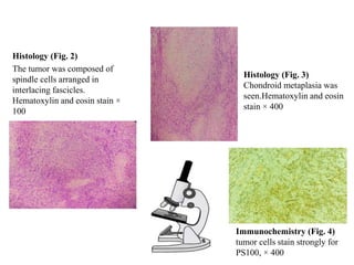
On histologic examination, the tumor is characterized by streams of elongated
spindle cells, with the elongated nuclei often arrayed in a palisade pattern.
Schwannomas should be differentiated from other spindle cell tumors such as
neurofibroma, leiomyoma, and desmoplastic melanoma.
14. Neurofibromas are not encapsulated , usually multicentric , which is an
important clinical distinction from schwannomas, and may be accompanied by a
special entity called von Recklinghausen’s disease.
Radiologic imaging by CT shows schwannomas to be well-circumscribed,
homogenous masses.
A CT scan is very useful in making a decision about the extent of the lesion,
integrity of the tympanic membrane, and the type of surgical approach [5].
Treatment is complete excision of the tumor via either transmeatal or post-aural
approach.
The choice of approach will depend on tumor size, location, and relation to
surrounding structures [3].
When complete excision is performed local recurrence is rare [1].
A transmeatal approach was performed in the present case and a good cleavage
plane provided an en bloc resection (EBR) with preservation of surrounding
structures [5].
15. CONCLUSION
A differential diagnosis should be made with respect to a number of other soft
tissue neoplasms such as fibroma, chondroma, and leiomyoma.
A definitive diagnosis should be based on the histological and
immunohistochemical findings.
Schwannoma should be considered in the differential diagnosis of benign or
malignant tissue masses involving the external ear canal, although, in this location,
the clinical and radiological findings are somewhat nonspecific and rare.
16. REFERENCES
1. Gross M, et al. Schwannoma of the external auditory canal. Auris Nasus Larynx.
2005;32(1):77–9.
2. Morais D, et al. Schwannoma of the external auditory canal: an exceptional location.
Acta Otorrinolaringol (Engl Ed). 2007;58(4):169–70.
3. Bakshi SS, Shankar K, Parida PK. A large schwannoma of external auditory canal: an
unusual case. Kulak Burun Boğaz Ihtis Derg. 2015;25(4):229.
4. Jovanovic MB, Djeric D, Poljovka R, Milenkovic S. Obliterative external ear canal
schwannoma. Int Adv Otol. 2009;5:394–8.
5. Topal O, Erbek SS, Erbek S. Schwannoma of the external auditory canal: a case report.
Head Face Med. 2007;3(1):6.
6. Kumar D, Somavanshi S, Kumar H, Agrawal A, Singh H. Schwannoma of the External
Auditory Canal: A Rare Location. Otorhinolaryngol Clin Int J. 2015; 7(3):147–8.
7. Wu C-M, Hwang C-F, Lin CH, Su C-Y. External ear canal schwannoma: an unusual
case report. J Laryngol Otolaryngol. 1993;107:829–30.
8. Lewis WB, Mattucci KF, Smilari T. Schwannoma of external auditory canal: an
unusual finding. Int Surg. 1995;80:287–90.
17. 9. Harcourt JP, Tungekar MF. Schwannoma of the external auditory canal. J Laryngol
Otol. 1995;109:1016–8.
10. Galli J, d’Ecclesia A, La-Rocca LM, Almadori G. Giant schwannoma of external
auditory canal: a case report. Otolaryngol Head Neck Surg. 2001; 124:473–4.
11. Bennani. A, Karich. N, Kamaoui. I, Chraibi. M, Abbaoui. S. (2017). Schwannoma
with chondroid metaplasia of the external auditory canal – a rare finding in a rare
location: a case report. Journal of Medical Case Reports. Retrieved
fromhttps://doi.org/10.1186/s13256-018-1584-4
12. Whaley. T. (n,d). Incisional & Excisional Biopsy. Retrieved from
https://www.oncolink.org/cancer-treatment/procedures-diagnostic-tests/biopsy-
procedures/incisional-excisional-biopsy
13. Mayo Clinic Staff. (n,d). Cancer blood tests: Lab tests used in cancer diagnosis.
Retrieved from https://www.mayoclinic.org/diseases-conditions/cancer/in-depth/cancer-
diagnosis/art-20046459
14. Gross M, et al. Schwannoma of the external auditory canal. Auris Nasus Larynx.
2005;32(1):77–9.
15. Bakshi SS, Shankar K, Parida PK. A large schwannoma of external auditory canal: an
unusual case. Kulak Burun Boğaz Ihtis Derg. 2015;25(4):229.
16. Topal O, Erbek SS, Erbek S. Schwannoma of the external auditory canal: a case
report. Head Face Med. 2007;3(1):6.