Chondrocyte Cell Profile Report
•Download as DOCX, PDF•
2 likes•2,915 views

Chondrocytes are cells found in cartilage that secrete collagen and proteoglycans to form the cartilaginous matrix. There are three types of cartilage - elastic, fibrocartilage, and hyaline cartilage - containing different numbers of chondrocytes. Chondrocytes play a key role in endochondral ossification, a process by which bones grow and mature. During this process, chondrocytes proliferate, mature, and eventually die to be replaced by osteoblasts that lay down new bone. The differentiation of mesenchymal stem cells into chondrocytes is regulated by signaling factors such as BMP and Sox9. Chondrocytes have low regener

Recommended
More Related Content
What's hot
What's hot (20)
Viewers also liked
Viewers also liked (16)
Similar to Chondrocyte Cell Profile Report
Similar to Chondrocyte Cell Profile Report (20)
Chondrocyte Cell Profile Report
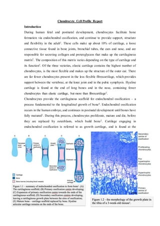
- 1. Chondrocyte Cell Profile Report Introduction During human fetal and postnatal development, chondrocytes facilitate bone formation via endochondral ossification, and continue to provide support, structure and flexibility in the adult1. These cells make up about 10% of cartilage, a loose connective tissue found in bone joints, bronchial tubes, the ears and nose, and are responsible for secreting collagen and proteoglycans that make up the cartilaginous matrix2. The composition of this matrix varies depending on the type of cartilage and its function3. Of the three varieties, elastic cartilage contains the highest number of chondrocytes, is the most flexible and makes up the structure of the outer ear. There are far fewer chondrocytes present in the less flexible fibrocartilage, which provides support between the vertebrae, at the knee joint and in the pubic symphysis. Hyaline cartilage is found at the end of long bones and in the nose, containing fewer chondrocytes than elastic cartilage, but more than fibrocartilage2. Chondrocytes provide the cartilaginous scaffold for endochondral ossification – a process fundamental to the longitudinal growth of bone4. Endochondral ossification occurs in the human embryo, and continues in postnatal development until bones have fully matured1. During this process, chondrocytes proliferate, mature and die, before they are replaced by osteoblasts, which build bone2. Cartilage engaging in endochondral ossification is referred to as growth cartilage, and is found at the
- 2. primary and secondary centres of ossification: the diaphysis and epiphyseal growth plates, seen in Figure 1.11. Chondrocytes in growth cartilage can be observed moving through separate zones (see Figure 1.2), indicating which stage of endochondral ossification they are in. Resting chondrocytes are round in shape, surrounded by extracellular matrix (ECM) and are most remote from the ossification centre. Bone morphogenic protein (BMP) signaling drives proliferation of chondrocytes1, which become flattened in shape, and can also be characterized by their expression of Sox5, Sox6 and Sox95. Adjacent to the zone of proliferation are pre-hypertrophic chondrocytes, which secrete Indian hedgehog (IHH) to aid both proliferation and hypertrophy1. These chondrocytes then increase about 20 times their original size6, and are referred to as hypertrophic, and are responsible for mineralizing the ECM surrounding them. Finally, the chondrocytes die (likely via apoptosis7) and part of the ECM is removed, allowing the entry of osteoblasts to construct new bone4. Figure 1.1 shows that hyaline articular cartilage remains at the end of long bones, aiding in lubrication, support and movement of joints. To initiate human embryonic development, two gametes (one sperm and one oocyte) must fuse to form a diploid cell – a process known as fertilization8. This single cell undergoes mitotic division, producing new cells referred to as blastomeres. By the 16- cell morula stage, there are already transcriptional differences between the cells, although they are still totipotent. 5 days post-fertilization, a blastocyst has been formed, showing a distinct structure called the inner cell mass. This delaminates into dorsal and ventral layers: the epiblast, giving rise to embryonic tissue; and hypoblast, which becomes extra-embryonic structures. All cells, with the exception of the germ line, are derived from the germ layers formed during gastrulation2. The formation of the primitive streak indicates the beginning of this process, and involves the condensation, proliferation and migration of epiblast cells in a linear fashion8. As cells move through the elongating primitive streak, they change shape and begin to form the endoderm, mesoderm and ectoderm germ layers. The paraxial mesoderm is located in the anterior region of the primitive streak, and gives rise to brick-shaped bundles of mesenchymal cells, referred to as somites. Within the somites, signaling molecules control gene expression, leading to the formation of dermatome, myotome and sclerotome layers. The sclerotome is created as a result of sonic hedgehog and noggin, released from the notochord. These molecules increase the expression of Pax1 and Pax9 in the somite’s ventral region, leading to cell proliferation, a loss of N-
- 3. cadherin and the conversion of proximal epithelial cells to a mesenchymal morphology8. These cells secrete molecules found in cartilaginous matrix, such as chondroitin sulfate proteoglycans, and aggregate near the notochord. Although a few key regulatory steps leading to chondrogenesis have been discovered, the cellular processes that influence the differentiation of mesenchymal cells to chondrocytes are mostly unknown5. Inhibition of retinoid signaling is required to stimulate Sox9 expression in prechondrogenic mesenchymal cells – a key step in chondroblast differentiation9. Chondroblasts are more spherical in shape, and begin to secrete the cartilaginous matrix. They become trapped in spaces filled with extracellular fluid, called lacunae. Once the chondroblast is surrounded by the lacuna, it is considered a mature chondrocyte. Depending on its location and the host organism’s stage of development, the chondrocyte can either become part of the permanent cartilage, or can undergo endochondral ossification8. The linage of the chondrocyte from mesenchymal cells, and the signaling factors involved in differentiation, is described in Figure 1.3. Chondrocytes have low reparative and mitotic capabilities, as cartilage is avascular3. The exchange of nutrients and waste products is therefore slow, and occurs by diffusion from blood vessels and synovial fluid10. It is consequently critical to understand the processes involved in chondrogenesis, so that disease of the cartilage may be prevented or treated appropriately. Osteoarthritis, for example, is a degenerative disease of the joints, caused by overuse or injury of the cartilage11. Globally, it is the most widespread musculoskeletal disease12, extremely prevalent in the elderly and presenting a huge economic burden3.
- 4. Results Figure 2.1 shows the complete section of a mouse embryo, including the vertebral body and ribs undergoing endochondral ossification. Bone shown in the limb has already undergone this process. Cartilage is found in different locations in the adult.
- 5. Figure 2.2 illustrates a closer view of the vertebral body (stained blue) undergoing endochondral ossification. At this magnification the different zones of endochondral ossification at the vertebral body can be observed. From the zone of resting cartilage, chondrocytes move towards the zone of ossification as they proliferate and hypertrophy.
- 6. From 20x magnification it’s possible to discern the hypertrophic chondrocytes from those undergoing apoptosis. The ECM is becoming mineralized to allow osteocytes to migrate to this region and lay down new bone. A secondary site of ossification can also be seen, in which chondrocytes are undergoing hypertrophy but have not yet undertaken cell death. In figure 2.5 the nuclei (purple) and lacunae (white) of the chondrocytes are visible. The cells are suspended in the ECM (blue), composed of collagen, proteoglycans, fibres and water.
- 7. Discussion In the macromolecular justice system, the cells are represented and protected by two separate but equally important groups: the B-cells, who investigate pathogens; and the T-cells, who prosecute the offenders. This, however, is not their story. (Click here for rad interactive reader experience). In the distant district of hyaline cartilage, the leukocyte law and order team rarely show their faces. In the beginning, fibroblast growth factor 2, vascular endothelial growth factors and angiopoietins cared not to build roads for chondrocytes. They rely on diffusion (an ancient and inefficient mode of transport) for the delivery of nutrients and waste disposal10. In this low oxygen environment, lived one peculiar chondrocyte, known as Chonelius. As a young mesenchymal cell, Chonelius grew up listening to stories conserved from ages past, remembered by the those who had not gone on to differentiate. They told the bleak history of the cartilaginous scaffold gang war, which sadly ended in endochondral ossification4. Countless chondrocytes fearlessly hypertrophied, but their bodies were left scattered, and the osteoblasts were victorious. Some chondrocytes were in the right place at the right time, and managed to survive, but those who were left carried a larger burden. The bones have been in power ever since, their density supported by the tireless work of chondrocytes. They didn’t seem to care that bone and cartilage were distant brothers8. Sox9 eventually contacted Chonelius. Accepting his fate, he underwent a morphological change and became a chondroblast13. He knuckled down and began to secrete fibres, collagen and proteoglycans, which were continually compressed under the weight of bone. Soon he had surrounded himself in his very own lacuna8, and all who knew him celebrated his coming of age. He was named a chondrocyte; a tough cog in the machine, built for survival. Chonelius, however, was not an ordinary chondrocyte. He noticed a shift in the matrix, feeling there were not quite as many of his kind as there once was14. It wasn’t always easy to talk to his friends about it, as so much cartilage came between them, but when he managed to mention it, they were skeptical. “Lacuna matata”, they told him, “times have been worse”. Chonelius was not reassured. He was sure chondrocytes were disappearing, and suspected the bones had a hand in it, but it was near impossible to communicate with the feds3. It was up to him to get to the bottom of this.
- 8. Word on the street had it that Chonelius’ suspicions were sensed in other cartilaginous hoods. He smelt death in the air. Over the months, some of Chonelius’ friends became distant, while others disappeared. The matrix was wearing thinner, slowly losing the capacity to support the ominous femur. The world began to creak and groan in despair. Rogue cytokines were becoming more common in these parts, inflaming the already dire situation15. This was more than a simple murder mystery case. This was genocide, and the bones were getting away with it. After a few days dealing with cytokines, Chonelius noticed some strange molecules he’d never seen before. They seemed to know the pro-inflammatory cytokines well. Chonelius recognized his chance to solve the mystery. Slowly, he crept behind one of the fresh faces, cuffed his hands and took him in for questioning. Chonelius was no bad cop. Thankfully he didn’t have much trouble getting the molecule to talk. His name was Chondroitin Sulfate, a drug lord from another universe, who was apparently trying to help. He’d had quite a journey from “the lab”, to packaging and through the gastrointestinal district to get here. Chondroitin Sulfate didn’t seem to have much more information, so Chonelius let him go. Chondroitin Sulfate and his friends seemed to help for a couple of weeks, but Chonelius heard the gut was nauseous and the brain was experiencing vertigo16, so they, too, dissipated. Chonelius ramped up his global networking. He heard skeletal muscle was atrophying, while adipocytes proliferated and sat greedily idle, exacerbating the inflammation they all endured. Astonishingly, there was a rumour that even the bones had begun to lose their density. Chonelius was in shock. How could his prime suspect be suffering? If this were true, the case was much larger than Chonelius could handle alone. He pushed aside his skepticism, and turned to the powers that be to put an end to this epidemic. His eyes closed, and his prayers went unanswered. The slow rumble of the bones roused Chonelius the next morning. He had barely opened his eyes, when there erupted an earsplitting sound he had never heard before; osteoblasts and osteoclasts screaming in their thousands, grinding against each other17 as the matrix
- 9. was pushed to the side. Then, from the heavens above, all the cells heard a majestic voice … “Ouch!” it said. Chonelius furrowed his brow. He had hoped for a little more clarity. The screaming endured for months. Chonelius and the other survivors were working overtime, but were unable to produce enough cartilage. The mesenchymal cells were doing their best to migrate12, but could not proliferate and differentiate at the rate they once did18. Chonelius began to brainstorm. Perhaps there was a way to develop more chondrocytes in that “lab” Chondroitin Sulfate spoke of, and transplant them here19? What if he could get a message to the powers that be to send more mesenchymal stem cells20? If the cartilage cannot be repaired or replaced, was there are least a way to calm the nociceptors down? Just as Chonelius was pondering about the nervous system, there were sounds of distant confusion, then silence. A chemical had entered the system, and caused the neurons to decrease their activity. The skeletal muscle stopped contracting, easing the pressure on the cartilage, but a steady thudding carrying from the cardiomyocytes to the smooth arteriolar muscle could still be heard. It was like a strange, unexpected sleep; the world was eerily still. The cells were uneasy, and Chonelius couldn’t help but feel something big was on the way. He nestled in his lacuna, feeling more alone than ever. The screams from the epithelial cells started first. Suddenly, a wave of erythrocytes, leukocytes and plasma came flooding into the cartilage, and for the first time since becoming a chondrocyte, Chonelius was mobile. A chaotic crowd of panicking cells surrounded him, many of them injured or dead from the fallout. A whirring sound began overhead, terrifying the cells into silence. A giant piece of spinning metal appeared, grinding through the top of the almighty tibia beneath them. The metal made its way from one side, all the way to the other, until the top of the bone was completely removed from the rest. Chonelius could barely watch the bloody massacre. He prayed for it to be over, for it to be a dream. But instead, gravity shifted, as rubber grasped the butchered bone and pulled upward and outward. Photons whizzed past, bouncing off Chonelius and back to the iris of a giant. Masked, goggled, her rubber hands moved Chonelius’ home to a table full of instruments. Chonelius watched her push the metal through the tip of the femur, remove it, and measure up her loot17. On the table next to him were two more pieces of metal, one similar in shape to the piece of femur. The giant took the metal, turned her back and inserted it where the bone once was. Chonelius couldn’t believe what he was seeing.
- 10. His gaze adjusted, affording him more perspective. There, on the table, lay another giant. The femur, the tibia, belonged to this giant! God almighty! All wrinkly3! Chonelius realized he had met his maker, and he was at war with a masked giant. The end was nigh, but the case was out of little Chonelius’ hands. He closed his tired eyes, hoping at long last justice would be served. References 1. Mackie EJ, Tatarczuch L, Mirams M (2011), The skeleton: a multi-functional complex organ. The growth plate chondrocyte and endochondral ossification, Journal of Endocrinology, Vol.211(2), pp.109-121, doi: 10.1530/JOE-11-0048 2. Kerr JB, (2010), Functional Histology – 2nd ed., Elsevier Australia, pp. 61-67, 238-251. 3. Jackson A, Gu W, (2009), Transport Properties of Cartilaginous Tissues, Curr Rheumatol Rev, Vol(5)1: 40, doi: 10.2174/157339709787315320 4. Mackie EJ, Ahmed YA, Tatarczuch L, Chen KS, Mirams M. (2008) Endochondral ossification: how cartilage is converted into bone in the developing skeleton. Int J Biochem Cell Biol. 40(1):46-62. 5. Kozhemyakina E, Lassar AB, Zelzer B. (2015), A pathway to bone: signaling molecules and transcription factors involved in chondrocyte development and maturation, Development (Cambridge), Vol.142(5), pp.817-831, doi: 10.1242/dev.105536 6. Cooper KL, Tabin CJ, Oh S, Kirschner MW, Sung Y, Dasari RR, (2013), Multiple phases of chondrocyte enlargement underlie differences in skeletal proportions, Nature, Vol.495(7441), pp.375-378, doi: 10.1038/nature11940 7. Correa D, Hesse E, Seriwatanachai D, Kiviranta R, Saito H, Yamana K, Neff L, Atfi A, Coillard L, Sitara D, Maeda Y, Warming S, Jenkins Na, Copeland Ng, Horne Wc, Lanske B, Baron R, (2010), Zfp521 Is a Target Gene and Key Effector of Parathyroid Hormone-Related Peptide Signaling in Growth Plate Chondrocytes, Developmental Cell, Vol.19(4), pp.533-546 8. Carlson BM, (2014), Human Embryology and Developmental Biology – 5th ed., Philadelphia: Elsevier, pp. 75-80, 99-101.
- 11. 9. Hoffman LM, Weston AD, Underhill TM, (2003), Molecular mechanisms regulating chondroblast differentiation, Journal Of Bone And Joint Surgery- American Vol.85(2), pp.124-132 10. Wang Y, Wei L, Zeng L, He D, Wei X, (2013), Nutrition and Degeneration of Articular Cartilage, Knee Surg Sports Trumatol Arthrosc, Vol.21(8), 1751- 1762. 11. Seol D, Yu Y, Choe H, Jang K, Brouillette MJ, Zheng H, Lim TH, Buckwalter JA, Martin JA, (2014), Effect of Short-Term Enzymatic Treatment on Cell Migration and Cartilage Regeneration: In Vitro Organ Culture of Bovine Articular Cartilage, Tissue Engineering, Vol.20(13), pp.1807-1814, doi: 10.1089/ten.tea.2013.0444 12. Jiang Y, Tuan RS, (2015), Origin and Function of Cartilage Stem/Progenitor Cells in Osteoarthritis, Nat. Rev. Rheumatol. Vol.11(1), 206–212, doi:10.1038/nrrheum.2014.200 13. Lefebvre V, Smits P, (2005), Transcriptional Control of Chondrocyte Fate and Differentiation, Birth Defects Research Part C: Embryo Today, Vol.75(3), pp.200-212, doi: 10.1002/bdrc.20048 14. Bobacz K, Erlacher L, Smolen J, Soleiman A, Graninger, Wb, (2004), Chondrocyte number and proteoglycan synthesis in the aging and osteoarthritic human articular cartilage, Annals Of The Rheumatic Diseases, Vol.63(12), pp.1618-1622, doi: 10.1136/ard.2002.002162 15. Levinger I, Levinger P, Trenerry Mk, Feller Ja, Bartlett Jr, Bergman N, Mckenna Mj, Cameron-Smith D, (2011), Increased Inflammatory Cytokine Expression in the Vastus Lateralis of Patients With Knee Osteoarthritis, Arthritis And Rheumatism, Vol.63(5), pp.1343-1348, doi: 10.1002/art.30287 16. Uebelhart D, Malaise M, Marcolongo R, Devathaire F, Piperno M, Mailleux E, Fioravanti A, Matoso L, Vignon E, (2004), Intermittent treatment of knee osteoarthritis with oral chondroitin sulfate: a one-year, randomized, double- blind, multicenter study versus placebo, Osteoarthritis And Cartilage, Vol.12(4), pp.269-276, doi: 10.1016/j.joca.2004.01.004 17. Riddle DL, Jiranek WA, Neff RS, Whitaker D, Hull JR, (2012), Extent of tibiofemoral osteoarthritis before knee arthroplasty: multicenter data from the osteoarthritis initiative, Clinical orthopaedics and related research, Vol.470(10), pp.2836-42, doi: 10.1007/s11999-012-2328-1
- 12. 18.Wei JP, Nawata M, Wakitani S, Kametani K, Ota M, Toda A, Konishi I, Ebara S, Nikaido T, Human amniotic mesenchymal cells differentiate into chondrocytes, Cloning and Stem Cells, Vol.11(1), pp.19-25 19. Brittberg, M, (1999), Autologous chondrocyte transplantation, Clin Orthop Relat Res, Issue 367S, pp.147-155 20. Orozco L, Munar A, Soler R, Alberca M, Sánchez A, García-Sancho J, Soler F, Huguet M, Sentís J, (2013), Treatment of knee osteoarthritis with autologous mesenchymal stem cells: A pilot study, Transplantation, Vol.95(12), pp.1535-1541, doi: 10.1097/TP.0b013e318291a2da