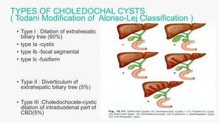
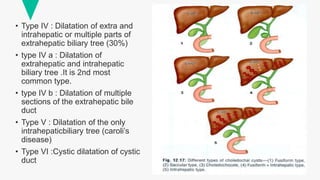
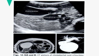
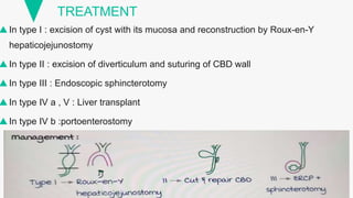
CHOLEDOCHAL CYSTS
V.S.S.M.ROHITH KUMAR
FINAL YR MBBS
DEFINITION
• ISOLATED/ FOCAL or COMBINED/DIFFUSE congenital
dialation of extra or intrahepatic biliary tree.
• More common in asia. Its incidence is 1 in 1000 hospital
admissions.
• Type I : Dilation of extraheoatic
biliary tree (60%)
• type Ia -cystic
• type Ib -focal segmental
• type Ic -fusiform
• Type II : Diverticulum of
extrahepatic biliary tree (5%)
• Type III :Choledochocele-cystic
dilation of intraduodenal part of
CBD(5%)
TYPES OF CHOLEDOCHAL CYSTS
( Todani Modification of Alonso-Lej Classification )
• Type IV : Dilatation of extra and
intrahepatic or multiple parts of
extrahepatic biliary tree (30%)
• type IV a : Dilatation of
extrahepatic and intrahepatic
biliary tree .It is 2nd most
common type.
• type IV b : Dilatation of multiple
sections of the extrahepatic bile
duct
• Type V : Dilatation of the only
intrahepaticbiliary tree (caroli’s
disease)
• Type VI :Cystic dilatation of cystic
duct
PRESENTATIONS
• common in females
• obstructive jaundice (80%)
• pain in right hypochondrium
• mass per abdomen(30%)
• failure to thrive
• TRAID OF CHOLEDOCHAL CYST
RIGHT UPPER QUADRANT PAIN
PALPABLE ABDOMINAL MASS
JAUNDICE
COMPLICATIONS
• Pancreatitis
• suppurative cholangitis
• Gall stone and CBD stone formation
• Biliary cirrhosis
• Rupture of cyst and peritonitis
• Cholangiocarcinoma in CBD
• Malignancy is common in posterior wall
INVESTIGATIONS
 US abdomen
 CT scan
 hepatobiliary nuclide scanning
 ERCP(endoscopic retrograde cholangiopancreatography) , CHOLANGIOGRAPHY-
to see ductal anatomy
 MRCP(Magnetic Resonance Cholangiopancreatography) status of pancreatic and
biliary system and pancreaticobiliary maljunction
 Liver function tests
 PTC (percutaneous transhepatic cholangiography)to see intrahepatic biliary tree
TREATMENT
In type I : excision of cyst with its mucosa and reconstruction by Roux-en-Y
hepaticojejunostomy
In type II : excision of diverticulum and suturing of CBD wall
In type III : Endoscopic sphincterotomy
In type IV a , V : Liver transplant
In type IV b :portoenterostomy
CHOLEDOCHAL CYSTS. Description done pptx

CHOLEDOCHAL CYSTS. Description done pptx

  • 1.
  • 2.
    DEFINITION • ISOLATED/ FOCALor COMBINED/DIFFUSE congenital dialation of extra or intrahepatic biliary tree. • More common in asia. Its incidence is 1 in 1000 hospital admissions.
  • 3.
    • Type I: Dilation of extraheoatic biliary tree (60%) • type Ia -cystic • type Ib -focal segmental • type Ic -fusiform • Type II : Diverticulum of extrahepatic biliary tree (5%) • Type III :Choledochocele-cystic dilation of intraduodenal part of CBD(5%) TYPES OF CHOLEDOCHAL CYSTS ( Todani Modification of Alonso-Lej Classification )
  • 4.
    • Type IV: Dilatation of extra and intrahepatic or multiple parts of extrahepatic biliary tree (30%) • type IV a : Dilatation of extrahepatic and intrahepatic biliary tree .It is 2nd most common type. • type IV b : Dilatation of multiple sections of the extrahepatic bile duct • Type V : Dilatation of the only intrahepaticbiliary tree (caroli’s disease) • Type VI :Cystic dilatation of cystic duct
  • 5.
    PRESENTATIONS • common infemales • obstructive jaundice (80%) • pain in right hypochondrium • mass per abdomen(30%) • failure to thrive • TRAID OF CHOLEDOCHAL CYST RIGHT UPPER QUADRANT PAIN PALPABLE ABDOMINAL MASS JAUNDICE
  • 6.
    COMPLICATIONS • Pancreatitis • suppurativecholangitis • Gall stone and CBD stone formation • Biliary cirrhosis • Rupture of cyst and peritonitis • Cholangiocarcinoma in CBD • Malignancy is common in posterior wall
  • 7.
    INVESTIGATIONS  US abdomen CT scan  hepatobiliary nuclide scanning  ERCP(endoscopic retrograde cholangiopancreatography) , CHOLANGIOGRAPHY- to see ductal anatomy  MRCP(Magnetic Resonance Cholangiopancreatography) status of pancreatic and biliary system and pancreaticobiliary maljunction  Liver function tests  PTC (percutaneous transhepatic cholangiography)to see intrahepatic biliary tree
  • 9.
    TREATMENT In type I: excision of cyst with its mucosa and reconstruction by Roux-en-Y hepaticojejunostomy In type II : excision of diverticulum and suturing of CBD wall In type III : Endoscopic sphincterotomy In type IV a , V : Liver transplant In type IV b :portoenterostomy