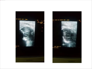
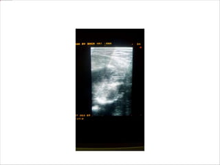
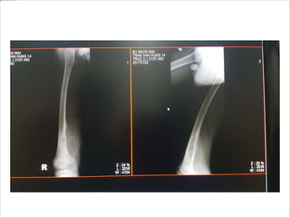
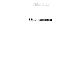
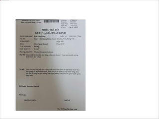
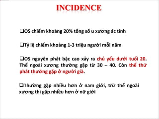
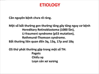
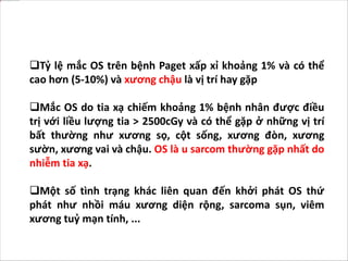
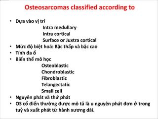
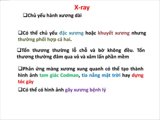
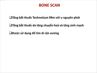
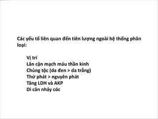
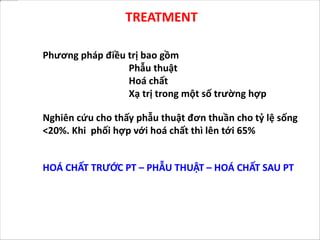
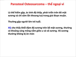
11. OS là u xương tạo xương ác tính, là khối u
nguyên phát thường gặp thứ 2 sau đa u tuỷ
xương
12. INCIDENCE
OS chiếm khoảng 20% tổng số u xương ác tính
Tỷ lệ chiếm khoảng 1-3 triệu người mỗi năm
Thường gặp nhiều hơn ở nam giới, trừ thể ngoài
xương thì gặp nhiều hơn ở nữ giới
OS nguyên phát bậc cao xảy ra chủ yếu dưới tuổi 20.
Thể ngoài xương thường gặp từ 30 – 40. Còn thể thứ
phát thường gặp ở người già.
13. Mọi xương đều có khả năng gặp osteosarcom, nhưng
thường gặp ở vị trí xương tăng trưởng mạnh.
Thường gặp ở đầu xa xương đùi, đầu gần xương chày và
đầu gần xương cánh tay
Hơn 50% trường hợp gặp ở quanh gối
Vị trí của OS thể thứ phát thay đổi, gặp ở mọi xương nhưng
chủ yếu xương chậu, là vị trí tổn thương hay gặp trong bệnh
Paget
Vị trí hay gặp của xương là: Hành xương 91%, thân xương
<9%. Tổn thương đầu xương rất hiếm gặp
14. ETIOLOGY
Căn nguyên bệnh chưa rõ ràng.
Một số bất thường gen thường tăng gây tăng nguy cơ bệnh
Hereditary Retinoblastoma (1000 lần),
Li-fraumeni syndrome (p53 mutation),
Rothmund-Thomson syndrome.
Bất thường liên quan đến 3q, 13q, 17p and 18q
OS thứ phát thường gặp trong một số TH:
Pagets
Chiếu xạ
Loạn sản xơ xương
15. Tỷ lệ mắc OS trên bệnh Paget xấp xỉ khoảng 1% và có thể
cao hơn (5-10%) và xương chậu là vị trí hay gặp
Mắc OS do tia xạ chiếm khoảng 1% bệnh nhân được điều
trị với liều lượng tia > 2500cGy và có thể gặp ở những vị trí
bất thường như xương sọ, cột sống, xương đòn, xương
sườn, xương vai và chậu. OS là u sarcom thường gặp nhất do
nhiễm tia xạ.
Một số tình trạng khác liên quan đến khởi phát OS thứ
phát như nhồi máu xương diện rộng, sarcoma sụn, viêm
xương tuỷ mạn tính, ...
16. Osteosarcomas classified according to
• Dựa vào vị trí
Intra medullary
Intra cortical
Surface or Juxtra cortical
• Mức độ biệt hoá: Bậc thấp và bậc cao
• Tính đa ổ
• Biến thể mô học
Osteoblastic
Chondroblastic
Fibroblastic
Telangectatic
Small cell
• Nguyên phát và thứ phát
• OS cổ điển thường được mô tả là u nguyên phát đơn ở trong
tuỷ và xuất phát từ hành xương dài.
18. PATHOGENESIS
Khối u điển hình phát triển từ trong khoang tuỷ xương, gần
hành xương xâm lấn và phá huỷ vỏ xương phá huỷ
màng xương, xâm lấn mô mềm.
19. Sụn khớp đóng vai trò như rào cản ngăn chận xâm lấn
vào khớp
Khối u có thể xâm lấn vào khớp tuy nhất thường qua dây
chằng và cấu trúc bao
Toward the diaphyseal end, the advancing tumor
presents as a conical plug that marks the limit of growth
of the lesion lengthwise along the shaft.
Di căn kiểu nhảy cóc có thể gặp
20.
21. CLINICAL FEATURES
Đau tiên triển là triệu chứng thường gặp nhất và là
triệu chứng đầu tiên
Đau ban đầu thường cải thiện bằng việc giảm vận
động.
Đau về ban đêm có thể là một dấu hiệu để chẩn
đoán, tuy nhiên chỉ 25% trường hợp
Đôi khi bệnh nhân đến vì bướu phần mềm
Gãy xương bệnh lý là triệu chứng có thể gặp
22. DIAGNOSIS
Trong đa phần các trường hợp, phim XQ là đủ để chẩn đoán
Tuy nhiên CT, MRI, xạ hình xương, CT ngực có ích trong việc
phân độ giai đoạn và tham gia kế hoạch điều trị
Ngoài xét nghiệm máu thông thường thì phosphatase kiềm
huyết thanh, LDH có giá trị trong việc tiên lượng.
23. Có thể chủ yếu đặc xương hoặc khuyết xương nhưng
thường phối hợp cả hai.
Tổn thương thường lỗ chỗ và bờ không đều. Tổn
thương thường đâm qua vỏ và xâm lấn phần mềm
X-ray
Chủ yếu hành xương dài
Phản ứng màng xương xung quanh có thể tạo thành
hình ảnh tam giác Codman, tia nắng mặt trời hay dựng
tóc gáy
Có thể có hình ảnh gãy xương bệnh lý
28. X Quang phim thẳng 1/2 dưới
xương đùi
Mô tả: Tam giác Codman, đặc
xương, h/a dựng tóc gáy
Chẩn đoán: sarcoma xương
tạo xương
Phân biệt: viêm xương tủy
xương, sarco ewing
29. X Quang phim thẳng đầu
trên cẳng chân và khớp
gối
Mô tả: tiêu xương đầu
xương mác, phá hủy
màng xương, h/a tia mặt
trời
Chẩn đoán: sarcoma
xương tạo xương
32. MRI
Có thể thấy di căn nhảy cóc
MRI cũng giúp đánh giá xâm lấn khớp lân cận.
MRI chỉ ra mức độ xâm lấn phần mềm
Giá trị lớn nhất của MRI là đánh giá chính xác mức độ
xâm lấn ống tuỷ của u
33.
34. CT Scan
Khoảng 10-20% bệnh nhân OS có di căn phát hiện được
trên CT tại thời điểm chẩn đoán. Đa số ở phổi.
Những di căn >10mm thấy được trên XQ. 3-7mm có thể
thấy trên CT
35. Tăng bắt thuốc Technetium 99m với u nguyên phát
Tăng bắt thuốc do tăng chuyển hoá và tăng sinh mạch
Được sử dụng để tìm di căn xương
BONE SCAN
38. Các yếu tố liên quan đến tiên lượng ngoài hệ thống phân
loại:
Vị trí
Lân cận mạch máu thần kinh
Chủng tộc (da đen > da trắng)
Thứ phát > nguyên phát
Tăng LDH và AKP
Di căn nhảy cóc
39. TREATMENT
Phương pháp điều trị bao gồm
Phẫu thuật
Hoá chất
Xạ trị trong một số trường hợp
Nghiên cứu cho thấy phẫu thuật đơn thuần cho tỷ lệ sống
<20%. Khi phối hợp với hoá chất thì lên tới 65%
HOÁ CHẤT TRƯỚC PT – PHẪU THUẬT – HOÁ CHẤT SAU PT
40. Hoá chất
Hoá chất giúp ngăn chặn tổn thương tiến triển
Tỷ lệ sống của hoá chất đơn thuần thấp, chỉ <25%
Gồm HC trước và sau phẫu thuật
41. Hoá chất tiền phẫu giúp ngăn chặn u tiến triển, giúp phẫu
thuật bảo tồn chi dễ dàng hơn
Hoá chất tiền phẫu có thể được chỉ định ngay khi phát
hiện, để tránh tiến triển ngay trong thời gian chờ phẫu
thuật
NEOADJUVANT CHEMOTHERAPY
42. Phẫu thuật
Mục đích của phẫu thuật là loại bỏ được rộng vùng tổn
thương
Lấy ít tổn thương nguy cơ tái phát tại chỗ PT
50. Periosteal Osteosarcoma - Thể màng xương
U ác tính phát triển từ bề mặt xương
Thường 30 – 50 tuổi
Điều trị như OS cổ điển
51.
52. Parosteal Osteosarcoma – thể ngoại vi
Là thể hiếm gặp, ác tính độ thấp, phát triển trên bề mặt
xương và chỉ xâm lấn khoang tuỷ trong giai đoạn muộn.
Thường gặp người lớn trẻ tuổi.
XQ cho thấy khối đậm độ xương trên bề mặt xương, thường
có khoảng sáng mỏng nằm giữa u và vỏ xương. Vỏ xương
thường không bị ăn mòn
53.
54. Chẩn đoán phân biệt với
Viêm cơ cốt hoá
Sarcoma sụn
Trong viêm cơ cốt hoá thì xương nằm ngoài tổn thương,
còn trong sarcoma xương thể ngoại vi thì trung tâm tổn
thương có cấu trúc xương trưởng thành hơn
Sarcom xương có khoang tuỷ thông với khoang tuỷ của
xương tổn thương.
CT và MRI có ích trong việc phân biệt tổn thương
55.
56. Là thể rất ác tính
Nam gấp 2 lần nữ, thường từ 20-30 tuổi
Đặc điểm là rất tăng sinh mạch và có các khoang tiêu
xương chứa máu, thường không có hình ảnh đặc xương
Telangiectatic osteosarcoma – OS thể giãn mạch
57. Chẩn đoán phân biệt với nang xương, nang xương phình
mạch.
Điều trị giống OS cổ điển
58.
59. Là thể hiếm gặp, có thể giống với Sarcoma Ewing hay
Lymphoma.
Thường đầu xa xương đùi và đầu gần xương cánh tay
Small cell osteosarcoma - thể tế bào nhỏ
60. Thường đi với các bệnh lý
Pagets
Chiếu xạ
Loạn sản xơ xương
Thường ở người già > 60 tuổi
Secondary osteosarcomas
Cystic + sclerotic/ blastic lesions in the epiphysial of the right femur, both in the medullary cânl and the cortex
Codman’s triangle sign
nearby soft tissue thickening
Osteosarcomas are malignant bone forming tumours and the second most common primary bone tumour after multiple myeloma .
Osteosarcoma accounts for approximately 20% of primary malignancies of bone.
The incidence is approximately 1 to 3 per 1 million per year.
Primary high-grade osteosarcoma occurs most commonly in the second decade of life. Parosteal osteosarcoma has a peak incidence in the third and fourth decades, and secondary osteosarcomas are more common in older individuals.
The incidence is slightly higher in males (with the exception that parosteal osteosarcoma is more common in females).
No bone is immune to develop osteosarcoma, commonly develops at sites of rapid bone growth where mitotic activity is high.
The most common sites involved are distal femur, proximal tibia, proximal humerus.
More than 50% lesions occurs around knee
Secondary osteosarcomas are equally distributed in all bones
Metaphysis 91%
diaphysis <9%
Epiphyseal involvement is extremely rare
No specific etiologic cause developing osteosarcoma is not established till now.
But some genetic abnormalities are associated with
increased incidence of these tumors
Hereditary Retinoblastoma (1000 times),
Li-fraumeni syndrome (p53 mutation),
Rothmund-Thomson syndrome.
3q, 13q, 17p and 18q are most commonly Involved.
Secondary osteosarcomas are commonly associated
with Pagets disease
Irradiation
Fibrous dysplasia
The incidence of osteosarcoma in Paget disease is approximately 1% and may be higher (5% to 10%) and the pelvis is the most common location.
Radiation-induced osteosarcoma occurs in approximately 1% of patients who have been treated with greater than 2500 cGy and can occur in unusual locations, such as the skull, spine, clavicle, ribs, scapula, and pelvis. osteosarcoma is the most common radiation-induced sarcoma.
Other conditions that have been reported to be associated with secondary osteosarcomas include bone infarcts, osteochondromas, chronic osteomyelitis, dedifferentiated chondrosarcomas, melorheostosis, and osteogenesis imperfecta.
Phân loại
Site of origin in bone
Degree of differentiation: High or Low grade
Multicentricity
Histological variant
Primary or Secondary
Classic osteosarcoma is a primary Intramedullary solitary high grade osteoblastic variant arising from metaphysis of long bones
The tumor ordinarily begins developing in the medullary cavity of a long bone near the metaphysis,
Then, they penetrated and extended through the cortex to raising the periosteum
In more advanced cases the periosteal barrier may be broken, and a soft tissue tumor mass may be seen invading the adjacent muscle tissue
The articular hyaline cartilage serves as barrier to prevent the extension of the neoplasm into the joint
The tumor may enter the joint, however, by extending along ligamentous and capsular structures
Skip metastases may occur.
Progressive pain is the first and most common presenting symptom.
Pain initially may improve with conservative measures and activity modifications.
Night pain may be an important clue to the true diagnosis; however, only about 25% of patients experience this phenomenon.
Some times patient presents with a lump
Pathological fracture is very unusual presentation
Plain radiograph is sufficient to diagnose osteosarcoma most of the times.
But CT, MRI, Bone scan, CT Chest
are useful in staging of the tumor and planning of
treatment.
Apart from routine blood investigations Serum
alkaline phosphatase, LDH are valuable and indicates poor prognosis.
Osteosarcoma predominantly osteoblastic lesion at
metaphyseal region of long bone.
Although the lesion can be either predominantly blastic or predominantly lytic, more commonly areas of bone production and bone destruction are present.
The lesion usually is quite permeative, and the borders are ill defined. Tumor pierces the cortex, and extend into adjucent soft-tissue.
Periosteal reaction may take the form of “Codman's triangle,” or it may have a “sunburst” or “hair on end” appearance
Pathological fracture may be seen.
Hair on end, Sunray, Codman’s triangle
Codman, Sunray
metaphysial Blastic lesion
metaphysial and diaphysial
Pathological fracture
MRI demonstrates the degree of soft tissue extension and the relationship of the extracompartmental tumor to fascial planes and neurovascular structures.
The best feature of MRI is its ability to precisely evaluate the extent of tumor in the medullary cavity.
Occult skip metastases of 2 mm or more in long bones are well seen on MRI.
MRI is also useful in evaluating the adjacent joint for tumor spread.
Approximately 10% to 20% of patients with osteosarcoma present with radiographically detectable metastases at diagnosis. Most of these are in the lungs.
Conventional radiographs of the chest show metastatic nodules 10 mm or greater in diameter. Pulmonary metastases 3 to 7 mm or greater in diameter are identified with CT.
Bone scan with technetium 99m shows a marked increase in the uptake of the radionuclide in the primary tumor.
The increased uptake is due to active formation of new tumor and host bone as well as the vascularity of the lesion.
Radionuclide bone scintigraphy is used to look for bony metastases in the involved bone (skip metastases)and at other skeletal sites.
Mineralized metastases are more likely to be detected by bone scans than are nonmineralized ones at extrapulmonary sites.
The factors which influence prognosis other than staging system are
Location of the tumor
Proximity to neurovascular structures
Race (blacks > whites )
Secondary > Primary
Elevated LDH and Alkaline phosphatase
Presence of skip lesions
Treatment options for osteosarcoma are
Surgery
Chemotherapy
Radiotherapy in some cases
Historical data suggests that survival rate with ablative surgery alone is <20%. when combined with chemotherapy it is almost 65%.
Chemotherapy has excellent role in terminating these
lesions.
The survival rate with chemotherapy alone is very
disappointing <25%
Chemotherapy applied in two modalities
Neoadjuvant chemotherapy
Adjuvant chemotherapy
Preoperative chemotherapy causes regression of the primary tumor, making a limb salvage operation easier.
Neoadjuvant chemotherapy usually can be started immediately to prevent tumor progression, which may occur during any delay before surgery
This allows time to plan the operation properly, including the possible manufacturing of a custom implant.
The aim of tumor surgery is to achieve wide margins whenever possible.
Main hazard of narrow margins of excision is local recurrence, which requires further surgery even amputation.
It is an intermediate-grade malignancy that arises on the surface of the bone
The most common locations are the diaphyses of the femur and tibia, and common in a age group of around 30-5oyrs.
It presents with swelling and local pain.
Histological examination of periosteal osteosarcoma shows strands of osteoid-producing spindle cells radiating between lobules of cartilage.
Treatment is same as classic osteosarcoma.
It is a rare, low-grade malignancy, arising on the surface of the bone and invades the medullary cavity only at a late stage.
It common in young adults
X-ray shows bony mass on the surface of the bone, usually thin gap remain between cortex and tumor.
cortex is not eroded
Differential diagnosis is
Myositis ossificans
Osteochondroma
CT and MRI helpful in differentiating parosteal osteosarcoma from these lesions
In myositis ossificans mature bone is at the periphery of the lesion, whereas in parosteal osteosarcoma centre of the lesion is more heavily matured.
Parosteal osteosarcoma can be easily differentiated from an osteochondroma by showing medullary cavity containing marrow in continuity with the medullary canal of the involved bone.
Telangiectatic osteosarcoma is a highly malignant variant of osteosarcoma .
It is twice as common in males than in females and common in 20-30 age group.
It is characterized by a high degree of vascularity and large cystic spaces filled with blood.
It is an osteolytic destructive lesion with an almost complete absence of sclerotic changes; a soft-tissue mass may also be present
Differential diagnosis is cystic bone lesions like simple bone cyst, aneurysmal bone cyst
Treatment is same as classic osteosarcoma
It is rare variant of osteosarcoma which is a high-grade lesion that consists of small blue cells that may resemble Ewing sarcoma or lymphoma.
Distal femur, proximal humerus are sites of prediliction.
Cytogenetic and immunohistochemistry studies sometimes are needed to differentiate these lesions
Secondary osteosarcomas are commonly associated
with
Pagets disease
Irradiation
Fibrous dysplasia
Common in elderly people after age 60