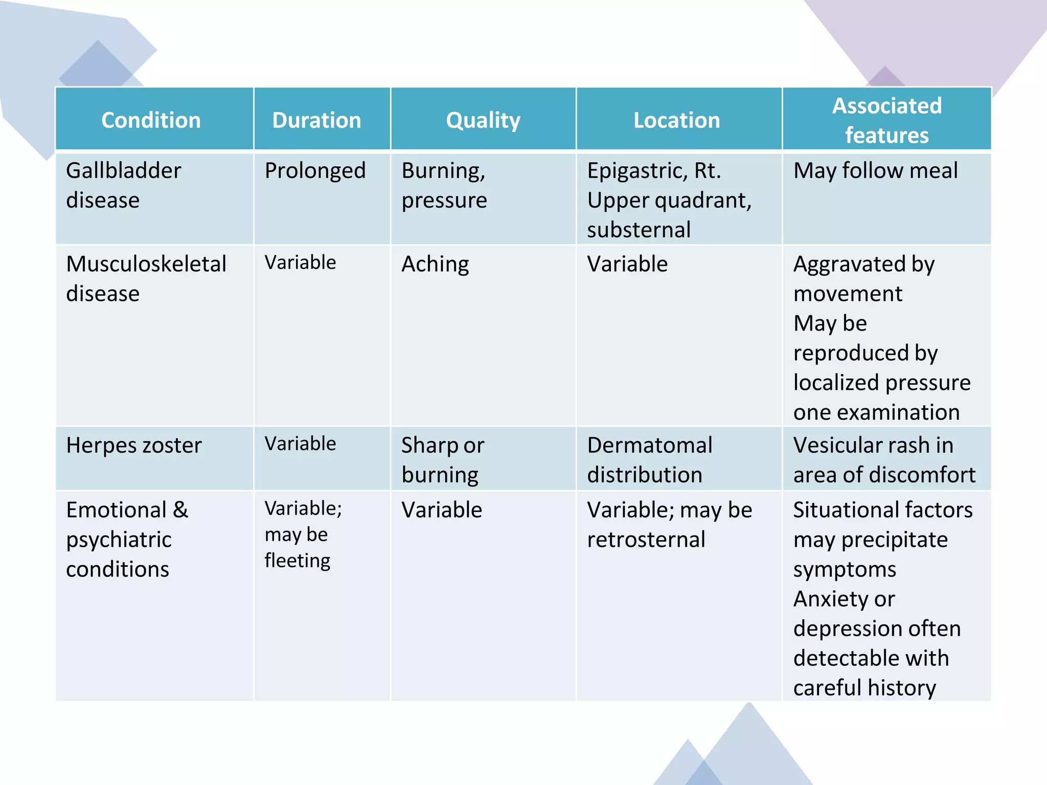
Chest pain can be cardiac or non-cardiac in origin. Cardiac causes include myocardial ischemia, angina, and acute coronary syndromes which present with characteristic dull, squeezing pain in the center of the chest that may radiate to the arms and is exacerbated by exertion. Non-cardiac causes include pulmonary embolism, pneumonia, gallbladder disease and musculoskeletal disorders which typically cause localized, sharp pains. A thorough history and physical exam is needed to differentiate cardiac from non-cardiac chest pain and determine appropriate treatment or need for further testing.
Chest pain can be cardiac or non-cardiac in origin. Cardiac causes include angina, myocardial infarction, and pericarditis while non-cardiac causes include pulmonary embolism, pneumonia, gallbladder disease, and musculoskeletal disorders. Characteristics of ischemic cardiac chest pain include central location, radiation to the jaw/arm, tight or squeezing quality, and precipitation by exertion. Differential diagnosis of chest pain involves evaluating for life-threatening causes, chronic serious conditions, treatable acute conditions, and other chronic treatable conditions. Distinguishing ischemic cardiac chest pain from non-cardiac pain relies on characteristics such as location, quality, precipitating/relieving factors, and associated symptoms.
Chest pain can be cardiac or non-cardiac in origin. Cardiac causes include angina, myocardial infarction, and pericarditis while non-cardiac causes include pulmonary embolism, pneumonia, gallbladder disease, and musculoskeletal disorders. Characteristics of ischemic cardiac chest pain include a central, pressing or squeezing quality that radiates to the jaw/arm and is provoked by exertion. Differential diagnosis of chest pain requires evaluating characteristics like location, quality, duration and associated features to determine cardiac vs. non-cardiac etiology.
This document defines and discusses chest pain, including its potential cardiac and non-cardiac causes. It outlines how to evaluate chest pain by determining if it could be due to an acute life-threatening condition, chronic condition likely to lead to complications, acute treatable condition, or other chronic treatable condition. It describes characteristics of ischemic cardiac pain and provides a differential diagnosis for chest pain that may be due to anxiety, cardiac, aortic, esophageal, lung/pleural, musculoskeletal, or neurological issues.
Chest pain is a common presenting complaint that can be caused by many cardiac and non-cardiac conditions. A thorough history, physical exam, EKG, biomarkers, imaging and stress testing are often needed to determine the underlying cause. The differential diagnosis includes acute coronary syndrome, pulmonary embolism, aortic dissection, pneumonia, gastroesophageal reflux disease, musculoskeletal disorders and anxiety. Location, radiation, relieving/worsening factors, associated symptoms and risk factors can help distinguish between potential life-threatening versus more benign causes of chest pain.
Cardiology 1.1. Chest pain - by Dr. Farjad IkramFarjad Ikram
Introduction to one of the most common symptoms that can represent a wide range of diseases, from benign to life-threatening, covering number of systems including gastrointestinal, cardiovascular, pulmonary, musculoskeletal and psychiatric. Includes a brief explanation of anti-anginal therapy.
Template design credits - http://www.slidescarnival.com
This document discusses the evaluation and diagnosis of chest pain. It notes that while chest pain can be caused by many life-threatening and non-life-threatening conditions, it is important for patients to seek medical evaluation due to the difficulty in distinguishing between causes without testing. A medical history and physical exam are important for evaluating potential causes, which can include conditions affecting the heart, lungs, chest wall, esophagus and abdomen. Based on the history and exam, targeted testing like EKGs, blood tests, imaging and other diagnostics may be used to determine the underlying cause.
Chest pain can have many potential causes. A thorough history and physical exam are important to help determine the likely diagnosis and guide appropriate testing. Key aspects of the history include characteristics of the pain such as location, radiation, onset and nature. The physical exam focuses on identifying potential causes of the pain or associated symptoms. Initial tests may include an ECG, blood tests, chest x-ray and echocardiogram to help differentiate causes such as heart disease, pulmonary embolism, pneumonia or musculoskeletal issues. Further tests are guided by the initial findings.
Angina pectoris is chest pain or discomfort that occurs when the heart muscle does not get enough oxygen. It is usually caused by narrowed coronary arteries that cannot supply enough blood and oxygen to meet increased demands. The symptoms include chest pain or pressure that can radiate to the arms, neck or jaw, brought on by activity or stress, and relieved by rest. Diagnosis involves evaluating symptoms, risk factors, and electrocardiogram changes during episodes to confirm ischemia as the cause of symptoms.
This document provides an overview of the approach to evaluating and diagnosing chest pain. It discusses how chest pain can be caused by conditions affecting the heart, lungs, esophagus and other organs. The evaluation involves taking a detailed history of the quality, location, timing and relieving/worsening factors of the pain and performing a physical exam. Key investigations include electrocardiography, chest x-ray, cardiac biomarkers and imaging tests like CT angiography to identify potential cardiac, pulmonary, aortic or gastrointestinal causes of the chest pain. A systematic approach considering the history, exam and test results is needed to diagnose the underlying condition causing the chest pain.
This document discusses the differential diagnosis of chest pain by listing common cardiac, respiratory, gastrointestinal, and musculoskeletal causes. Key cardiac causes mentioned are angina, myocardial infarction, mitral valve prolapse, and pericarditis. Common respiratory causes listed are pleurisy, pneumothorax, and pulmonary embolism. Gastrointestinal causes such as reflux esophagitis and diffuse esophageal spasm are also summarized. The document provides brief descriptions of symptoms, signs, and investigations for each of these differential diagnoses of chest pain.
The document provides information on evaluating and managing patients presenting with chest pain and acute coronary syndrome. It discusses how to take a clinical history to determine the nature and cause of chest pain. Differential diagnoses are provided for cardiac and non-cardiac causes. Acute coronary syndrome is described as encompassing unstable angina, NSTEMI, and STEMI, which present with chest pain at rest or minimal exertion due to plaque rupture or erosion in coronary arteries. Immediate management in the first 12 hours and long-term management are outlined.
This document provides guidance on assessing symptoms related to cardiac issues in geriatric patients. It discusses how to evaluate dyspnea, cough, palpitations, chest pain, leg swelling, and other symptoms by asking about duration, onset, precipitating/relieving factors, and associated symptoms. Specific questions are provided to assess orthopnea, paroxysmal nocturnal dyspnea, hemoptysis, edema, and different types of chest pain. Causes and characteristics of different conditions like pulmonary venous congestion, acute pulmonary edema, and low cardiac output are also summarized.
Angina pectoris is chest pain caused by inadequate blood supply to the heart muscle. There are several types of angina including stable angina brought on by exertion and relieved by rest, unstable angina which occurs at rest, and variant angina caused by coronary artery spasms. Factors that can trigger angina include increased cardiac demand from exercise or stress as well as reduced blood supply from conditions like atherosclerosis. Diagnosis involves evaluating symptoms, ECG, cardiac enzymes, and imaging tests like angiography. Treatment focuses on lifestyle changes, medications to relieve symptoms and improve blood flow, and potentially revascularization through procedures like angioplasty.
The document provides guidance on evaluating and diagnosing the causes of chest pain, including cardiac conditions like acute coronary syndrome (ACS) and pulmonary embolism. It describes approaches to obtaining a history, performing a physical exam, and ordering diagnostic tests like electrocardiograms (ECGs) and cardiac biomarkers. The goal is to identify high-risk conditions and accurately diagnose the underlying cause of the patient's chest discomfort.
This document discusses cardiac emergencies including angina pectoris, myocardial infarction, and congestive cardiac failure. It defines each condition, lists causes and risk factors, describes signs and symptoms, outlines diagnostic tests and treatment options, discusses complications, and provides nursing management guidelines. Angina is chest pain due to decreased blood flow to the heart while myocardial infarction and congestive cardiac failure involve the heart's inability to pump sufficiently due to disease or damage. Prompt recognition and treatment are important to save lives during these deadly emergencies.
This document discusses the evaluation and diagnosis of chest pain. It notes that chest pain is a common reason for medical attention and may be caused by cardiovascular, pulmonary, gastrointestinal, chest wall or psychological conditions. The history, physical exam, electrocardiogram and cardiac biomarkers can help diagnose causes like acute coronary syndrome, pericarditis, pulmonary embolism and pneumonia. Serial cardiac troponin testing can distinguish between unstable angina and non-ST-segment elevation myocardial infarction. The document provides details on symptoms and exam findings for various conditions that can cause chest pain.
This document discusses the pathogenesis of atherosclerosis and myocardial ischemia, as well as the presentation and treatment of acute myocardial infarction (AMI). It describes the development of atherosclerotic plaques, factors that increase oxygen demand and decrease supply leading to angina, and common symptoms of AMI such as chest pain and shortness of breath. The treatment principles of AMI focus on restoring blood flow to the heart and preventing complications. Medications like nitrates can help by reducing oxygen requirements and increasing supply to the heart.
Angina pectoris is a clinical syndrome characterized by chest pain or pressure due to insufficient blood flow to the heart. It occurs when oxygen demand of the heart exceeds the oxygen supply. The main causes are atherosclerosis and coronary artery spasm. Diagnosis is made based on symptoms, ECG, stress test, and coronary angiography. Treatment involves lifestyle changes, medications to reduce oxygen demand or increase supply like nitroglycerin, beta blockers, and calcium channel blockers. The goal is to manage symptoms and prevent further heart issues.
1. The document discusses the approach to evaluating and categorizing different types of chest pain, including acute coronary syndrome.
2. Key factors in the initial evaluation of chest pain include obtaining a detailed history, performing a physical exam, 12-lead EKG, and cardiac biomarker testing to help determine if the chest pain is caused by life-threatening conditions like myocardial infarction.
3. Patients are categorized as having possible acute ischemia, probable acute ischemia, or myocardial infarction based on their history, symptoms, physical exam findings, EKG results, and cardiac enzyme levels in order to determine the urgency of further testing and treatment.
Central chest pain can be caused by cardiac issues like angina or myocardial infarction, or non-cardiac issues like pulmonary embolism or esophageal disorders. A thorough history of the characteristics of the pain, including location, duration, aggravating/relieving factors, and associated symptoms can help determine the underlying cause. Key factors that help differentiate cardiac from non-cardiac chest pain include relationship to exertion and response to nitroglycerin.
The document discusses various cardiac emergencies including acute coronary syndrome, myocardial infarction, shock, heart failure, mitral regurgitation, ventricular septal rupture, pericarditis, atypical angina, cardiac arrest, hypertensive emergency, aortic aneurysm, aortic dissection, air embolism, cardiac tamponade, and cardiac arrhythmias. It provides details on symptoms, signs, diagnosis and management of these conditions. The goal of treatment is rapid stabilization of vital functions and prompt transfer to a facility capable of definitive care.
Angina pectoris is the medical term for chest pain or discomfort due to coronary heart disease. It occurs when the heart muscle doesn't get as much blood as it needs. This usually happens because one or more of the heart's arteries is narrowed or blocked, also called ischemia.
This document provides an overview of chest pain differentials (DDx). It begins by defining chest pain and discussing acute coronary syndrome. It then covers risk factors, symptoms, and physical exam findings for conditions like aortic dissection, acute pericarditis, cardiac tamponade, pulmonary embolism, tension pneumothorax, and esophageal rupture. Investigations, treatments, and management are described for each potential cause. The goal is to educate on evaluating and distinguishing between cardiac and non-cardiac causes of chest pain.
Chest pain or discomfort
Common presenting symptom of cardiovascular disease
May be cardiac or noncardiac in origin.
Cardiac – angina, MI, pericarditis, mitral valve prolapse, dissecting aortic aneurysm
Non cardiac – anemia (physical exertion), cervical disc disease, anxiety, trigger points etc
Follows pattern of ulnar nerve distribution (heart supplied by C3-T4 spinal segments)
Radiating pain to neck, jaw, upper trapezius, upper back, shoulder or arms (commonly left
Cardiovascular disease is the leading cause of death in the United States. The document discusses signs and symptoms of various cardiovascular emergencies like myocardial infarction, angina, cardiogenic shock, and congestive heart failure. It also covers assessment of chest pain, including gathering a history and performing a physical exam to determine the likely cause and guide treatment and transport. The goal is to identify life threats and provide care to stabilize the patient while expediting transport to a hospital.
SEMINAR ON Myocardial infarction FOR NURSING STUDENT.pptxApurva Dwivedi
Look for signs of heart failure,
peripheral vascular disease, or other
abnormalities.
3. Palpate:
- Neck for carotid upstrokes
- Chest for thrills, heaves, lifts
- Abdomen for organomegaly, pulsations
- Extremities for pulses, edema
Feel for thrills, lifts, pulses to assess for
murmurs, heaves, peripheral pulses.
4. Percuss:
- Chest for dullness, hyper-resonance
Percuss chest to assess for pleural
effusions or other abnormalities.
5. Auscultate:
- Heart sounds and rhythm
- L
This document outlines key concepts related to healthcare supply chain and inventory management. It discusses the typical players in the supply chain including manufacturers, distributors, group purchasing organizations, and e-distributors. It also describes how materials flow through the supply chain and some contemporary issues in medical inventory management like just-in-time systems and single vs multiple vendors. Key aspects of effective inventory management are identified such as inventory accounting systems, lead times, costs including holding, ordering and shortage costs, and the economic order quantity model.
Introduction to quantitative decision-making methods in healthcare managementMUKESH SUNDARARAJAN
This document provides an introduction to quantitative decision-making methods in healthcare management. It discusses the historical development of management techniques from scientific management to modern approaches like operations research, management information systems, and total quality management. The document also outlines the nature of the healthcare industry, the healthcare process, and the types of decisions made by healthcare managers at strategic, tactical, and operational levels. These decisions involve areas like capacity, personnel, inventory, scheduling, and quality assurance. The document concludes by noting some distinctive characteristics of healthcare services like the simultaneous production and consumption of care.
This document defines health and productivity management (HPM) as a systematic approach for companies to quantify, evaluate, and optimize their investment in employee health. HPM aims to reduce health-related costs like medical expenses and lost productivity from absenteeism and presenteeism. The history of HPM shows early studies linking health factors like iron deficiency to lost productivity. Common health risks like poor diet and smoking drive chronic diseases responsible for most health costs. Implementing strategies to improve lifestyle behaviors and reduce health risks through workplace programs can help control costs and improve employee health.
The document discusses the need for healthcare project management training and the benefits it provides. It notes that recent US legislation and industry trends have led to an increased number of healthcare projects. Good project management is required to implement projects successfully and achieve goals like improved quality and reduced costs. However, healthcare workers often lack project management skills since they are more familiar with operational versus project work. The document advocates for training clinical leaders in project management principles and provides suggestions for developing effective training programs.
This document discusses various decision making tools and techniques that can be used in healthcare facilities. It outlines the typical decision making process and some common causes of poor decisions. It then covers different types of decisions based on certainty, uncertainty and risk. Various decision tools are presented, including payoff tables, expected value, decision trees, and techniques for decisions with non-monetary attributes. Examples are provided to illustrate how to apply these tools and techniques to make optimal decisions under different conditions.
Inductive transducers work by changing the inductance of a coil due to changes in a measured quantity like displacement, force, or pressure. There are two main types - self-generating transducers that produce a voltage signal from relative motion in a magnetic field, and non-self generating transducers that require an external power source to energize coils whose inductance changes. Inductive transducers work by changing the self-inductance of a coil, producing eddy currents in a nearby conductor, or altering the mutual inductance between two coils due to the measured quantity. They are used for proximity sensing, touchpads, and detecting metal objects or movement.
The document discusses different types of ultrasound transducers. The essential element of each transducer is a piezoelectric crystal that generates and receives ultrasound waves. Transducers come in various shapes, sizes, and features depending on the body part being imaged. The main types of transducers discussed are linear, convex, phased array, pencil, endocavitary, transesophageal, and 4D transducers. Each type has a different piezoelectric crystal arrangement, aperture, frequency, and intended medical applications.
This document discusses three main types of photoelectric cells: photoemissive cells, photovoltaic cells, and photoconductive cells. Photoemissive cells contain a photosensitive cathode and anode inside an evacuated glass bulb, and emit electrons when light hits the cathode. Photovoltaic cells, also called true cells, generate their own voltage when light hits a semiconductor-metal junction without needing an external voltage. Photoconductive cells have a semiconductor like selenium between two electrodes, and their resistance decreases when light hits, allowing current to flow in an external circuit. All three types of cells convert light energy into electrical energy or signals based on the photoelectric effect.
Piezoelectric transducers work by exploiting the piezoelectric effect, where certain materials generate an electric voltage when subjected to mechanical stress. They can be used to measure dynamic changes by converting mechanical energy into electrical signals. Common piezoelectric materials used in transducers include barium titanate, lead zirconate titanate, Rochelle salt, and quartz. While offering high frequency response and transient response, piezoelectric transducers also have low output and high impedance. Applications include dynamic measurement, studying high-speed phenomena, vibration detection, and use in medical devices, printers, lighters, and phone screens.
A scintillation counter detects and measures ionizing radiation by using a scintillator material that produces light pulses when excited by incident radiation. It consists of a scintillator that generates photons when hit by radiation, a photomultiplier tube that converts the light to an electrical signal, and electronics to process the signal. Scintillation counters are used in applications like radiation monitoring, medical imaging, and nuclear security to detect radioactive contamination. Variants mounted on vehicles can provide rapid response in security situations involving dirty bombs or radioactive waste.
Spectrophotometry involves measuring the intensity of light at selected wavelengths to analyze substances. It relies on substances absorbing light at characteristic wavelengths. A spectrophotometer uses light sources, monochromators, and detectors to isolate wavelengths and measure light intensity. Key concepts include Beer's Law which states absorbance is directly proportional to concentration. Spectrophotometers are used in applications like measuring analyte concentration, detecting impurities, and studying chemical kinetics through observation of absorbance changes over time.
This document discusses different types of photoelectric transducers, including photoemissive, photoconductive, and photovoltaic devices. It focuses on photomultiplier tubes, describing their construction and working principle of electron multiplication through secondary emission at dynode stages. Photomultiplier tubes can amplify current by 105 to 109 times, achieving high luminous sensitivity down to 10-5 lumens. The document also covers photoconductive cells, whose resistance varies with light intensity, allowing their use in light-controlled circuits.
Photodiodes are semiconductor devices that convert light into an electrical current. When light strikes the photodiode's PN junction, electrons are excited creating free charge carriers. This results in a photocurrent that can be measured. Photodiodes operate in reverse bias and can be used as solar cells, photodetectors, or LEDs. Common materials used include silicon, germanium, indium gallium arsenide, and lead sulfide, depending on the desired wavelength detection range. Photodiodes find applications in devices like optical mice, smoke detectors, and infrared remote controls.
Inductive transducers work by changing the inductance of a coil due to changes in a measured quantity like displacement, force, or pressure. There are two main types - self-generating transducers that produce a voltage signal from relative motion in a magnetic field, and non-self generating transducers that require an external power source to energize coils whose inductance changes with motion. Inductive transducers work by changing the self-inductance of a coil, producing eddy currents in a nearby conductor, or altering the mutual inductance between two coils as a function of the measured quantity.
This document discusses training, promotion, and transfer in an organizational context. It defines training as a systematic process aimed at developing employee skills, abilities, and attitudes. The purpose of training includes improving performance, reducing waste, and developing manpower. Promotion is defined as a change to a better job with higher status and responsibilities, and is used to reward performance, boost morale, and retain skilled employees. Both seniority and merit are discussed as bases for promotion, as well as a combination of the two. Transfer is defined as a lateral shift between jobs, departments, or locations with no change in pay or status. Different types of transfers are outlined, including production, replacement, remedial, and versatility transfers.
The document describes the different departments found in hospitals and provides brief descriptions of their functions. It discusses 20 departments including casualty, cardiology, critical care, ENT, geriatrics, gastroenterology, general surgery, gynecology, hematology, maternal/neonatal/pediatrics, neurology, oncology, ophthalmology, orthopedics, urology, psychiatry, outpatient, inpatient, central sterilization unit, housekeeping, catering and food services, medical social work, physiotherapy, pharmacy, nutrition and dietetics, microbiology, diagnostic imaging, medical records, medical maintenance and engineering, information technology and communication, human resources, finance, and administration.
This document provides an overview of human resource management. It defines HRM and discusses its key functions such as acquisition, development, compensation, and industrial relations. It also outlines the nature and scope of HRM, highlighting that it is a pervasive, future-oriented, and people-focused function. The document further discusses human resource planning, recruitment, selection, and placement processes. It compares personnel management with HRM and lists the objectives and importance of effective HRM. Finally, it notes some emerging issues and the role of HR professionals in adapting to changing business environments.
1. The document discusses measurement systems and instrumentation. It covers topics like order of instruments, instrument classification, units of measurement, standards of measurement, dimensions of measurement, and errors in measurement.
2. Instruments can be classified as mechanical, electrical, or electronic. They can also be categorized as absolute, secondary, digital, or analogue instruments.
3. The seven base SI units are meter, kilogram, second, Kelvin, mole, candela, and ampere. Derived units are formed by combining base units.
4. Standards provide defined relationships to measurement units and are used to calibrate other instruments. Primary standards define measurement units while secondary and working standards are calibrated against primary standards.
This document provides first aid guidelines for insect, scorpion, snake, and animal bites. It describes symptoms of bites and stings from bees, wasps, ants, scorpions, centipedes, snakes, and animals. Treatment recommendations include washing the affected area, applying antiseptic or cold compress, keeping it elevated, and seeking medical help for snake or scorpion bites. Guidelines are given for proper tick removal and when to see a doctor after insect bites depending on rash or disease risk in the region.
This document provides guidance on first aid for animal bites, insect stings, and snake bites. It advises remaining calm, cleaning and dressing wounds, immobilizing affected areas as needed, and seeking medical help for serious injuries or allergic reactions. Precautions are described such as giving animals space, not provoking them, and calling for assistance if a situation cannot be handled alone.
A Free 200-Page eBook ~ Brain and Mind Exercise.pptxOH TEIK BIN
(A Free eBook comprising 3 Sets of Presentation of a selection of Puzzles, Brain Teasers and Thinking Problems to exercise both the mind and the Right and Left Brain. To help keep the mind and brain fit and healthy. Good for both the young and old alike.
Answers are given for all the puzzles and problems.)
With Metta,
Bro. Oh Teik Bin 🙏🤓🤔🥰
This document provides an overview of wound healing, its functions, stages, mechanisms, factors affecting it, and complications.
A wound is a break in the integrity of the skin or tissues, which may be associated with disruption of the structure and function.
Healing is the body’s response to injury in an attempt to restore normal structure and functions.
Healing can occur in two ways: Regeneration and Repair
There are 4 phases of wound healing: hemostasis, inflammation, proliferation, and remodeling. This document also describes the mechanism of wound healing. Factors that affect healing include infection, uncontrolled diabetes, poor nutrition, age, anemia, the presence of foreign bodies, etc.
Complications of wound healing like infection, hyperpigmentation of scar, contractures, and keloid formation.
This presentation was provided by Racquel Jemison, Ph.D., Christina MacLaughlin, Ph.D., and Paulomi Majumder. Ph.D., all of the American Chemical Society, for the second session of NISO's 2024 Training Series "DEIA in the Scholarly Landscape." Session Two: 'Expanding Pathways to Publishing Careers,' was held June 13, 2024.
A Visual Guide to 1 Samuel | A Tale of Two HeartsSteve Thomason
These slides walk through the story of 1 Samuel. Samuel is the last judge of Israel. The people reject God and want a king. Saul is anointed as the first king, but he is not a good king. David, the shepherd boy is anointed and Saul is envious of him. David shows honor while Saul continues to self destruct.
Andreas Schleicher presents PISA 2022 Volume III - Creative Thinking - 18 Jun...EduSkills OECD
Andreas Schleicher, Director of Education and Skills at the OECD presents at the launch of PISA 2022 Volume III - Creative Minds, Creative Schools on 18 June 2024.
Philippine Edukasyong Pantahanan at Pangkabuhayan (EPP) CurriculumMJDuyan
(𝐓𝐋𝐄 𝟏𝟎𝟎) (𝐋𝐞𝐬𝐬𝐨𝐧 𝟏)-𝐏𝐫𝐞𝐥𝐢𝐦𝐬
𝐃𝐢𝐬𝐜𝐮𝐬𝐬 𝐭𝐡𝐞 𝐄𝐏𝐏 𝐂𝐮𝐫𝐫𝐢𝐜𝐮𝐥𝐮𝐦 𝐢𝐧 𝐭𝐡𝐞 𝐏𝐡𝐢𝐥𝐢𝐩𝐩𝐢𝐧𝐞𝐬:
- Understand the goals and objectives of the Edukasyong Pantahanan at Pangkabuhayan (EPP) curriculum, recognizing its importance in fostering practical life skills and values among students. Students will also be able to identify the key components and subjects covered, such as agriculture, home economics, industrial arts, and information and communication technology.
𝐄𝐱𝐩𝐥𝐚𝐢𝐧 𝐭𝐡𝐞 𝐍𝐚𝐭𝐮𝐫𝐞 𝐚𝐧𝐝 𝐒𝐜𝐨𝐩𝐞 𝐨𝐟 𝐚𝐧 𝐄𝐧𝐭𝐫𝐞𝐩𝐫𝐞𝐧𝐞𝐮𝐫:
-Define entrepreneurship, distinguishing it from general business activities by emphasizing its focus on innovation, risk-taking, and value creation. Students will describe the characteristics and traits of successful entrepreneurs, including their roles and responsibilities, and discuss the broader economic and social impacts of entrepreneurial activities on both local and global scales.
How to Manage Reception Report in Odoo 17Celine George
A business may deal with both sales and purchases occasionally. They buy things from vendors and then sell them to their customers. Such dealings can be confusing at times. Because multiple clients may inquire about the same product at the same time, after purchasing those products, customers must be assigned to them. Odoo has a tool called Reception Report that can be used to complete this assignment. By enabling this, a reception report comes automatically after confirming a receipt, from which we can assign products to orders.
Leveraging Generative AI to Drive Nonprofit InnovationTechSoup
In this webinar, participants learned how to utilize Generative AI to streamline operations and elevate member engagement. Amazon Web Service experts provided a customer specific use cases and dived into low/no-code tools that are quick and easy to deploy through Amazon Web Service (AWS.)
How to Download & Install Module From the Odoo App Store in Odoo 17Celine George
Custom modules offer the flexibility to extend Odoo's capabilities, address unique requirements, and optimize workflows to align seamlessly with your organization's processes. By leveraging custom modules, businesses can unlock greater efficiency, productivity, and innovation, empowering them to stay competitive in today's dynamic market landscape. In this tutorial, we'll guide you step by step on how to easily download and install modules from the Odoo App Store.
This presentation was provided by Rebecca Benner, Ph.D., of the American Society of Anesthesiologists, for the second session of NISO's 2024 Training Series "DEIA in the Scholarly Landscape." Session Two: 'Expanding Pathways to Publishing Careers,' was held June 13, 2024.
6. Evaluate a chest pain
1. Could the chest discomfort be due to an acute, potentially life-threatening
condition that warrants immediate hospitalization and aggressive
evaluation?
-Acute ischemic heart disease
-Aortic dissection
-Pulmonary embolism
-Spontaneous pneumothorax
2. If not, could the discomfort be due to a chronic condition likely to lead to
serious complication?
-Stable angina -Aortic stenosis -Pulmonary hypertension
3. If not, could the discomfort be due to an acute condition that warrants
specific treatment?
-Pericarditis -Pneumonia/pleuritis -Herpes zoster
4. If not, could the discomfort be due to another treatable chronic condition?
-Oesophagel reflux, oesophageal spasm, peptic ulcer disease, other GI
condition, cervical disc disease, arthritis of the shoulder or spine,
costochondritis, other musculoskeletal disorders, anxiety state
7. Initial Evaluation of Suspected Cardiac
Pain
Importance of initial evaluation:-
• Crucial process
• Determine the:-
– Nature and extent of any underlying heart disease
– Risk of serious adverse event
– Management
8. Characteristics of ISCHAEMIC cardiac
Pain
• Characteristic of pain
• Site
• Radiation
• Provocation
• Onset
• Associated features
9. Character
• Dull, constricting, choking or heavy
• Squeezing, crushing, burning or aching
• Breathlessness
• Discomfort > pain
10. Site
• Centre of the chest
• Derivation of the nerve supply to the heart & mediastinum
(sensory sympathetic cardiac nerves; T1-T5, mostly dorsal root
ganglion Lt.)
Radiation
• Radiate to neck, jaw & upper or even lower arms
• Occasionally, at the sites of radiation or in the back
11.
12. Provocation
• Angina pain: during exertion and promptly
relieved by rest (<5 minutes), pain may
exacerbated by emotion but occur more
readily by exertion; large meal, cold wind
• Crescendo/Unstable angina: similar pain can
be precipitated by minimal exertion or at rest
• Decubitus angina: increase venous
return/preload by lying down can provoke
pain in vulnerable patients
13. Onset
• Myocardial infarction (MI): Pain
of MI takes several minutes or
longer to develop
• Angina: Pain builds up gradually in
proportion to the intensity of
exertion
• Aortic dissection, massive
pulmonary embolism or
pneumothorax : Pain is very
sudden or instantaneous
• Musculoskeletal or psychological:
Pain occur after exertion
14. Associated features
• Autonomic disturbance;
sweating, nausea, vomiting
• Breathlessness: pulmonary
congestion from transient
ischaemic Lt. ventricular
dysfunction
15. CHARACTERISTIC
ISCHAEMIC CARDIAC
CHEST PAIN
NON-CARDIAC CHEST
PAIN
LOCATION Central, diffuse Peripheral, localised
RADIATION
Jaw/neck/shoulder/arm
(occasionally back) Other or no radiation
CHARACTER Tight, squeezing, choking Sharp, stabbing, catching
PRECIPITATION Exertion and/or emotion
Spontaneous, provoked by
posture,respiration or
palpitation
RELIEVING FACTOR
Rest, quick response to
nitrates
Not relieved by rest, slow
or no response to nitrates
ASSOCIATED FEATURES Breathlessness
Respiratory, gastrointestinal,
locomotor or psychological
17. Anxiety
• Common cause for atypical chest pain
• Lack of relationship with exercise
• Receiving bad news
Cardiac
• Myocardial ishaemia (angina), MI, myocarditis,
pericarditis, mitral valve prolapse
• Myocarditis & pericarditis:
– Pain felt retrosternally, to the Lt. of the sternum, or in
the Lt./Rt. Shoulder
– Intensity varies with movement and phase of
respiration. ‘sharp’ and may ‘catch’ during inspiration,
coughing or lying flat.
– Occasionally, history of prodromal viral illness
18. Aortic
• Aortic dissection, aortic aneurysm
• Aortic dissection:
– Pain is severe, sharp and tearing
– Penetrating through to the back
– Abrupt in onset
– Pain follows path of the dissection
20. Oesophageal
• Oesophagitis, oesophageal spasm,
Mallory-Weiss syndrome
• Pressure, tightness, burning
• Retrosternal
• Mimic angina very closely
– Sometimes precipitated by exercise
– Sometimes relieved by nitrates
• Elicit history of chest pain to supine posture or
eating, drinking or oesophageal reflux
• Radiates to the back
21. Lungs/Pleura
• Bronchospasm, pulmonary infarct,
pneumonia, tracheitis, pneumothorax,
pulmonary embolism, malignancy,
tuberculosis
• Bronchospasm:
– Reversible airways obstruction (e.g. asthma):
exertional chest tightness that is relieved by rest.
Difficult to distinguish from ischaemic chest
tightness
• Pneumonia, pleuritis and pulmonary
embolism:
– Pleuritic pain (sharp pain when breathing)
22. Musculoskeletal
• Osteoarthritis, rib fracture/injury, costochondritis
(Tietze’s syndrome), intercostal muscle injury,
epidemic myalgia (Bornholm disease-by
coxsackievirus)
• Aching
• Very variable in site and intensity
• Vary with posture and movement of upper body
• Can be accompanied by local tenderness over a
rib or costal cartilage
• Injuries related to everyday activities or viral
infection
25. STABLE ANGINA ACUTE CORONARY
SYNDROMES
(unstable angina, STEMI,
NSTEMI)
•Effort-related chest or
‘choking in the chest’
•Relationship to physical
exertion (and occasionally
emotion) of the chest pain
•The duration of symptoms
should be noted because
patients with recent-onset
angina are at greater risk
• Urgent evaluation
•Prolonged, severe cardiac
chest pain
26. STABLE ANGINA ACUTE CORONARY SYNDROMES
(unstable angina, STEMI, NSTEMI)
•Physical examination: often
normal but may reveal evidence of
risk factors (eg xanthoma indicate
hyperlipidaemia),
Lt. ventricular dysfunction
(dyskinetic, apex beat, gallop
rhythm), other manifestations
of arterial disease (eg bruits,
signs of peripheral vascular
disease) and unrelated
conditions that may
exacerbate angina (eg
anaemia, thyroid disease)
•Physical examination: signs of
important comorbidity, such as
peripheral or cerebrovascular
disease, autonomic disturbance
(pallor or sweating) and
complications (arrhythmia or heart
failure)
27. STABLE ANGINA ACUTE CORONARY SYNDROMES
(unstable angina, STEMI, NSTEMI)
•Coronary artery disease, aortic
valve disease and hypertrophic
cardiomyopathy
•Angina+murmur=
echocardiography
•A full blood count, fasting blood
glucose, lipids, TFT, 12-lead ECG,
exercise testing
• CT Coronary angiography
•Signs of haemodynamic
compromise (hypotension,
pulmonary oedema)
•ECG changes: ST segment
elevation or depression)
•Biochemical markers: elevated
troponin I or T (short-term)
•A 12-lead ECG
•New ECG changes or
an elevated plasma troponin
concentration confirm the
diagnosis of an acute coronary
syndrome. exercise test or CT
coronary angiogram to diagnose
underlying coronary artery
disease.
31. Condition Duration Quality Location
Associated
features
Angina 2 min <t< 10 min Pressure, Retrosternal, Precipitated by
tightness, often with exertion,
heaviness, radiation to or exposure to cold,
burning isolated psychologic stress
discomfort in
neck, jaw,
S4 gallop or mitral
regurgitation
sholders, or murmur during
arms- freq. left pain
Unstable 10-20 min Similar to Similar to angina Similar to angina
angina angina but but occurs with
>severe low levels of
exertion or even
at rest
Acute MI Variable; often Similar to Similar to angina Unrelieved with
>30 min angina but nitroglycerin
>severe May be
associated with
heart failure or
arrhythmia
32. Condition Duration Quality Location Associated features
Aortic
stenosis
Recurrent
episodes
Same as angina Same as angina Late-peaking systolic
murmur radiating to
carotid arteries
Pericarditis Hours-days;
may be
episodic
Sharp Retrosternal or
toward cardiac apex;
may radiate to Lt.
shoulder
May be relieved by sitting
up and leaning forward
Pericardial friction rub
Aortic
dissection
Abrupt onset
of unrelenting
pain
Tearing or
ripping
sensation
; knifelike
Anterior chest offten
radiating to
back,between
shoulder blades
Hypertension and/or
underlying connective
tissue disorder,e.g.,
Marfan syndrome
Pulmonary
embolism
Abrupt onset;
several min-
few hours
Pleuritic Often lateral, on the
side of the
embolism
Dyspnea, tachypnea,
tachycardia and
hypotension
Pulmonary
hypertension
Variable Pressure Substernal Dyspnea,signs of increased venous
pressure including edema & jv
distension
33. Condition Duration Quality Location
Associated
features
Pneumonia/
pleuritis
Variable Pleuritic Unilateral,often
localized
Dyspnea, cough,
fever, rales,
occasional rub
Spontaneous
hypertension
Sudden
onset;
several
hours
Pleuritic Lateral to side of
pneumothorax
Dyspnea,
decreased breath
sounds on side of
pneumothorax
Esophageal
reflux
10-60 min Burning Substernal,
epigastric
Worsened by
postprandial
recumbency
Relieved by
antacids
Esophageal
spasm
2-30 min Pressure,
tightness,
burning
Retrosternal Can closely mimic
angina
Peptic ulcer Prolonged Burning Epigastric,
substernal
Relieved with
food or antacids
34. Condition Duration Quality Location
Associated
features
Gallbladder
disease
Prolonged Burning,
pressure
Epigastric, Rt.
Upper quadrant,
substernal
May follow meal
Musculoskeletal
disease
Variable Aching Variable Aggravated by
movement
May be
reproduced by
localized pressure
one examination
Herpes zoster Variable Sharp or
burning
Dermatomal
distribution
Vesicular rash in
area of discomfort
Emotional &
psychiatric
conditions
Variable;
may be
fleeting
Variable Variable; may be
retrosternal
Situational factors
may precipitate
symptoms
Anxiety or
depression often
detectable with
careful history
35. Conclusion
Topics which are covered:-
• Define chest pain
• Types of chest pain
• Characteristic of cardiac chest pain
• Ischaemic cardiac pain vs non-cardiac
chest pain
• Differential diagnosis
36. References
• Davidson’s Principles & Practice of Medicine
23rd Edition
• Harrison’s Internal Medicine 18th Edition
• Hutchinson’s Clinical Method 22nd Edition