Chemical and molecular basis of muscle contraction
The document discusses the structure and function of skeletal muscles, focusing on their components, including proteins like actin, myosin, and troponin. It explains the process of muscle contraction through the sliding filament theory, detailing how nerve signals trigger contractions via chemical interactions at the neuromuscular junction. Additionally, it covers the molecular changes that occur during contraction and relaxation, emphasizing the role of calcium ions and ATP in muscle physiology.
Chemical and molecular basis of muscle contraction
3.
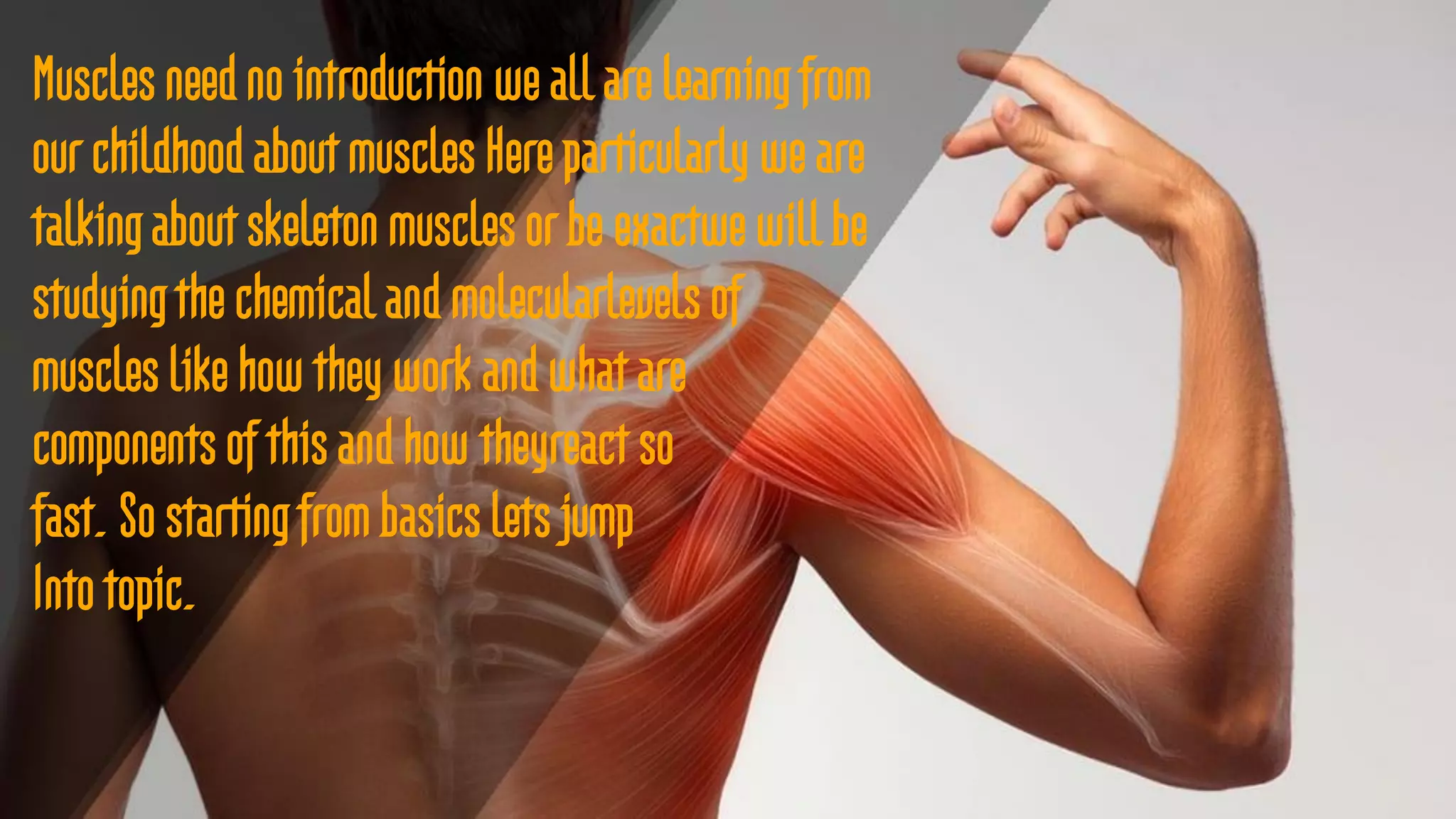
Muscles need nointroduction we all are learning from
our childhood about muscles Here particularly we are
talking about skeleton muscles or be exactwe will be
studying the chemical and molecularlevels of
muscles like how they work and what are
components of this and how theyreact so
fast. So starting from basics lets jump
Into topic.
4.
what are muscles?
Muscles are biological machines
that are made up of proteins
converts the chemical energy
from the ATP and CP
They are able to take receive and
respond to stimuli.
We can only control voluntary
muscles or Skeleton muscles.
First basics
Skeletal muscle
is amuscle tissue that is attached
to the bones and is involved in the
functioning of different parts of
the body.
These muscles are also called
voluntary muscles as they come
under the control of the nervous
system in the body.
Actinis a familyof globular multi-functional proteins that form microfilaments in the cytoskeleton, and the
thin filaments in muscle fibrils. It is found in essentially all eukaryotic cells, where it may be present at a
concentration of over 100 μM; its mass is roughly 42-kDa, with a diameter of 4 to 7 nm.
Myosinare motor proteins that interact with actin filaments and couple hydrolysis of ATP to conformational
changes that result in the movement of myosin and an actin filament relative to each other. ... During each cycle,
myosin moves 5 – 25 nm and one ATP is hydrolyzed.
Tropomyosin are a large family of integral components of actin filaments that play a critical role in
regulating the function of actin filaments in both muscle and nonmuscle cells. These proteins consist of rod-
shaped coiled-coil hetero- or homo-dimers that lie along the α-helical groove of most actin filaments.
Muscle proteins and Chemicals
9.
The Sarcomere isthe basic contractile unit for both striated and cardiac muscle and is made
up of a complex mesh of thick filaments, thin filaments, and a giant protein titin.
Troponinsare a group of proteins found in skeletal and heart (cardiac) muscle fibers that
regulate muscular contraction. Troponin tests measure the level of cardiac-specific troponin in the
blood to help detect heart injury.
A muscle sarcomere's borders are defined by the Z disc (or Z line). The borders of a single
sarcomere are marked by two neighbouring Z discs along the myofibril. The Z discs are where the
thin filaments are attached
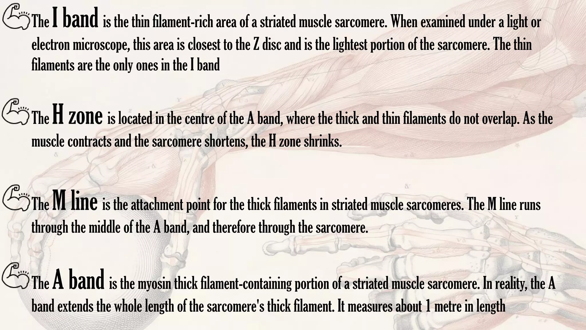
Muscle Fiber Regions
10.
The I bandis the thin filament-rich area of a striated muscle sarcomere. When examined under a light or
electron microscope, this area is closest to the Z disc and is the lightest portion of the sarcomere. The thin
filaments are the only ones in the I band
The A band is the myosin thick filament-containing portion of a striated muscle sarcomere. In reality, the A
band extends the whole length of the sarcomere's thick filament. It measures about 1 metre in length
The H zone is located in the centre of the A band, where the thick and thin filaments do not overlap. As the
muscle contracts and the sarcomere shortens, the H zone shrinks.
The M line is the attachment point for the thick filaments in striated muscle sarcomeres. The M line runs
through the middle of the A band, and therefore through the sarcomere.
11.
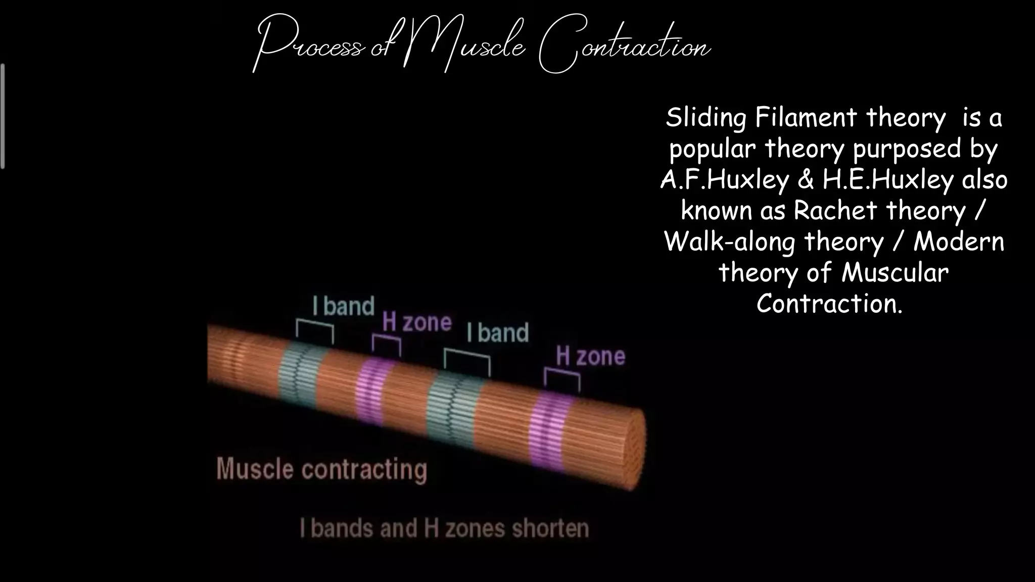
Process of MuscleContraction
Sliding Filament theory is a
popular theory purposed by
A.F.Huxley & H.E.Huxley also
known as Rachet theory /
Walk-along theory / Modern
theory of Muscular
Contraction.
12.
The very firstprocess of muscle contraction is starts in
brain . The nerves carries the signal in the form of
impulse of potential difference and leads to the
neuromuscular junction which is nearly touching the
muscle cell but separated by synaptic cliff . While
muscles have junctional folds within postsynaptic
membrane.
13.
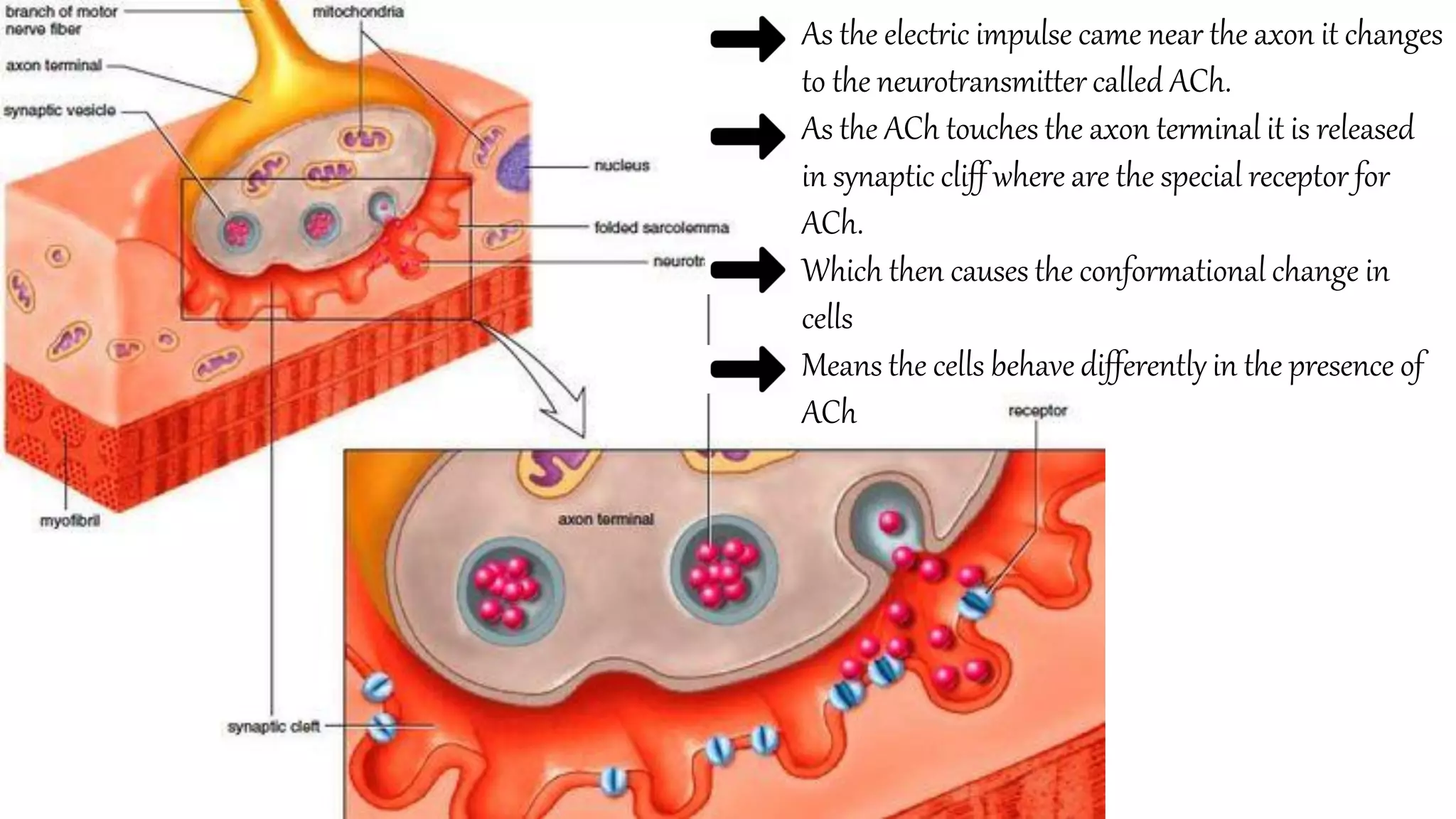
As the electricimpulse came near the axon it changes
to the neurotransmitter called ACh.
As the ACh touches the axon terminal it is released
in synaptic cliff where are the special receptor for
ACh.
Which then causes the conformational change in
cells
Means the cells behave differently in the presence of
ACh
14.
Which leads toan
membrane potential in
sodium and potassium
ions which leads to the
depolarization and
creates a chain reaction
and further increasing
the difference
Once the particular level (Action potential ) is reached an enzyme
releases to degrade the ACh preventing further sodium ions to entering.
But the Action potential continues to journey to T tubules which leads to
the calcium channels.
The action potential then causes the calcium levels to rise in the muscle
cells
15.
The calcium ionsthen binds with the troponin and this leads to a
change in shape in which binding sites available.
Now the myosin head will pivot and bend pulling the actin
filament along and using ATP in the process.
And the muscle contracts
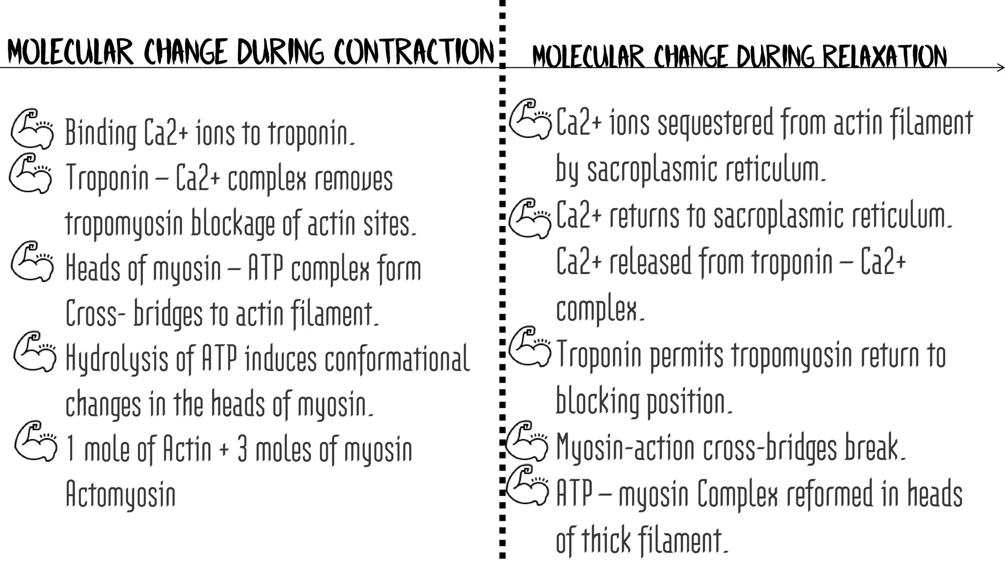
MOLECULAR CHANGE DURINGCONTRACTION MOLECULAR CHANGE DURING RELAXATION
Binding Ca2+ ions to troponin.
Troponin – Ca2+ complex removes
tropomyosin blockage of actin sites.
Heads of myosin – ATP complex form
Cross- bridges to actin filament.
Hydrolysis of ATP induces conformational
changes in the heads of myosin.
1 mole of Actin + 3 moles of myosin
Actomyosin
Ca2+ ions sequestered from actin filament
by sacroplasmic reticulum.
Ca2+ returns to sacroplasmic reticulum.
Ca2+ released from troponin – Ca2+
complex.
Troponin permits tropomyosin return to
blocking position.
Myosin-action cross-bridges break.
ATP – myosin Complex reformed in heads
of thick filament.


![Jaw suspension in vertebrates [autosaved]](https://cdn.slidesharecdn.com/ss_thumbnails/jawsuspensioninvertebratesautosaved-201219155254-thumbnail.jpg?width=640&height=640&fit=bounds)


