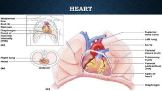
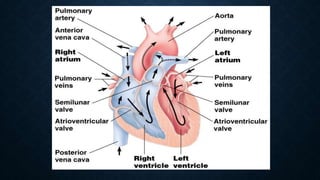
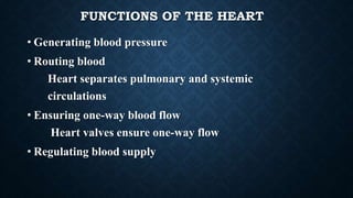
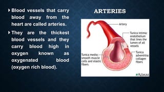
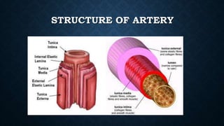
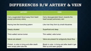
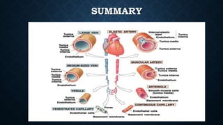
CARDIOVASCULAR SYSTEM
Dr. Pritesh menaria
INTRODUCTION
 The cardiovascular system is transport system of body
 The system supplies nutrients to and remove waste
products from various tissue of body.
 The conveying media is liquid in form of blood which
flows in close tubular system.
FUNCTIONS OF CARDIOVASCULAR SYSTEM
 Transport nutrients, hormones
 Remove waste products
 Gaseous exchange
 Heart pumps blood through blood vessels
COMPONENTS OF CARDIOVASCULAR SYSTEM
•BLOOD
•HEART
•BLOOD VESSELS
BLOOD
•The Blood: Blood cells & Plasma
•Blood cells
1- Erythrocytes - Red Blood Cells
2- Leucocytes
3- Thrombocytes
•Plasma is fluid portion
HEART
• Heart is a four chambered, hollow muscular organ
approximately the size of your fist
• Location:
• Superior surface of diaphragm
• Left of the midline
• Anterior to the vertebral column, posterior to the
sternum
Chapter 18, Cardiovascular System 6
HEART
Chapter 18, Cardiovascular System 7
Figure 18.1
FUNCTIONS OF THE HEART
• Generating blood pressure
• Routing blood
Heart separates pulmonary and systemic
circulations
• Ensuring one-way blood flow
Heart valves ensure one-way flow
• Regulating blood supply
BLOOD VESSELS
•Blood Vessels -A closed network of tubes
•These includes:
 Arteries
 Capillaries
 Veins
CLASSIFICATION OF BLOOD VESSELS
•Conducting Vessels – large arteries
•Distributing Vessels – medium & small arteries
•Resistance Vessels - arterioles
•Exchange Vessels - capillaries & sinusoids
•Capacitance / Reservoir Vessels – venules & vein
BLOOD VESSELS
• General structure
1.Tunica intima
2.Tunica media
3.Tunica adventitia
ARTERIES
 Blood vessels that carry
blood away from the
heart are called arteries.
 They are the thickest
blood vessels and they
carry blood high in
oxygen known as
oxygenated blood
(oxygen rich blood).
ARTERIES
•Accompanied by vein and
nerves
•Lumen is small
•No valves
•Repeated branching
STRUCTURE OF ARTERY
CLASSIFICATION OF ARTERIES
• Elastic / large /conducting arteries - aorta, pulmonary trunk etc.
• Muscular / distributing / medium & small sized arteries-
branches of femoral, brachial, popliteal, axillary carotid arteries
• Arterioles – terminal arterioles
meta arterioles
• Elastic arteries – the largest arteries
• Diameters range from 2.5 cm to 1 cm
• called conducting arteries
• High elastin content in tunica media.
• Muscular (distributing) arteries
• Lie distal to elastic arteries
• Diameters range from 1 cm to 0.3 mm
• Tunica media consist of more muscle fibers.
• Arterioles
• Smallest arteries
• Diameters range from 50 – 100 micron
• Larger arterioles possess all three tunics
• Terminal arterioles (15-20 micron) - devoid of internal elastic lamina &
covered by a continous coat of smooth muscle cells.
• Meta arterioles (10-15 micron) – these are side branches of terminal arterioles.
in this smooth muscles are replaced by discontinous non contractile cells cld
pericytes or rouget cells .
CAPILLARY BEDS
• Precapillary sphincters - regulate the flow of blood to
tissues
CAPILLARIES
• Smallest blood vessels-diameter 6-8 micron.
TYPE OF CAPILLARIES
• On the basis of endothelium
• Continuous
• Fenestrated
SINUSOIDS
• Large ,irregular vascular space which are closely
surrounded by the parenchyma of organ.
• Usually fenestrated
• Have large diameters
• Occur in bone marrow, liver, spleen and lymphoid
tissue
VEINS
• Conduct blood from capillaries toward the heart
• Blood pressure is much lower than in arteries
• Smallest veins – called venules
• Diameters from 8 – 100 µm
• Smallest venules – called post-capillary venules
• Venules join to form veins
VEINS
• Thin Walled
• Large irregular lumen
• Have valves
• Dead space around
• Types:
Large
Medium
Small
Structure Of Veins
Tunica intima
Little more than endothelium
Tunica media
Thin layer of smooth muscle
Tunica adventitia
Thickest layer
VEINS
•Factors responsible for venous return:
1.Muscle contraction
2.Negative intrathoracic pressure
3.Pulsation of arteries
4. Gravity
5. Valves
Skeletal Muscle Pump
DIFFERENCES B/W ARTERY & VEIN
ARTERY VEIN
Carry oxygenated blood away from heart
except pulmonary artery
Carry deoxygenated blood ,towards the
heart except pulmonary vein
Having tree like branching patern Like river they form by small tributaries
Deeply situated Superficial and deep
Thick walled narrow lumen Thin walled, wide lumen
Valves are absent Valve are present for antigravity blood flow
Pinkish in color in living and after death
seen empty pale tube like
Bluish in color in living and after death it get
filled up and seen grayish
ANASTOMOSIS
• Communication between the neighboring blood vessels
• Type – arterial, venous and arterio- venous anastomosis
ARTERIAL ANASTOMOSIS
• Communication between
arteries
• Provides alternate rout for the
blood to reach the tissue in case
if there is compression of
vessels
• Prevent the death of tissue
supplied by blocked artery
• Collateral circulation
PALMAR ARCHES
END ARTERIES
• Arteries which do not anastomose with neighboring arteries at
pre-capillary level
• Blockage of end arteries the area of the tissue supplied by end
artery will die
• e. g. central artery of retina, central branches of cerebral
arteries;vasa recta of mesenteric arteries, arteries of spleen,
kidney, lungs
VENOUS ANASTOMOSIS
Dorsal Venous Arches
ARTERIO- VENOUS ANASTOMOSIS
• Communication
between the arteriole
and venule
• e. g. tip of nose, lips
lobules of the ear,
finger tips etc.
TYPES OF CIRCULATION
• Pulmonary
• Systemic
• Portal
PULMONARY CIRCULATION
SYSTEMIC CIRCULATION
PORTAL CIRCULATION
• Circulation through the portal vein
• The vein present between two sets of capillary networks
is called as portal vein
• Carries blood from one location in the body to another
without passing through heart
• Sites: liver, kidney, anal canal, hypophysis cerebrii
HEPATIC PORTAL SYSTEM
FETAL CIRCULATION
BLOOD SUPPLY OF VESSELS
• By vasa vasorum- tunica adventitia and outer
2/3 part of tunica media
• By diffusion- inner 1/3 part of tunica media
and tunica intima
NERVE SUPPLY OF VESSELS
• Muscles of tunica media are innervated by
autonomic nerves
• Nervi- vascularis
• Vasomotor, vasodilator nerve, sensory nerves
APPLIED
• Blood pressure- arterial pressure exerted by the blood on the
arterial walls.
• Hemorrhage or bleeding - rupture of blood vessels, venous
hemorrhage causes oozing of blood and arterial hemorrhage
cause spurting of blood
• Arteriosclerosis- stiffness of arteries
• Arteritis- inflammation of arteries
• Phlebitis- inflammation of veins
• Aneurysm- swelling or dilation of blood vessels
Arteriosclerosis:
SUMMARY
SUMMARY
THANK YOU!

Cardio vascular system (1).pptx

  • 1.
  • 2.
    INTRODUCTION  The cardiovascularsystem is transport system of body  The system supplies nutrients to and remove waste products from various tissue of body.  The conveying media is liquid in form of blood which flows in close tubular system.
  • 3.
    FUNCTIONS OF CARDIOVASCULARSYSTEM  Transport nutrients, hormones  Remove waste products  Gaseous exchange  Heart pumps blood through blood vessels
  • 4.
    COMPONENTS OF CARDIOVASCULARSYSTEM •BLOOD •HEART •BLOOD VESSELS
  • 5.
    BLOOD •The Blood: Bloodcells & Plasma •Blood cells 1- Erythrocytes - Red Blood Cells 2- Leucocytes 3- Thrombocytes •Plasma is fluid portion
  • 6.
    HEART • Heart isa four chambered, hollow muscular organ approximately the size of your fist • Location: • Superior surface of diaphragm • Left of the midline • Anterior to the vertebral column, posterior to the sternum Chapter 18, Cardiovascular System 6
  • 7.
  • 9.
    FUNCTIONS OF THEHEART • Generating blood pressure • Routing blood Heart separates pulmonary and systemic circulations • Ensuring one-way blood flow Heart valves ensure one-way flow • Regulating blood supply
  • 10.
    BLOOD VESSELS •Blood Vessels-A closed network of tubes •These includes:  Arteries  Capillaries  Veins
  • 11.
    CLASSIFICATION OF BLOODVESSELS •Conducting Vessels – large arteries •Distributing Vessels – medium & small arteries •Resistance Vessels - arterioles •Exchange Vessels - capillaries & sinusoids •Capacitance / Reservoir Vessels – venules & vein
  • 12.
    BLOOD VESSELS • Generalstructure 1.Tunica intima 2.Tunica media 3.Tunica adventitia
  • 13.
    ARTERIES  Blood vesselsthat carry blood away from the heart are called arteries.  They are the thickest blood vessels and they carry blood high in oxygen known as oxygenated blood (oxygen rich blood).
  • 14.
    ARTERIES •Accompanied by veinand nerves •Lumen is small •No valves •Repeated branching
  • 16.
  • 17.
    CLASSIFICATION OF ARTERIES •Elastic / large /conducting arteries - aorta, pulmonary trunk etc. • Muscular / distributing / medium & small sized arteries- branches of femoral, brachial, popliteal, axillary carotid arteries • Arterioles – terminal arterioles meta arterioles
  • 18.
    • Elastic arteries– the largest arteries • Diameters range from 2.5 cm to 1 cm • called conducting arteries • High elastin content in tunica media.
  • 19.
    • Muscular (distributing)arteries • Lie distal to elastic arteries • Diameters range from 1 cm to 0.3 mm • Tunica media consist of more muscle fibers.
  • 20.
    • Arterioles • Smallestarteries • Diameters range from 50 – 100 micron • Larger arterioles possess all three tunics • Terminal arterioles (15-20 micron) - devoid of internal elastic lamina & covered by a continous coat of smooth muscle cells. • Meta arterioles (10-15 micron) – these are side branches of terminal arterioles. in this smooth muscles are replaced by discontinous non contractile cells cld pericytes or rouget cells .
  • 22.
    CAPILLARY BEDS • Precapillarysphincters - regulate the flow of blood to tissues
  • 23.
    CAPILLARIES • Smallest bloodvessels-diameter 6-8 micron.
  • 24.
    TYPE OF CAPILLARIES •On the basis of endothelium • Continuous • Fenestrated
  • 25.
    SINUSOIDS • Large ,irregularvascular space which are closely surrounded by the parenchyma of organ. • Usually fenestrated • Have large diameters • Occur in bone marrow, liver, spleen and lymphoid tissue
  • 27.
    VEINS • Conduct bloodfrom capillaries toward the heart • Blood pressure is much lower than in arteries • Smallest veins – called venules • Diameters from 8 – 100 µm • Smallest venules – called post-capillary venules • Venules join to form veins
  • 28.
    VEINS • Thin Walled •Large irregular lumen • Have valves • Dead space around • Types: Large Medium Small
  • 30.
    Structure Of Veins Tunicaintima Little more than endothelium Tunica media Thin layer of smooth muscle Tunica adventitia Thickest layer
  • 32.
    VEINS •Factors responsible forvenous return: 1.Muscle contraction 2.Negative intrathoracic pressure 3.Pulsation of arteries 4. Gravity 5. Valves
  • 33.
  • 34.
    DIFFERENCES B/W ARTERY& VEIN ARTERY VEIN Carry oxygenated blood away from heart except pulmonary artery Carry deoxygenated blood ,towards the heart except pulmonary vein Having tree like branching patern Like river they form by small tributaries Deeply situated Superficial and deep Thick walled narrow lumen Thin walled, wide lumen Valves are absent Valve are present for antigravity blood flow Pinkish in color in living and after death seen empty pale tube like Bluish in color in living and after death it get filled up and seen grayish
  • 36.
    ANASTOMOSIS • Communication betweenthe neighboring blood vessels • Type – arterial, venous and arterio- venous anastomosis
  • 37.
    ARTERIAL ANASTOMOSIS • Communicationbetween arteries • Provides alternate rout for the blood to reach the tissue in case if there is compression of vessels • Prevent the death of tissue supplied by blocked artery • Collateral circulation
  • 38.
  • 39.
    END ARTERIES • Arterieswhich do not anastomose with neighboring arteries at pre-capillary level • Blockage of end arteries the area of the tissue supplied by end artery will die • e. g. central artery of retina, central branches of cerebral arteries;vasa recta of mesenteric arteries, arteries of spleen, kidney, lungs
  • 40.
  • 41.
    ARTERIO- VENOUS ANASTOMOSIS •Communication between the arteriole and venule • e. g. tip of nose, lips lobules of the ear, finger tips etc.
  • 42.
    TYPES OF CIRCULATION •Pulmonary • Systemic • Portal
  • 44.
  • 45.
  • 46.
    PORTAL CIRCULATION • Circulationthrough the portal vein • The vein present between two sets of capillary networks is called as portal vein • Carries blood from one location in the body to another without passing through heart • Sites: liver, kidney, anal canal, hypophysis cerebrii
  • 47.
  • 48.
  • 49.
    BLOOD SUPPLY OFVESSELS • By vasa vasorum- tunica adventitia and outer 2/3 part of tunica media • By diffusion- inner 1/3 part of tunica media and tunica intima
  • 50.
    NERVE SUPPLY OFVESSELS • Muscles of tunica media are innervated by autonomic nerves • Nervi- vascularis • Vasomotor, vasodilator nerve, sensory nerves
  • 51.
    APPLIED • Blood pressure-arterial pressure exerted by the blood on the arterial walls. • Hemorrhage or bleeding - rupture of blood vessels, venous hemorrhage causes oozing of blood and arterial hemorrhage cause spurting of blood • Arteriosclerosis- stiffness of arteries • Arteritis- inflammation of arteries • Phlebitis- inflammation of veins • Aneurysm- swelling or dilation of blood vessels
  • 52.
  • 55.
  • 56.
  • 57.