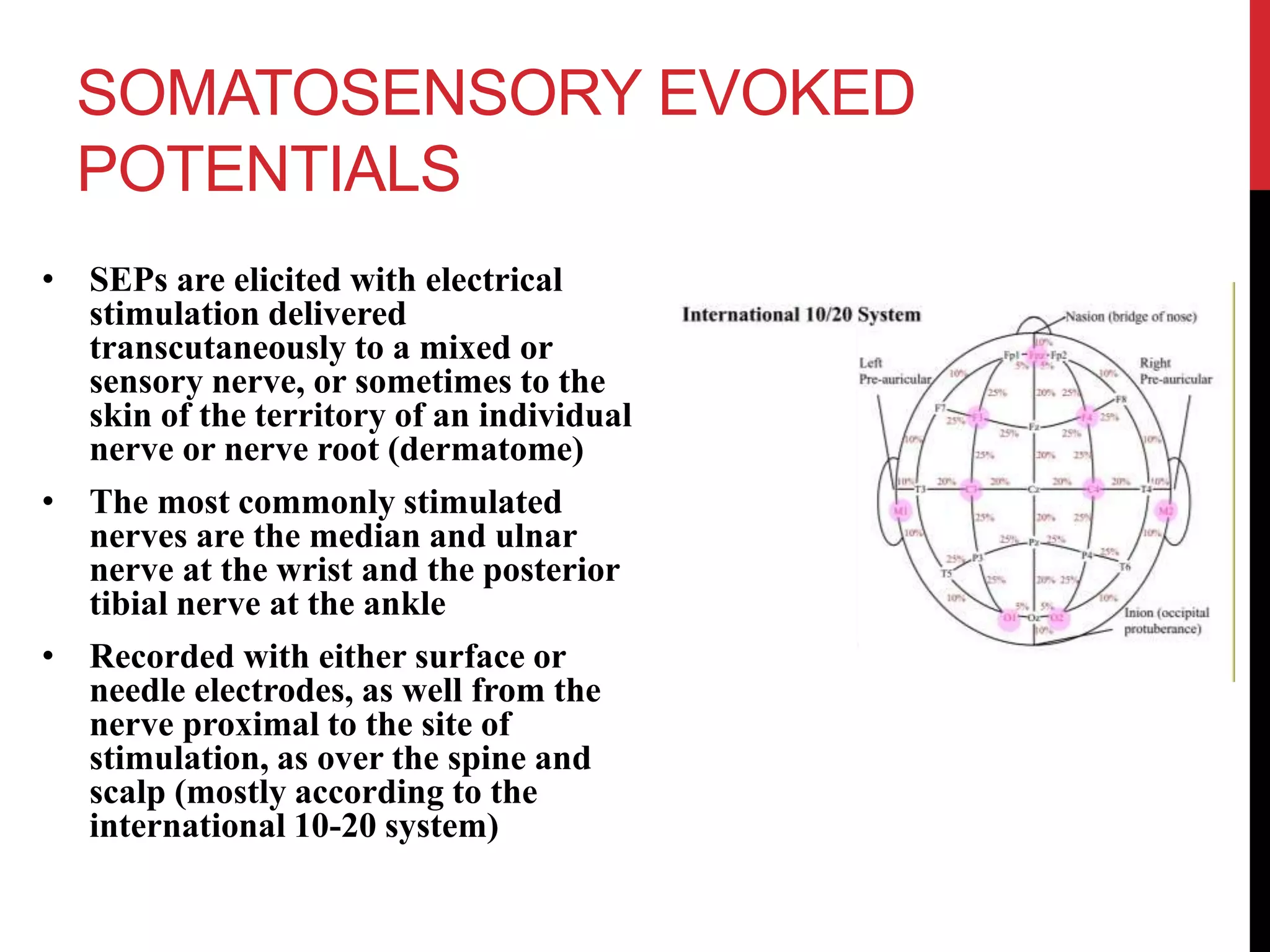
2. OBJECTIVES
- To provide a brief overview of somatosensory evoked potentials
(SEPs) and motor evoked potentials (MEPs)
- To discuss briefly the different clinical applications of SEPs and
MEPs
3. SOMATOSENSORY EVOKED
POTENTIALS
• SEPs are elicited with electrical
stimulation delivered
transcutaneously to a mixed or
sensory nerve, or sometimes to the
skin of the territory of an individual
nerve or nerve root (dermatome)
• The most commonly stimulated
nerves are the median and ulnar
nerve at the wrist and the posterior
tibial nerve at the ankle
• Recorded with either surface or
needle electrodes, as well from the
nerve proximal to the site of
stimulation, as over the spine and
scalp (mostly according to the
international 10-20 system)
4. SOMATOSENSORY
EVOKED POTENTIALS
• Clinical Applications
• It provides limited information on the exact location of the
lesions proximal to the dorsal root ganglion
• Helpful in diagnosis of numerous spinal disorders, determining
prognosis, evaluating treatment, and in following up patients
• SEPs seem to be rather sensitive in predicting outcome in the
acute phase of SCI and stroke (when SEPs are absent, prognosis
seems to be poor)
• Helpful in evaluation of the severity and level of the lesion in
cervical spondylytic myelopathy
• Patients in coma are unlikely to recover from their condition
when the cortical responses of SEPs are bilaterally absent
5. MOTOR EVOKED
POTENTIALS
• MEP procedure consists of transcranial stimulation followed by
measurement of the compound muscle action potential (CMAP)
from different limb and trunk muscles
• Can be elicited with magnetic brain stimulation or
transcutaneous electrical stimulation
6. MOTOR EVOKED
POTENTIALS
• Clinical Applications
• Multiple sclerosis – delayed CMCTs, absent MEPs in patient’s
with marked clinical disability
• ALS – prolonged or absent MEPs
• SCI – strong correlation seen between MEP findings and motor
function
• Decreased MEP amplitudes or absent MEP responses are more
frequently seen in neoplastic lesions
• MEP with more often increased latencies are seen in
inflammatory lesions
7. MOTOR EVOKED
POTENTIALS
• Clinical Applications
• Spondylotic myelopathy
• CMCT has been reported to correlate well with clinical and
radiological signs of cord compression
• MEP > SEP abnormality in cervical spondylosis
• Lumbosacral radiculopathies – electrical stimulation is useful
versus TMS
• Cervical radiculopathies – both electrical stimulation and TMS
are not so useful detectors
• Stroke – prognostic value (if MEPs are present during the acute
phase, outcome usually is favorable; however, absent MEPs are
not correlated with poor outcome)
8. SUMMARY
• Somatosensory evoked potentials and its clinical applications ✔
• Motor evoked potentials and its clinical applications ✔
Depending on the purpose of the test there are many important parameters that can be measured in cortical stimulation, such as stimulation threshold, MEP latency, MEP amplitude, response morphology, central motor conduction time, silent period duration, fatigue, intracortical inhibitory and excitatory pathways, etc.
The central motor conduction time can be calculated by substracting the latency in response to spinal root stimulation from the latency in response to cortical stimulation.
It may also be caluclated by using the F wave latency
CMCT = latency from cortex to muscle
= F latency – (motor terminal latency + 1) / 2
Central motor conducting time
In multiple sclerosis, most authors found clearly delayed CMCTs. Absent MEPs were seen in patients with makred clinical disability
MEPs in SCI can help differentiate between neoplastic versus inflammatory lesions.
Neoplastic lesions – decreased MEP amplitudes or absent MEP responses are seen
inflammatory lesions – MEPS more often have increased latencies
Central motor conducting time
In cervical spondylosis MEP abnormalities exceed those of SEPs, most probably because spondylosis and disc herniation are more likely to produce compression of the corticospinal tracts than the dorsal columns
SEP - They represent the function of the ascending sensory pathways using an afferent potential, which travels from the peripheral nerve to the plexus, root, spinal cord (posterior column), contralateral medial lemniscus, thalamus, to the somatosensory cortex.
SSEP monitors for problems such as: (a) peripheral nerve injuries, (b) CNS lesions such as multiple sclerosis (increased interpeak latency), or (c) intra-operative monitoring of spinal surgery.