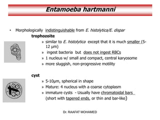
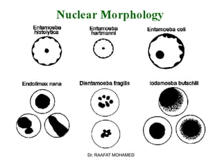
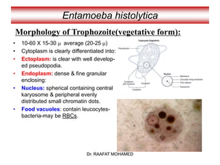
Amoebas
Introduction
• The most important feature that separates amebas from the other
groups of unicellular Protozoa is the means by which they move.
• Amebas are equipped with the ability to extend their cytoplasm in
the form of pseudopods (often referred to as false feet), which
allows them move within their environment.
• With one exception, there are two morphologic forms in the amebic
life cycle—trophozoites, the form that feeds, multiplies, and
possesses pseudopods, and cysts, the nonfeeding stage
characterized by a thick protective cell wall designed to protect the
parasite from the harsh outside environment when deemed
necessary.
Dr. RAAFAT MOHAMED
Entamoeba histolytica
Causes : Amoebiasis.
Geog.Distribution: cosmopolitan
Habitat: caecum and sigmoido-
rectal region of man.
Infective stage:
Quadrinucleate cyst.
Mode of infection:
Eating raw vegetables (salad)
Drinking water
Flies and food handlers (cyst passer)
Faeco-oral
Heteroinfection
Autoinfection
Trophozoite
Cyst
caecum
Dr. RAAFAT MOHAMED
Dr. RAAFAT MOHAMED
• 10-60 X 15-30 m average (20-25 m)
• Cytoplasm is clearly differentiated into:
• Ectoplasm: is clear with well develop-
ed pseudopodia.
• Endoplasm: dense & fine granular
enclosing:
• Nucleus: spherical containing central
karyosome & peripheral evenly
distributed small chromatin dots.
• Food vacuoles: contain leucocytes-
bacteria-may be RBCs.
Entamoeba histolytica
Morphology of Trophozoite(vegetative form):
Dr. RAAFAT MOHAMED
Dr. RAAFAT MOHAMED
Entamoeba histolytica
Dr. RAAFAT MOHAMED
Dr. RAAFAT MOHAMED
Entamoeba histolytica
Precyst stage:-
10-60 X 15-30 m average (15-20 m)
-Round or oval with a blunt pseudopodia.
-Absent cyst wall
-Single nucleus present.
Cyst stage:-
10-20 m average (15 m)
-Four nuclei are present in mature
quadrinucleated cyst
-Glycogen mass & chromatoid bodies are
present in immature cysts –disappear in
mature ones.
Dr. RAAFAT MOHAMED
Dr. RAAFAT MOHAMED
Entamoeba histolytica
Morphology of cyst:
Dr. RAAFAT MOHAMED
Dr. RAAFAT MOHAMED
Life Cycle of Entamoeba inside human colon
Mucosa of large intestine
In the
lumen
Quadrinucleate cyst
Enter with food
Pass out in stool
Precyst Uninucleate cyst Binucleate cyst
Binary fission
Attached
to
mucosa
trophozoite
Dr. RAAFAT MOHAMED
Lumen(non invasive) form
Dr. RAAFAT MOHAMED
Entamoeba histolytica
Dr. RAAFAT MOHAMED
Life cycle of
E. histolytica
Dr. RAAFAT MOHAMED
Pathogenesis
Depends on:
 Parasite virulence.
 Host resistance.
 Condition of the intestinal tract.
Non-pathogenic:
in the lumen.
OR Pathogenic:
trophozoites invade intestinal mucosa.
Trophozoites produce histolytic enzyme
that produce necrosis of mucosa
leading to the formation of
flask-shaped ulcer. Trophozoites exist in the base
of the ulcer
Trophozoite
Dr. RAAFAT MOHAMED
Tissue (invasive form)
Dr. RAAFAT MOHAMED
Dr. RAAFAT MOHAMED
Pathogenesis (Cont.)
Factors determining Pathogenicity
1- Strain E.dispar similar to E. histolytica differ in being non invasive
2- Virulence Virulent strains are capable of transformation into invasive due to:-
* Adherence and colonization * Enterotoxin production
* Contact dependent cell lysis * Lytic enzyme secretion (proteolysis)
* Phagocytic activity
3-Host factors
1- Immunity (secretory IgA) 2-Nutrition: carbohydrate rich diet ↑, protein ↓, change diet habit
3- Drugs: immunosuppressive. 4- Debilitating states (malignancy, pregnancy, etc),.
5- Intestine: bacteria & intestinal flora –hypermotility or stasis of the bowel.
Dr. RAAFAT MOHAMED
Pathogenesis & Complications
Dr. RAAFAT MOHAMED
Dr. RAAFAT MOHAMED
This is followed by:
• Proliferation of connective tissue.
• Intensive ulcerations.
• Extra-intestinal invasion to brain,
liver, lung or skin.
Brain
abscess
Liver
abscess
(common)
Lung
abscess
Skin
abscess
Blood vessel
Dr. RAAFAT MOHAMED
Dr. RAAFAT MOHAMED
Clinical Picture
Asymptomatic: parasite in lumen and cysts pass in stool.
(healthy cyst passer – most common – more than 75%)
Symptomatic: (gradual onset), fever (low grade), diarrhea,
dysentery, abdominal pain, localized abdominal tenderness,
tenesmus & strain, painful spasm of anal sphincter (indicates
rectal ulceration). Acute intestinal amoebiasis (colitis)
Recurrent attacks of dysentery with intervening periods of
constipation, abdominal distension & Flatulence, weight loss
and cachexia. Chronic intestinal amoebiasis.
Rare progressive disease of high mortality (high fever- severe
bloody diarrhea – diffuse tenderness – peritonitis)
(fulminating colitis)
Amoebic hepatitis or amoebic abscess, lung abscess, brain
abscess or skin abscess.
Extra-intestinal amoebiasis
Dr. RAAFAT MOHAMED
Dr. RAAFAT MOHAMED
Complications
• Amoeboma.
(localized granulomatous mass
misdiagnosed with carcinoma)
• Hemorrhage.
• Perforation of ulcer.
(secondary peritonitis --- rare but fatal)
• Stricture of colon.
(secondary to fibrosis)
• Appendicitis.
Dr. RAAFAT MOHAMED
Dr. RAAFAT MOHAMED
Diagnosis (Intestinal amoebiasis)
• Clinically: Dysentery: painful frequent evacuation of
small quantities of stool containing mucus tinged
with blood.
• Laboratory:
1- Direct stool examination: Trophozoites are found
in diarrhoeic stool. Cysts are found in formed stool.
- Wet preparation.
- Iodine stained.
- Permanent stain with iron haematoxylin or
trichrome.
2- Concentration techniques for cysts.
Dr. RAAFAT MOHAMED
Dr. RAAFAT MOHAMED
In the Lab
Typical amoebic
dysentery stool
Bulky.
Acidic.
Scanty exudate.
Pus cells +
Blood +
Charcot Leyden
Crystals : present.
Amoebae trophozoites :
present.
Typical bacillary
dysentery stool
Scanty.
Alkaline.
Massive exudate.
Pus cells +++
Blood +
Charcot Leyden
Crystals : absent.
Amoebae trophozoites :
absent.
Dr. RAAFAT MOHAMED
Dr. RAAFAT MOHAMED
Diagnosis (intestinal amoebiasis)
3- Indirect diagnosis:
Serological tests in chronic amoebiasis.
Detection of copro-antigen using
monoclonal antibodies.
Molecular techniques.
• Radiological examination:
using barium enema.
Coating the well with MAb and add patient’s stool to detect
Entamoeba Ag
wash wash wash
Dr. RAAFAT MOHAMED
Dr. RAAFAT MOHAMED
• Sigmoidoscopy: to visualize the ulcer, scrap,
aspirate or take biopsy to see the trophozoites.
Sigmoidoscope
Sigmoidoscopy
Dr. RAAFAT MOHAMED
Dr. RAAFAT MOHAMED
Diagnosis (Extraintestinal amoebiasis)
• Clinical: according to the organ affected.
• Laboratory:
1-Examination of aspirate from lung or liver abscesses
for trophozoites.
2-Liver scanning.
3-Radiology of diaphragm level and pulmonary lesions.
4-Serology.
5-Leucocytosis due to 2ry bacterial infection.
Coat with Ag and look for
Ab in patient’s serum
Coat with Ab and look for
Ag in patient’s serum
Dr. RAAFAT MOHAMED
Dr. RAAFAT MOHAMED
Treatment
• Metronidazole, Tinidazole.
Very effective in killing amoebas in the wall of the
intestine, in blood and in liver abscesses.
• Diluxanide furoate.
kills trophozoites and cysts in the lumen of the
intestine.
Luminal amoebicide
Asymptomatic patients: are given luminal
amoebicide as Diluxanide furoate.
Symptomatic patients: are given tissue amoebicide
as Metronidazole followed by luminal amoebicide
as Diluxanide furoate.
Tissue amoebicide
Dr. RAAFAT MOHAMED
Dr. RAAFAT MOHAMED
Treatment of Amoebic abscesses
by aspiration or open surgical drainage
Aspiration of abscess
Surgical
drainage of
abscess
Thick chocolate-
coloured or anchovy-
sauce pus with
trophozoites
Dr. RAAFAT MOHAMED
Dr. RAAFAT MOHAMED
Epidemiology
• Cyst passers are the main source of infection.
• Cysts remain viable in faeces for few days, in
water for longer periods.
• Cysts are killed by dryness, heat (over 55ºC)
and by chlorine.
Control
 Treatment of patients.
 Examination and treatment of food handlers.
 Environmental sanitation.
 Personal prophylaxis.
Human faeces should not be used as fertilizers.
Dr. RAAFAT MOHAMED
Dr. RAAFAT MOHAMED
Check for understanding
M.C.Q.
1- Entamoeba histolytica trophozoites are found in:
a- Duodenum of infected human.
b- Jejunum of infected human.
c- Caecum of infected human.
d- All of the above.
2- Infection with Entamoeba histolytica occurs
through eating green salad contaminated with:
a- Trophozoites of Entamoeba histolytica.
b- Cysts of Entamoeba histolytica.
c- Both trophozoites and cysts of Entamoeba
histolytica.
Dr. RAAFAT MOHAMED
Dr. RAAFAT MOHAMED
3- Pathogenicity of Entamoeba histolytica depends on:
4- Patients with chronic intestinal amoebiasis suffer from:
a- Dysentery, tenesmus with painful spasm of anal sphincter.
b- Recurrent attacks of dysentery alternating with constipation.
c- Fever, diarrhoea and tenesmus.
d- Non of the above.
a- Parasite virulence.
b- Host resistance.
c- Condition of intestinal tract.
d- All of the above.
5- The following findings in stool suggest amoebic not bacillary
dysentery infection:
a- Acidic pH and presence of Charcot Leyden Crystals.
b- Alkaine pH and presence of Charcot Leyden Crystals.
c- Massive exudate with presence of many pus cells.
Dr. RAAFAT MOHAMED
Dr. RAAFAT MOHAMED
State True Or False
• Cyst passers are the main source of Entamoeba
histolytica infection.
• Trophozoites of Entamoeba histolytica produce ulcers
with indurated margin in intestinal mucosa.
• Examination and treatment of food handlers is very
important to control Entamoeba histolytica infection.
• Infection with Entamoeba histolytica is totally
localized to the gastrointestinal tract.
• Both trophozoites and cysts of Entamoeba histolytica
are infective to man.
True
False
True
False
False
Dr. RAAFAT MOHAMED
Dr. RAAFAT MOHAMED
M.C.Q.
Rounded quadrinucleate cysts (20µ) seen in stool
of patients should be reported:
1- Entamoeba histolytica cyst
2- Entamoeba dispar cyst
3- Entamoeba histolytica / dispar cyst
4- Entamoeba coli cyst
Dr. RAAFAT MOHAMED
Dr. RAAFAT MOHAMED
Dr. RAAFAT MOHAMED
CASE STUDY
• A 28 year old male presented with
low grade fever, loss of appetite and
pain in right upper quadrant since 15
days.
• Three months back, he suffered
from dysentery.
• Physical examination revealed an
enlarged liver
• Blood picture showed leucocytosis
with raised eosinophils.
• CT scan showed the presence of
cavitatory lesion in the right side of
the liver.
• Aspirate from liver showed motile
trophozoites in wet mount
preparation.
Dr. RAAFAT MOHAMED
Entamoeba dispar
Dr. RAAFAT MOHAMED
• E. dispar - formerly designated as non-pathogenic E.
histolytica
• 9x more prevalent than E. histolytica
• Morphologically identical with E. histolytica
• their DNA and ribosomal RNA are different
• microscopic identification unreliable
• Dx: - negative serologic tests
- Trophozoite will have no ingested RBC
Dr. RAAFAT MOHAMED
Entamoeba hartmanni
Dr. RAAFAT MOHAMED
• Morphologically indistinguishable from E. histolytica/E. dispar
trophozoite
» similar to E. histolytica except that it is much smaller (5-
12 µm)
» ingest bacteria but does not ingest RBCs
» 1 nucleus w/ small and compact, central karyosome
» more sluggish, non-progressive motility
cyst
» 5-10µm, spherical in shape
» Mature: 4 nucleus with a coarse cytoplasm
» immature cysts - Usually have chromatoidal bars
(short with tapered ends, or thin and bar-like)
Dr. RAAFAT MOHAMED
Entamoeba hartmanni
Dr. RAAFAT MOHAMED
Dr. RAAFAT MOHAMED
Entamoeba coli
Dr. RAAFAT MOHAMED
*most common endocommensal of humans
* has a worldwide distribution
* 10-50% of the population can be infected worldwide
* Feeds on bacteria and any other cells available to it
* does not invade tissues
* common inhabitant of the lumen of the cecum and
colon of man and other animals
* Has the typical Entamoeba nucleus
Dr. RAAFAT MOHAMED
Entamoeba coli
Dr. RAAFAT MOHAMED
Trophozoites
• Usually 15-25 μm in diameter (range 10-50 µm)
• Cytoplasm:
– More vacuolated or granular endoplasm with
bacteria and debris but no RBCs
– Dirty-looking or honeycomb appearance
– Narrower, less differentiated ectoplasm
• Pseudopodia: broader and blunter
– function more to ingest food
– sluggish, non-directional motility
• Nucleus
– 1 nucleus
– Thicker, irregular, coarsely granular peripheral
chromatin with a large eccentric karyosome
(not compact, may/may not be eccentric)
Dr. RAAFAT MOHAMED
Entamoeba coli
Dr. RAAFAT MOHAMED
Cysts
• size: 10-35 μm
• Nucleus
– Usually spherical
– mature cyst: 8 nuclei
– Immature cyst: 2 or more nuclei
– Karyosome is large, may/may not
be compact and/or eccentric
• Cytoplasm: coarsely granular
• chromatoidal bodies: Splinter-shaped
or broom-shaped
with rough, pointed end
Dr. RAAFAT MOHAMED
Entamoeba coli
Dr. RAAFAT MOHAMED
Dr. RAAFAT MOHAMED
Entamoeba coli
Dr. RAAFAT MOHAMED
Dr. RAAFAT MOHAMED
Entamoeba gingivalis
Dr. RAAFAT MOHAMED
• a common inhabitant of the mouth of man
• lives on the surface of teeth and gums, in gum pockets and
sometimes in the tonsillar crypts
• Organisms are more common in persons with pyorrhea
(gum disease) but they are not the cause of the condition
• Hosts: Humans, other primates, dogs and cats
• Prevalence is from 50 to 95%
• Transmission:
– kissing
– droplet spray
– sharing eating utensils
Dr. RAAFAT MOHAMED
Entamoeba gingivalis
Dr. RAAFAT MOHAMED
Trophozoite
• 10-20µm
• Moves quickly
• Has numerous blunt pseudopodia
• Has numerous food vacuoles that
contain cellular debris and bacteria
and ingested leukocytes
• no cyst stage
Dr. RAAFAT MOHAMED
Entamoeba gingivalis
Dr. RAAFAT MOHAMED
Dr. RAAFAT MOHAMED
Endolimax nana
Dr. RAAFAT MOHAMED
• Second most common endocommensal of humans
• worldwide distribution 30%
• Lives in the large intestine mainly near the cecum
• feed on bacteria
• non pathogenic
• Outstanding feature:
– larger karyosome than those of the genus Entamoeba
– absent peripheral chromatin
Dr. RAAFAT MOHAMED
Endolimax nana
Dr. RAAFAT MOHAMED
Trophozoites
• small size of 6 to 15 µm
• Cytoplasm is granular and vacuolated
• nucleus exhibits a large irregularly shaped karyosome
(may appear “blot-like”) with no peripheral chromatin
on the nuclear membrane
• Pseudopodia are blunt and hyaline
• Sluggish, non-progressive motility
• Cyst
• Spherical or ovoid in shape
• Mature cyst
– 5 – 14 μm in diameter
– 4 nuclei when mature
– A refractile cyst wall present
– Chromatoidal bodies are not usually found
Dr. RAAFAT MOHAMED
Endolimax nana
Dr. RAAFAT MOHAMED
Dr. RAAFAT MOHAMED
Iodamoeba bϋtschlii
Dr. RAAFAT MOHAMED
• Not very common endocommensal in people
• non-pathogenic
• Lives in the large intestine, predominantly in the cecal areas
• Has a very high prevalence in pigs
– 50% of pigs are infected with this ameba in France and Egypt
– pigs are probably its normal host
Dr. RAAFAT MOHAMED
Iodamoeba bϋtschlii
Dr. RAAFAT MOHAMED
Trophozoites
• Usually 9-14 μm long (range 6-20 μm)
• Single large vesicular nucleus with large chromatin- rich
karyosome , surrounded by a layer
of achromatic granules globules and anchored to
nuclear membrane by achromatic fibrils
( described as “basket nuclei”)
• Cytoplasm appears granular, containing vacuoles
with ingested bacteria and debris
• Hyaline pseudopodia with sluggish progressive
motility
Cysts
• 5-20 µm , oval-round in shape
• uninucleated “basket nuclei”
• Prominent characteristic: large glycogen vacuole
which stains deeply w/ iodine
Dr. RAAFAT MOHAMED
Iodomoeba butschlii
Dr. RAAFAT MOHAMED
Dr. RAAFAT MOHAMED
Dientamoeba fragilis
Dr. RAAFAT MOHAMED
Trophozoite No Cyst Nucleus
LIFE CYCLE - it does not form cysts and trophozoites cannot survive
passage through the small intestine.
Humans probably get infected by this endocommensal when they
ingest pinworm eggs!
Dr. RAAFAT MOHAMED
Dr. RAAFAT MOHAMED
Dr. RAAFAT MOHAMED
Dr. RAAFAT MOHAMED

Chapter 3 - THE AMOEBAS.ppt

  • 1.
  • 2.
    Introduction • The mostimportant feature that separates amebas from the other groups of unicellular Protozoa is the means by which they move. • Amebas are equipped with the ability to extend their cytoplasm in the form of pseudopods (often referred to as false feet), which allows them move within their environment. • With one exception, there are two morphologic forms in the amebic life cycle—trophozoites, the form that feeds, multiplies, and possesses pseudopods, and cysts, the nonfeeding stage characterized by a thick protective cell wall designed to protect the parasite from the harsh outside environment when deemed necessary.
  • 9.
    Dr. RAAFAT MOHAMED Entamoebahistolytica Causes : Amoebiasis. Geog.Distribution: cosmopolitan Habitat: caecum and sigmoido- rectal region of man. Infective stage: Quadrinucleate cyst. Mode of infection: Eating raw vegetables (salad) Drinking water Flies and food handlers (cyst passer) Faeco-oral Heteroinfection Autoinfection Trophozoite Cyst caecum Dr. RAAFAT MOHAMED
  • 10.
    Dr. RAAFAT MOHAMED •10-60 X 15-30 m average (20-25 m) • Cytoplasm is clearly differentiated into: • Ectoplasm: is clear with well develop- ed pseudopodia. • Endoplasm: dense & fine granular enclosing: • Nucleus: spherical containing central karyosome & peripheral evenly distributed small chromatin dots. • Food vacuoles: contain leucocytes- bacteria-may be RBCs. Entamoeba histolytica Morphology of Trophozoite(vegetative form): Dr. RAAFAT MOHAMED
  • 11.
    Dr. RAAFAT MOHAMED Entamoebahistolytica Dr. RAAFAT MOHAMED
  • 12.
    Dr. RAAFAT MOHAMED Entamoebahistolytica Precyst stage:- 10-60 X 15-30 m average (15-20 m) -Round or oval with a blunt pseudopodia. -Absent cyst wall -Single nucleus present. Cyst stage:- 10-20 m average (15 m) -Four nuclei are present in mature quadrinucleated cyst -Glycogen mass & chromatoid bodies are present in immature cysts –disappear in mature ones. Dr. RAAFAT MOHAMED
  • 13.
    Dr. RAAFAT MOHAMED Entamoebahistolytica Morphology of cyst: Dr. RAAFAT MOHAMED
  • 14.
    Dr. RAAFAT MOHAMED LifeCycle of Entamoeba inside human colon Mucosa of large intestine In the lumen Quadrinucleate cyst Enter with food Pass out in stool Precyst Uninucleate cyst Binucleate cyst Binary fission Attached to mucosa trophozoite Dr. RAAFAT MOHAMED Lumen(non invasive) form
  • 15.
    Dr. RAAFAT MOHAMED Entamoebahistolytica Dr. RAAFAT MOHAMED Life cycle of E. histolytica
  • 16.
    Dr. RAAFAT MOHAMED Pathogenesis Dependson:  Parasite virulence.  Host resistance.  Condition of the intestinal tract. Non-pathogenic: in the lumen. OR Pathogenic: trophozoites invade intestinal mucosa. Trophozoites produce histolytic enzyme that produce necrosis of mucosa leading to the formation of flask-shaped ulcer. Trophozoites exist in the base of the ulcer Trophozoite Dr. RAAFAT MOHAMED Tissue (invasive form)
  • 17.
    Dr. RAAFAT MOHAMED Dr.RAAFAT MOHAMED Pathogenesis (Cont.) Factors determining Pathogenicity 1- Strain E.dispar similar to E. histolytica differ in being non invasive 2- Virulence Virulent strains are capable of transformation into invasive due to:- * Adherence and colonization * Enterotoxin production * Contact dependent cell lysis * Lytic enzyme secretion (proteolysis) * Phagocytic activity 3-Host factors 1- Immunity (secretory IgA) 2-Nutrition: carbohydrate rich diet ↑, protein ↓, change diet habit 3- Drugs: immunosuppressive. 4- Debilitating states (malignancy, pregnancy, etc),. 5- Intestine: bacteria & intestinal flora –hypermotility or stasis of the bowel.
  • 18.
    Dr. RAAFAT MOHAMED Pathogenesis& Complications Dr. RAAFAT MOHAMED
  • 19.
    Dr. RAAFAT MOHAMED Thisis followed by: • Proliferation of connective tissue. • Intensive ulcerations. • Extra-intestinal invasion to brain, liver, lung or skin. Brain abscess Liver abscess (common) Lung abscess Skin abscess Blood vessel Dr. RAAFAT MOHAMED
  • 20.
    Dr. RAAFAT MOHAMED ClinicalPicture Asymptomatic: parasite in lumen and cysts pass in stool. (healthy cyst passer – most common – more than 75%) Symptomatic: (gradual onset), fever (low grade), diarrhea, dysentery, abdominal pain, localized abdominal tenderness, tenesmus & strain, painful spasm of anal sphincter (indicates rectal ulceration). Acute intestinal amoebiasis (colitis) Recurrent attacks of dysentery with intervening periods of constipation, abdominal distension & Flatulence, weight loss and cachexia. Chronic intestinal amoebiasis. Rare progressive disease of high mortality (high fever- severe bloody diarrhea – diffuse tenderness – peritonitis) (fulminating colitis) Amoebic hepatitis or amoebic abscess, lung abscess, brain abscess or skin abscess. Extra-intestinal amoebiasis Dr. RAAFAT MOHAMED
  • 21.
    Dr. RAAFAT MOHAMED Complications •Amoeboma. (localized granulomatous mass misdiagnosed with carcinoma) • Hemorrhage. • Perforation of ulcer. (secondary peritonitis --- rare but fatal) • Stricture of colon. (secondary to fibrosis) • Appendicitis. Dr. RAAFAT MOHAMED
  • 22.
    Dr. RAAFAT MOHAMED Diagnosis(Intestinal amoebiasis) • Clinically: Dysentery: painful frequent evacuation of small quantities of stool containing mucus tinged with blood. • Laboratory: 1- Direct stool examination: Trophozoites are found in diarrhoeic stool. Cysts are found in formed stool. - Wet preparation. - Iodine stained. - Permanent stain with iron haematoxylin or trichrome. 2- Concentration techniques for cysts. Dr. RAAFAT MOHAMED
  • 23.
    Dr. RAAFAT MOHAMED Inthe Lab Typical amoebic dysentery stool Bulky. Acidic. Scanty exudate. Pus cells + Blood + Charcot Leyden Crystals : present. Amoebae trophozoites : present. Typical bacillary dysentery stool Scanty. Alkaline. Massive exudate. Pus cells +++ Blood + Charcot Leyden Crystals : absent. Amoebae trophozoites : absent. Dr. RAAFAT MOHAMED
  • 24.
    Dr. RAAFAT MOHAMED Diagnosis(intestinal amoebiasis) 3- Indirect diagnosis: Serological tests in chronic amoebiasis. Detection of copro-antigen using monoclonal antibodies. Molecular techniques. • Radiological examination: using barium enema. Coating the well with MAb and add patient’s stool to detect Entamoeba Ag wash wash wash Dr. RAAFAT MOHAMED
  • 25.
    Dr. RAAFAT MOHAMED •Sigmoidoscopy: to visualize the ulcer, scrap, aspirate or take biopsy to see the trophozoites. Sigmoidoscope Sigmoidoscopy Dr. RAAFAT MOHAMED
  • 26.
    Dr. RAAFAT MOHAMED Diagnosis(Extraintestinal amoebiasis) • Clinical: according to the organ affected. • Laboratory: 1-Examination of aspirate from lung or liver abscesses for trophozoites. 2-Liver scanning. 3-Radiology of diaphragm level and pulmonary lesions. 4-Serology. 5-Leucocytosis due to 2ry bacterial infection. Coat with Ag and look for Ab in patient’s serum Coat with Ab and look for Ag in patient’s serum Dr. RAAFAT MOHAMED
  • 27.
    Dr. RAAFAT MOHAMED Treatment •Metronidazole, Tinidazole. Very effective in killing amoebas in the wall of the intestine, in blood and in liver abscesses. • Diluxanide furoate. kills trophozoites and cysts in the lumen of the intestine. Luminal amoebicide Asymptomatic patients: are given luminal amoebicide as Diluxanide furoate. Symptomatic patients: are given tissue amoebicide as Metronidazole followed by luminal amoebicide as Diluxanide furoate. Tissue amoebicide Dr. RAAFAT MOHAMED
  • 28.
    Dr. RAAFAT MOHAMED Treatmentof Amoebic abscesses by aspiration or open surgical drainage Aspiration of abscess Surgical drainage of abscess Thick chocolate- coloured or anchovy- sauce pus with trophozoites Dr. RAAFAT MOHAMED
  • 29.
    Dr. RAAFAT MOHAMED Epidemiology •Cyst passers are the main source of infection. • Cysts remain viable in faeces for few days, in water for longer periods. • Cysts are killed by dryness, heat (over 55ºC) and by chlorine. Control  Treatment of patients.  Examination and treatment of food handlers.  Environmental sanitation.  Personal prophylaxis. Human faeces should not be used as fertilizers. Dr. RAAFAT MOHAMED
  • 30.
    Dr. RAAFAT MOHAMED Checkfor understanding M.C.Q. 1- Entamoeba histolytica trophozoites are found in: a- Duodenum of infected human. b- Jejunum of infected human. c- Caecum of infected human. d- All of the above. 2- Infection with Entamoeba histolytica occurs through eating green salad contaminated with: a- Trophozoites of Entamoeba histolytica. b- Cysts of Entamoeba histolytica. c- Both trophozoites and cysts of Entamoeba histolytica. Dr. RAAFAT MOHAMED
  • 31.
    Dr. RAAFAT MOHAMED 3-Pathogenicity of Entamoeba histolytica depends on: 4- Patients with chronic intestinal amoebiasis suffer from: a- Dysentery, tenesmus with painful spasm of anal sphincter. b- Recurrent attacks of dysentery alternating with constipation. c- Fever, diarrhoea and tenesmus. d- Non of the above. a- Parasite virulence. b- Host resistance. c- Condition of intestinal tract. d- All of the above. 5- The following findings in stool suggest amoebic not bacillary dysentery infection: a- Acidic pH and presence of Charcot Leyden Crystals. b- Alkaine pH and presence of Charcot Leyden Crystals. c- Massive exudate with presence of many pus cells. Dr. RAAFAT MOHAMED
  • 32.
    Dr. RAAFAT MOHAMED StateTrue Or False • Cyst passers are the main source of Entamoeba histolytica infection. • Trophozoites of Entamoeba histolytica produce ulcers with indurated margin in intestinal mucosa. • Examination and treatment of food handlers is very important to control Entamoeba histolytica infection. • Infection with Entamoeba histolytica is totally localized to the gastrointestinal tract. • Both trophozoites and cysts of Entamoeba histolytica are infective to man. True False True False False Dr. RAAFAT MOHAMED
  • 33.
    Dr. RAAFAT MOHAMED M.C.Q. Roundedquadrinucleate cysts (20µ) seen in stool of patients should be reported: 1- Entamoeba histolytica cyst 2- Entamoeba dispar cyst 3- Entamoeba histolytica / dispar cyst 4- Entamoeba coli cyst Dr. RAAFAT MOHAMED
  • 34.
    Dr. RAAFAT MOHAMED Dr.RAAFAT MOHAMED CASE STUDY • A 28 year old male presented with low grade fever, loss of appetite and pain in right upper quadrant since 15 days. • Three months back, he suffered from dysentery. • Physical examination revealed an enlarged liver • Blood picture showed leucocytosis with raised eosinophils. • CT scan showed the presence of cavitatory lesion in the right side of the liver. • Aspirate from liver showed motile trophozoites in wet mount preparation.
  • 35.
    Dr. RAAFAT MOHAMED Entamoebadispar Dr. RAAFAT MOHAMED • E. dispar - formerly designated as non-pathogenic E. histolytica • 9x more prevalent than E. histolytica • Morphologically identical with E. histolytica • their DNA and ribosomal RNA are different • microscopic identification unreliable • Dx: - negative serologic tests - Trophozoite will have no ingested RBC
  • 36.
    Dr. RAAFAT MOHAMED Entamoebahartmanni Dr. RAAFAT MOHAMED • Morphologically indistinguishable from E. histolytica/E. dispar trophozoite » similar to E. histolytica except that it is much smaller (5- 12 µm) » ingest bacteria but does not ingest RBCs » 1 nucleus w/ small and compact, central karyosome » more sluggish, non-progressive motility cyst » 5-10µm, spherical in shape » Mature: 4 nucleus with a coarse cytoplasm » immature cysts - Usually have chromatoidal bars (short with tapered ends, or thin and bar-like)
  • 37.
    Dr. RAAFAT MOHAMED Entamoebahartmanni Dr. RAAFAT MOHAMED
  • 38.
    Dr. RAAFAT MOHAMED Entamoebacoli Dr. RAAFAT MOHAMED *most common endocommensal of humans * has a worldwide distribution * 10-50% of the population can be infected worldwide * Feeds on bacteria and any other cells available to it * does not invade tissues * common inhabitant of the lumen of the cecum and colon of man and other animals * Has the typical Entamoeba nucleus
  • 39.
    Dr. RAAFAT MOHAMED Entamoebacoli Dr. RAAFAT MOHAMED Trophozoites • Usually 15-25 μm in diameter (range 10-50 µm) • Cytoplasm: – More vacuolated or granular endoplasm with bacteria and debris but no RBCs – Dirty-looking or honeycomb appearance – Narrower, less differentiated ectoplasm • Pseudopodia: broader and blunter – function more to ingest food – sluggish, non-directional motility • Nucleus – 1 nucleus – Thicker, irregular, coarsely granular peripheral chromatin with a large eccentric karyosome (not compact, may/may not be eccentric)
  • 40.
    Dr. RAAFAT MOHAMED Entamoebacoli Dr. RAAFAT MOHAMED Cysts • size: 10-35 μm • Nucleus – Usually spherical – mature cyst: 8 nuclei – Immature cyst: 2 or more nuclei – Karyosome is large, may/may not be compact and/or eccentric • Cytoplasm: coarsely granular • chromatoidal bodies: Splinter-shaped or broom-shaped with rough, pointed end
  • 41.
    Dr. RAAFAT MOHAMED Entamoebacoli Dr. RAAFAT MOHAMED
  • 42.
    Dr. RAAFAT MOHAMED Entamoebacoli Dr. RAAFAT MOHAMED
  • 43.
    Dr. RAAFAT MOHAMED Entamoebagingivalis Dr. RAAFAT MOHAMED • a common inhabitant of the mouth of man • lives on the surface of teeth and gums, in gum pockets and sometimes in the tonsillar crypts • Organisms are more common in persons with pyorrhea (gum disease) but they are not the cause of the condition • Hosts: Humans, other primates, dogs and cats • Prevalence is from 50 to 95% • Transmission: – kissing – droplet spray – sharing eating utensils
  • 44.
    Dr. RAAFAT MOHAMED Entamoebagingivalis Dr. RAAFAT MOHAMED Trophozoite • 10-20µm • Moves quickly • Has numerous blunt pseudopodia • Has numerous food vacuoles that contain cellular debris and bacteria and ingested leukocytes • no cyst stage
  • 45.
    Dr. RAAFAT MOHAMED Entamoebagingivalis Dr. RAAFAT MOHAMED
  • 46.
    Dr. RAAFAT MOHAMED Endolimaxnana Dr. RAAFAT MOHAMED • Second most common endocommensal of humans • worldwide distribution 30% • Lives in the large intestine mainly near the cecum • feed on bacteria • non pathogenic • Outstanding feature: – larger karyosome than those of the genus Entamoeba – absent peripheral chromatin
  • 47.
    Dr. RAAFAT MOHAMED Endolimaxnana Dr. RAAFAT MOHAMED Trophozoites • small size of 6 to 15 µm • Cytoplasm is granular and vacuolated • nucleus exhibits a large irregularly shaped karyosome (may appear “blot-like”) with no peripheral chromatin on the nuclear membrane • Pseudopodia are blunt and hyaline • Sluggish, non-progressive motility • Cyst • Spherical or ovoid in shape • Mature cyst – 5 – 14 μm in diameter – 4 nuclei when mature – A refractile cyst wall present – Chromatoidal bodies are not usually found
  • 48.
    Dr. RAAFAT MOHAMED Endolimaxnana Dr. RAAFAT MOHAMED
  • 49.
    Dr. RAAFAT MOHAMED Iodamoebabϋtschlii Dr. RAAFAT MOHAMED • Not very common endocommensal in people • non-pathogenic • Lives in the large intestine, predominantly in the cecal areas • Has a very high prevalence in pigs – 50% of pigs are infected with this ameba in France and Egypt – pigs are probably its normal host
  • 50.
    Dr. RAAFAT MOHAMED Iodamoebabϋtschlii Dr. RAAFAT MOHAMED Trophozoites • Usually 9-14 μm long (range 6-20 μm) • Single large vesicular nucleus with large chromatin- rich karyosome , surrounded by a layer of achromatic granules globules and anchored to nuclear membrane by achromatic fibrils ( described as “basket nuclei”) • Cytoplasm appears granular, containing vacuoles with ingested bacteria and debris • Hyaline pseudopodia with sluggish progressive motility Cysts • 5-20 µm , oval-round in shape • uninucleated “basket nuclei” • Prominent characteristic: large glycogen vacuole which stains deeply w/ iodine
  • 51.
    Dr. RAAFAT MOHAMED Iodomoebabutschlii Dr. RAAFAT MOHAMED
  • 52.
    Dr. RAAFAT MOHAMED Dientamoebafragilis Dr. RAAFAT MOHAMED Trophozoite No Cyst Nucleus LIFE CYCLE - it does not form cysts and trophozoites cannot survive passage through the small intestine. Humans probably get infected by this endocommensal when they ingest pinworm eggs!
  • 53.
    Dr. RAAFAT MOHAMED Dr.RAAFAT MOHAMED
  • 54.
    Dr. RAAFAT MOHAMED Dr.RAAFAT MOHAMED