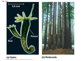
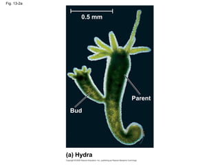
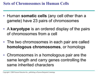
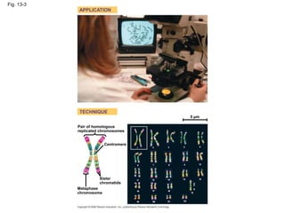
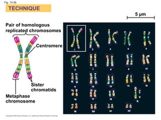
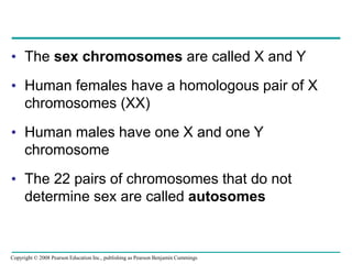
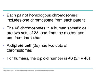
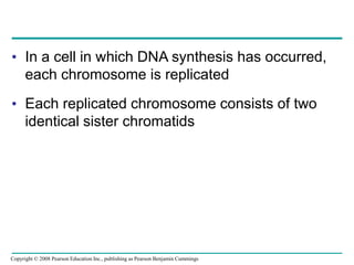
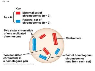
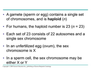
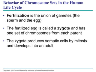
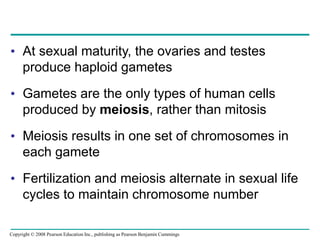
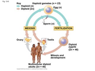
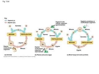
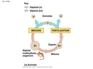
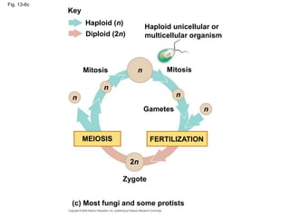
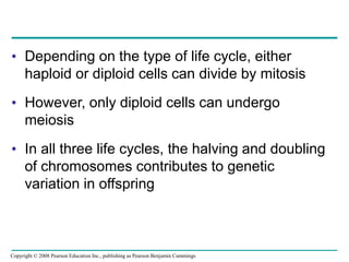
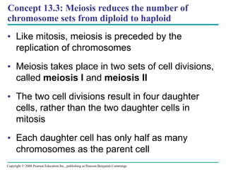
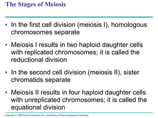
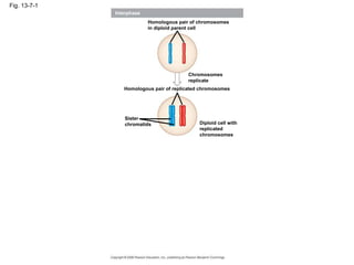
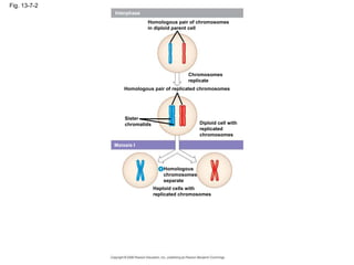
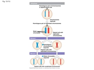
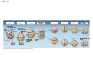
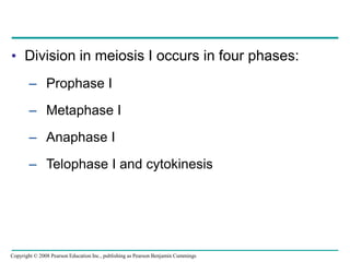
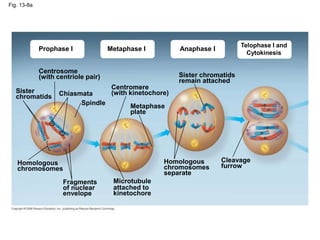
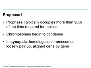
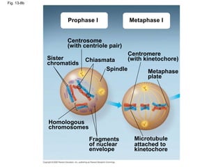
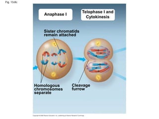
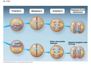
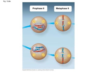
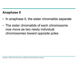
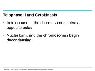
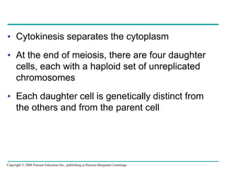
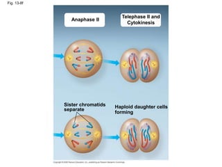
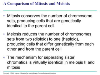
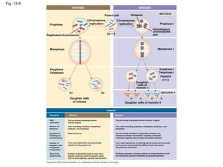
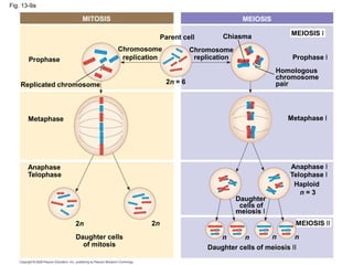
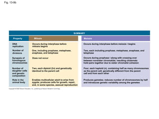
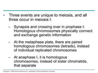
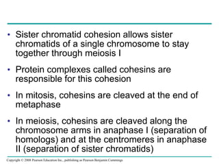
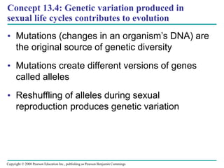
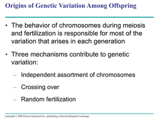
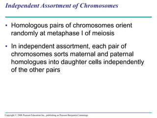
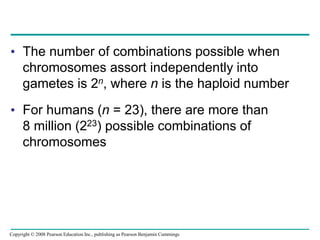
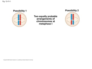
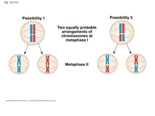
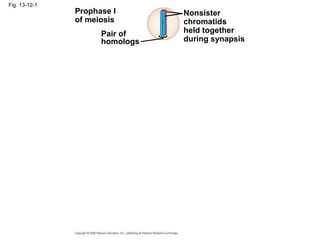
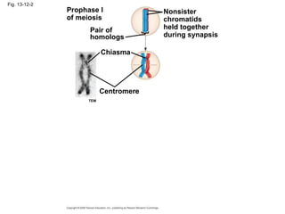
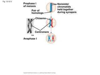
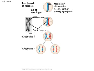
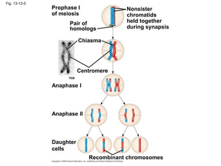
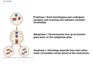
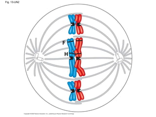
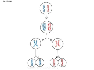
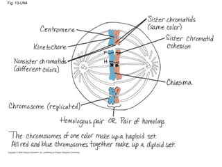
Copyright © 2008 Pearson Education, Inc., publishing as Pearson Benjamin Cummings
PowerPoint® Lecture Presentations for
Biology
Eighth Edition
Neil Campbell and Jane Reece
Lectures by Chris Romero, updated by Erin Barley with contributions from Joan Sharp
Chapter 13
Meiosis and Sexual
Life Cycles
Overview: Variations on a Theme
• Living organisms are distinguished by their
ability to reproduce their own kind
• Genetics is the scientific study of heredity and
variation
• Heredity is the transmission of traits from one
generation to the next
• Variation is demonstrated by the differences in
appearance that offspring show from parents
and siblings
Copyright © 2008 Pearson Education Inc., publishing as Pearson Benjamin Cummings
Fig. 13-1
Concept 13.1: Offspring acquire genes from
parents by inheriting chromosomes
• In a literal sense, children do not inherit
particular physical traits from their parents
• It is genes that are actually inherited
Copyright © 2008 Pearson Education Inc., publishing as Pearson Benjamin Cummings
Inheritance of Genes
• Genes are the units of heredity, and are made
up of segments of DNA
• Genes are passed to the next generation
through reproductive cells called gametes
(sperm and eggs)
• Each gene has a specific location called a
locus on a certain chromosome
• Most DNA is packaged into chromosomes
• One set of chromosomes is inherited from each
parent
Copyright © 2008 Pearson Education Inc., publishing as Pearson Benjamin Cummings
Comparison of Asexual and Sexual Reproduction
• In asexual reproduction, one parent produces
genetically identical offspring by mitosis
• A clone is a group of genetically identical
individuals from the same parent
• In sexual reproduction, two parents give rise
to offspring that have unique combinations of
genes inherited from the two parents
Video: Hydra Budding
Copyright © 2008 Pearson Education Inc., publishing as Pearson Benjamin Cummings
Fig. 13-2
(a) Hydra (b) Redwoods
Parent
Bud
0.5 mm
Fig. 13-2a
(a) Hydra
0.5 mm
Bud
Parent
Fig. 13-2b
(b) Redwoods
Concept 13.2: Fertilization and meiosis alternate
in sexual life cycles
• A life cycle is the generation-to-generation
sequence of stages in the reproductive history
of an organism
Copyright © 2008 Pearson Education Inc., publishing as Pearson Benjamin Cummings
Sets of Chromosomes in Human Cells
• Human somatic cells (any cell other than a
gamete) have 23 pairs of chromosomes
• A karyotype is an ordered display of the pairs
of chromosomes from a cell
• The two chromosomes in each pair are called
homologous chromosomes, or homologs
• Chromosomes in a homologous pair are the
same length and carry genes controlling the
same inherited characters
Copyright © 2008 Pearson Education Inc., publishing as Pearson Benjamin Cummings
Fig. 13-3
APPLICATION
TECHNIQUE
Pair of homologous
replicated chromosomes
5 µm
Centromere
Sister
chromatids
Metaphase
chromosome
Fig. 13-3a
APPLICATION
Fig. 13-3b
TECHNIQUE
Pair of homologous
replicated chromosomes
Centromere
Sister
chromatids
Metaphase
chromosome
5 µm
• The sex chromosomes are called X and Y
• Human females have a homologous pair of X
chromosomes (XX)
• Human males have one X and one Y
chromosome
• The 22 pairs of chromosomes that do not
determine sex are called autosomes
Copyright © 2008 Pearson Education Inc., publishing as Pearson Benjamin Cummings
• Each pair of homologous chromosomes
includes one chromosome from each parent
• The 46 chromosomes in a human somatic cell
are two sets of 23: one from the mother and
one from the father
• A diploid cell (2n) has two sets of
chromosomes
• For humans, the diploid number is 46 (2n = 46)
Copyright © 2008 Pearson Education Inc., publishing as Pearson Benjamin Cummings
• In a cell in which DNA synthesis has occurred,
each chromosome is replicated
• Each replicated chromosome consists of two
identical sister chromatids
Copyright © 2008 Pearson Education Inc., publishing as Pearson Benjamin Cummings
Fig. 13-4
Key
Maternal set of
chromosomes (n = 3)
Paternal set of
chromosomes (n = 3)
2n = 6
Centromere
Two sister chromatids
of one replicated
chromosome
Two nonsister
chromatids in
a homologous pair
Pair of homologous
chromosomes
(one from each set)
• A gamete (sperm or egg) contains a single set
of chromosomes, and is haploid (n)
• For humans, the haploid number is 23 (n = 23)
• Each set of 23 consists of 22 autosomes and a
single sex chromosome
• In an unfertilized egg (ovum), the sex
chromosome is X
• In a sperm cell, the sex chromosome may be
either X or Y
Copyright © 2008 Pearson Education Inc., publishing as Pearson Benjamin Cummings
• Fertilization is the union of gametes (the
sperm and the egg)
• The fertilized egg is called a zygote and has
one set of chromosomes from each parent
• The zygote produces somatic cells by mitosis
and develops into an adult
Behavior of Chromosome Sets in the Human
Life Cycle
Copyright © 2008 Pearson Education Inc., publishing as Pearson Benjamin Cummings
• At sexual maturity, the ovaries and testes
produce haploid gametes
• Gametes are the only types of human cells
produced by meiosis, rather than mitosis
• Meiosis results in one set of chromosomes in
each gamete
• Fertilization and meiosis alternate in sexual life
cycles to maintain chromosome number
Copyright © 2008 Pearson Education Inc., publishing as Pearson Benjamin Cummings
Fig. 13-5
Key
Haploid (n)
Diploid (2n)
Haploid gametes (n = 23)
Egg (n)
Sperm (n)
MEIOSIS FERTILIZATION
Ovary Testis
Diploid
zygote
(2n = 46)
Mitosis and
development
Multicellular diploid
adults (2n = 46)
The Variety of Sexual Life Cycles
• The alternation of meiosis and fertilization is
common to all organisms that reproduce
sexually
• The three main types of sexual life cycles differ
in the timing of meiosis and fertilization
Copyright © 2008 Pearson Education Inc., publishing as Pearson Benjamin Cummings
• In animals, meiosis produces gametes, which
undergo no further cell division before
fertilization
• Gametes are the only haploid cells in animals
• Gametes fuse to form a diploid zygote that
divides by mitosis to develop into a multicellular
organism
Copyright © 2008 Pearson Education Inc., publishing as Pearson Benjamin Cummings
Fig. 13-6
Key
Haploid (n)
Diploid (2n)
n n
Gametes
n
n
n
Mitosis
MEIOSIS FERTILIZATION
MEIOSIS
2n 2n
Zygote
2n
Mitosis
Diploid
multicellular
organism
(a) Animals
Spores
Diploid
multicellular
organism
(sporophyte)
(b) Plants and some algae
2n
Mitosis
Gametes
Mitosis
n
n
n
Zygote
FERTILIZATION
n
n
n
Mitosis
Zygote
(c) Most fungi and some protists
MEIOSIS FERTILIZATION
2n
Gametes
n
n
Mitosis
Haploid multi-
cellular organism
(gametophyte)
Haploid unicellular or
multicellular organism
Fig. 13-6a
Key
Haploid (n)
Diploid (2n)
Gametes
n
n
n
2n 2n
Zygote
MEIOSIS FERTILIZATION
Mitosis
Diploid
multicellular
organism
(a) Animals
• Plants and some algae exhibit an alternation
of generations
• This life cycle includes both a diploid and
haploid multicellular stage
• The diploid organism, called the sporophyte,
makes haploid spores by meiosis
Copyright © 2008 Pearson Education Inc., publishing as Pearson Benjamin Cummings
• Each spore grows by mitosis into a haploid
organism called a gametophyte
• A gametophyte makes haploid gametes by
mitosis
• Fertilization of gametes results in a diploid
sporophyte
Copyright © 2008 Pearson Education Inc., publishing as Pearson Benjamin Cummings
Fig. 13-6b
Key
Haploid (n)
Diploid (2n)
n
n
n
n
n
2n
2n
Mitosis
Mitosis
Mitosis
Zygote
Spores
Gametes
MEIOSIS FERTILIZATION
Diploid
multicellular
organism
(sporophyte)
Haploid multi-
cellular organism
(gametophyte)
(b) Plants and some algae
• In most fungi and some protists, the only
diploid stage is the single-celled zygote; there
is no multicellular diploid stage
• The zygote produces haploid cells by meiosis
• Each haploid cell grows by mitosis into a
haploid multicellular organism
• The haploid adult produces gametes by mitosis
Copyright © 2008 Pearson Education Inc., publishing as Pearson Benjamin Cummings
Fig. 13-6c
Key
Haploid (n)
Diploid (2n)
Mitosis Mitosis
Gametes
Zygote
Haploid unicellular or
multicellular organism
MEIOSIS FERTILIZATION
n
n
n n
n
2n
(c) Most fungi and some protists
• Depending on the type of life cycle, either
haploid or diploid cells can divide by mitosis
• However, only diploid cells can undergo
meiosis
• In all three life cycles, the halving and doubling
of chromosomes contributes to genetic
variation in offspring
Copyright © 2008 Pearson Education Inc., publishing as Pearson Benjamin Cummings
Concept 13.3: Meiosis reduces the number of
chromosome sets from diploid to haploid
• Like mitosis, meiosis is preceded by the
replication of chromosomes
• Meiosis takes place in two sets of cell divisions,
called meiosis I and meiosis II
• The two cell divisions result in four daughter
cells, rather than the two daughter cells in
mitosis
• Each daughter cell has only half as many
chromosomes as the parent cell
Copyright © 2008 Pearson Education Inc., publishing as Pearson Benjamin Cummings
The Stages of Meiosis
• In the first cell division (meiosis I), homologous
chromosomes separate
• Meiosis I results in two haploid daughter cells
with replicated chromosomes; it is called the
reductional division
• In the second cell division (meiosis II), sister
chromatids separate
• Meiosis II results in four haploid daughter cells
with unreplicated chromosomes; it is called the
equational division
Copyright © 2008 Pearson Education Inc., publishing as Pearson Benjamin Cummings
Fig. 13-7-1
Interphase
Homologous pair of chromosomes
in diploid parent cell
Chromosomes
replicate
Homologous pair of replicated chromosomes
Sister
chromatids Diploid cell with
replicated
chromosomes
Fig. 13-7-2
Interphase
Homologous pair of chromosomes
in diploid parent cell
Chromosomes
replicate
Homologous pair of replicated chromosomes
Sister
chromatids Diploid cell with
replicated
chromosomes
Meiosis I
Homologous
chromosomes
separate
1
Haploid cells with
replicated chromosomes
Fig. 13-7-3
Interphase
Homologous pair of chromosomes
in diploid parent cell
Chromosomes
replicate
Homologous pair of replicated chromosomes
Sister
chromatids Diploid cell with
replicated
chromosomes
Meiosis I
Homologous
chromosomes
separate
1
Haploid cells with
replicated chromosomes
Meiosis II
2 Sister chromatids
separate
Haploid cells with unreplicated chromosomes
• Meiosis I is preceded by interphase, in which
chromosomes are replicated to form sister
chromatids
• The sister chromatids are genetically identical
and joined at the centromere
• The single centrosome replicates, forming two
centrosomes
BioFlix: Meiosis
Copyright © 2008 Pearson Education Inc., publishing as Pearson Benjamin Cummings
Fig. 13-8
Prophase I Metaphase I Anaphase I
Telophase I and
Cytokinesis
Prophase II Metaphase II Anaphase II Telophase II and
Cytokinesis
Centrosome
(with centriole pair)
Sister
chromatids
Chiasmata
Spindle
Homologous
chromosomes
Fragments
of nuclear
envelope
Centromere
(with kinetochore)
Metaphase
plate
Microtubule
attached to
kinetochore
Sister chromatids
remain attached
Homologous
chromosomes
separate
Cleavage
furrow
Sister chromatids
separate Haploid daughter cells
forming
• Division in meiosis I occurs in four phases:
– Prophase I
– Metaphase I
– Anaphase I
– Telophase I and cytokinesis
Copyright © 2008 Pearson Education Inc., publishing as Pearson Benjamin Cummings
Metaphase I
Fig. 13-8a
Prophase I Anaphase I
Telophase I and
Cytokinesis
Centrosome
(with centriole pair)
Sister
chromatids Chiasmata
Spindle
Homologous
chromosomes
Fragments
of nuclear
envelope
Centromere
(with kinetochore)
Metaphase
plate
Microtubule
attached to
kinetochore
Sister chromatids
remain attached
Homologous
chromosomes
separate
Cleavage
furrow
Prophase I
• Prophase I typically occupies more than 90%
of the time required for meiosis
• Chromosomes begin to condense
• In synapsis, homologous chromosomes
loosely pair up, aligned gene by gene
Copyright © 2008 Pearson Education Inc., publishing as Pearson Benjamin Cummings
• In crossing over, nonsister chromatids
exchange DNA segments
• Each pair of chromosomes forms a tetrad, a
group of four chromatids
• Each tetrad usually has one or more
chiasmata, X-shaped regions where crossing
over occurred
Copyright © 2008 Pearson Education Inc., publishing as Pearson Benjamin Cummings
Metaphase I
• In metaphase I, tetrads line up at the
metaphase plate, with one chromosome facing
each pole
• Microtubules from one pole are attached to the
kinetochore of one chromosome of each tetrad
• Microtubules from the other pole are attached
to the kinetochore of the other chromosome
Copyright © 2008 Pearson Education Inc., publishing as Pearson Benjamin Cummings
Fig. 13-8b
Prophase I Metaphase I
Centrosome
(with centriole pair)
Sister
chromatids
Chiasmata
Spindle
Centromere
(with kinetochore)
Metaphase
plate
Homologous
chromosomes
Fragments
of nuclear
envelope
Microtubule
attached to
kinetochore
Anaphase I
• In anaphase I, pairs of homologous
chromosomes separate
• One chromosome moves toward each pole,
guided by the spindle apparatus
• Sister chromatids remain attached at the
centromere and move as one unit toward the
pole
Copyright © 2008 Pearson Education Inc., publishing as Pearson Benjamin Cummings
Telophase I and Cytokinesis
• In the beginning of telophase I, each half of the
cell has a haploid set of chromosomes; each
chromosome still consists of two sister
chromatids
• Cytokinesis usually occurs simultaneously,
forming two haploid daughter cells
Copyright © 2008 Pearson Education Inc., publishing as Pearson Benjamin Cummings
• In animal cells, a cleavage furrow forms; in
plant cells, a cell plate forms
• No chromosome replication occurs between
the end of meiosis I and the beginning of
meiosis II because the chromosomes are
already replicated
Copyright © 2008 Pearson Education Inc., publishing as Pearson Benjamin Cummings
Fig. 13-8c
Anaphase I
Telophase I and
Cytokinesis
Sister chromatids
remain attached
Homologous
chromosomes
separate
Cleavage
furrow
• Division in meiosis II also occurs in four
phases:
– Prophase II
– Metaphase II
– Anaphase II
– Telophase II and cytokinesis
• Meiosis II is very similar to mitosis
Copyright © 2008 Pearson Education Inc., publishing as Pearson Benjamin Cummings
Fig. 13-8d
Prophase II Metaphase II Anaphase II
Telophase II and
Cytokinesis
Sister chromatids
separate Haploid daughter cells
forming
Prophase II
• In prophase II, a spindle apparatus forms
• In late prophase II, chromosomes (each still
composed of two chromatids) move toward the
metaphase plate
Copyright © 2008 Pearson Education Inc., publishing as Pearson Benjamin Cummings
Metaphase II
• In metaphase II, the sister chromatids are
arranged at the metaphase plate
• Because of crossing over in meiosis I, the two
sister chromatids of each chromosome are no
longer genetically identical
• The kinetochores of sister chromatids attach to
microtubules extending from opposite poles
Copyright © 2008 Pearson Education Inc., publishing as Pearson Benjamin Cummings
Fig. 13-8e
Prophase II Metaphase II
Anaphase II
• In anaphase II, the sister chromatids separate
• The sister chromatids of each chromosome
now move as two newly individual
chromosomes toward opposite poles
Copyright © 2008 Pearson Education Inc., publishing as Pearson Benjamin Cummings
Telophase II and Cytokinesis
• In telophase II, the chromosomes arrive at
opposite poles
• Nuclei form, and the chromosomes begin
decondensing
Copyright © 2008 Pearson Education Inc., publishing as Pearson Benjamin Cummings
• Cytokinesis separates the cytoplasm
• At the end of meiosis, there are four daughter
cells, each with a haploid set of unreplicated
chromosomes
• Each daughter cell is genetically distinct from
the others and from the parent cell
Copyright © 2008 Pearson Education Inc., publishing as Pearson Benjamin Cummings
Fig. 13-8f
Anaphase II
Telephase II and
Cytokinesis
Sister chromatids
separate
Haploid daughter cells
forming
A Comparison of Mitosis and Meiosis
• Mitosis conserves the number of chromosome
sets, producing cells that are genetically
identical to the parent cell
• Meiosis reduces the number of chromosomes
sets from two (diploid) to one (haploid),
producing cells that differ genetically from each
other and from the parent cell
• The mechanism for separating sister
chromatids is virtually identical in meiosis II and
mitosis
Copyright © 2008 Pearson Education Inc., publishing as Pearson Benjamin Cummings
Fig. 13-9
MITOSIS MEIOSIS
MEIOSIS I
Prophase I
Chiasma
Homologous
chromosome
pair
Chromosome
replication
Parent cell
2n = 6
Chromosome
replication
Replicated chromosome
Prophase
Metaphase Metaphase I
Anaphase I
Telophase I
Haploid
n = 3
Daughter
cells of
meiosis I
Anaphase
Telophase
2n 2n
Daughter cells
of mitosis
n n n n
MEIOSIS II
Daughter cells of meiosis II
SUMMARY
Meiosis
Occurs during interphase before meiosis I begins
Two, each including prophase, metaphase, anaphase, and
telophase
Occurs during prophase I along with crossing over
between nonsister chromatids; resulting chiasmata
hold pairs together due to sister chromatid cohesion
Four, each haploid (n), containing half as many chromosomes
as the parent cell; genetically different from the parent
cell and from each other
Produces gametes; reduces number of chromosomes by half
and introduces genetic variability amoung the gametes
Mitosis
Occurs during interphase before
mitosis begins
One, including prophase, metaphase,
anahase, and telophase
Does not occur
Two, each diploid (2n) and genetically
identical to the parent cell
Enables multicellular adult to arise from
zygote; produces cells for growth, repair,
and, in some species, asexual reproduction
Property
DNA
replication
Number of
divisions
Synapsis of
homologous
chromosomes
Number of
daughter cells
and genetic
composition
Role in the
animal body
Fig. 13-9a
MITOSIS MEIOSIS
MEIOSIS I
Prophase I
Chiasma
Chromosome
replication
Homologous
chromosome
pair
Chromosome
replication
2n = 6
Parent cell
Prophase
Replicated chromosome
Metaphase Metaphase I
Anaphase I
Telophase I
Haploid
n = 3
Daughter
cells of
meiosis I
MEIOSIS II
Daughter cells of meiosis II
n
n
n
n
2n
2n
Daughter cells
of mitosis
Anaphase
Telophase
Fig. 13-9b
SUMMARY
Meiosis
Mitosis
Property
DNA
replication
Number of
divisions
Occurs during interphase before
mitosis begins
One, including prophase, metaphase,
anaphase, and telophase
Synapsis of
homologous
chromosomes
Does not occur
Number of
daughter cells
and genetic
composition
Two, each diploid (2n) and genetically
identical to the parent cell
Role in the
animal body
Enables multicellular adult to arise from
zygote; produces cells for growth, repair,
and, in some species, asexual reproduction
Occurs during interphase before meiosis I begins
Two, each including prophase, metaphase, anaphase, and
telophase
Occurs during prophase I along with crossing over
between nonsister chromatids; resulting chiasmata
hold pairs together due to sister chromatid cohesion
Four, each haploid (n), containing half as many chromosomes
as the parent cell; genetically different from the parent
cell and from each other
Produces gametes; reduces number of chromosomes by half
and introduces genetic variability among the gametes
• Three events are unique to meiosis, and all
three occur in meiosis l:
– Synapsis and crossing over in prophase I:
Homologous chromosomes physically connect
and exchange genetic information
– At the metaphase plate, there are paired
homologous chromosomes (tetrads), instead
of individual replicated chromosomes
– At anaphase I, it is homologous
chromosomes, instead of sister chromatids,
that separate
Copyright © 2008 Pearson Education Inc., publishing as Pearson Benjamin Cummings
• Sister chromatid cohesion allows sister
chromatids of a single chromosome to stay
together through meiosis I
• Protein complexes called cohesins are
responsible for this cohesion
• In mitosis, cohesins are cleaved at the end of
metaphase
• In meiosis, cohesins are cleaved along the
chromosome arms in anaphase I (separation of
homologs) and at the centromeres in anaphase
II (separation of sister chromatids)
Copyright © 2008 Pearson Education Inc., publishing as Pearson Benjamin Cummings
Fig. 13-10
EXPERIMENT
RESULTS
Shugoshin+ (normal)+
Spore case Fluorescent label
Metaphase I
Shugoshin–
Anaphase I
Metaphase II
Anaphase II
Mature
spores
OR
Spore Two of three possible arrange-
ments of labeled chromosomes
Shugoshin+
Shugoshin–
Spore
cases
(%)
100
80
60
40
20
0
? ?
?
?
? ?
?
?
Fig. 13-10a
EXPERIMENT
Shugoshin+ (normal)
Spore case Fluorescent label
Metaphase I
Anaphase I
Metaphase II
Anaphase II
Mature
spores
Spore
OR
Two of three possible arrange-
ments of labeled chromosomes
Shugoshin–
? ? ?
?
?
?
?
?
Fig. 13-10b
RESULTS
Shugoshin+ Shugoshin–
Spore
cases
(%)
100
80
60
40
20
0
Concept 13.4: Genetic variation produced in
sexual life cycles contributes to evolution
• Mutations (changes in an organism’s DNA) are
the original source of genetic diversity
• Mutations create different versions of genes
called alleles
• Reshuffling of alleles during sexual
reproduction produces genetic variation
Copyright © 2008 Pearson Education Inc., publishing as Pearson Benjamin Cummings
Origins of Genetic Variation Among Offspring
• The behavior of chromosomes during meiosis
and fertilization is responsible for most of the
variation that arises in each generation
• Three mechanisms contribute to genetic
variation:
– Independent assortment of chromosomes
– Crossing over
– Random fertilization
Copyright © 2008 Pearson Education Inc., publishing as Pearson Benjamin Cummings
Independent Assortment of Chromosomes
• Homologous pairs of chromosomes orient
randomly at metaphase I of meiosis
• In independent assortment, each pair of
chromosomes sorts maternal and paternal
homologues into daughter cells independently
of the other pairs
Copyright © 2008 Pearson Education Inc., publishing as Pearson Benjamin Cummings
• The number of combinations possible when
chromosomes assort independently into
gametes is 2n, where n is the haploid number
• For humans (n = 23), there are more than
8 million (223) possible combinations of
chromosomes
Copyright © 2008 Pearson Education Inc., publishing as Pearson Benjamin Cummings
Fig. 13-11-1
Possibility 1 Possibility 2
Two equally probable
arrangements of
chromosomes at
metaphase I
Fig. 13-11-2
Possibility 1 Possibility 2
Two equally probable
arrangements of
chromosomes at
metaphase I
Metaphase II
Fig. 13-11-3
Possibility 1 Possibility 2
Two equally probable
arrangements of
chromosomes at
metaphase I
Metaphase II
Daughter
cells
Combination 1 Combination 2 Combination 3 Combination 4
Crossing Over
• Crossing over produces recombinant
chromosomes, which combine genes
inherited from each parent
• Crossing over begins very early in prophase I,
as homologous chromosomes pair up gene by
gene
Copyright © 2008 Pearson Education Inc., publishing as Pearson Benjamin Cummings
• In crossing over, homologous portions of two
nonsister chromatids trade places
• Crossing over contributes to genetic variation
by combining DNA from two parents into a
single chromosome
Copyright © 2008 Pearson Education Inc., publishing as Pearson Benjamin Cummings
Fig. 13-12-1
Prophase I
of meiosis
Pair of
homologs
Nonsister
chromatids
held together
during synapsis
Fig. 13-12-2
Prophase I
of meiosis
Pair of
homologs
Nonsister
chromatids
held together
during synapsis
Chiasma
Centromere
TEM
Fig. 13-12-3
Prophase I
of meiosis
Pair of
homologs
Nonsister
chromatids
held together
during synapsis
Chiasma
Centromere
Anaphase I
TEM
Fig. 13-12-4
Prophase I
of meiosis
Pair of
homologs
Nonsister
chromatids
held together
during synapsis
Chiasma
Centromere
Anaphase I
Anaphase II
TEM
Fig. 13-12-5
Prophase I
of meiosis
Pair of
homologs
Nonsister
chromatids
held together
during synapsis
Chiasma
Centromere
Anaphase I
Anaphase II
Daughter
cells
Recombinant chromosomes
TEM
Random Fertilization
• Random fertilization adds to genetic variation
because any sperm can fuse with any ovum
(unfertilized egg)
• The fusion of two gametes (each with 8.4
million possible chromosome combinations
from independent assortment) produces a
zygote with any of about 70 trillion diploid
combinations
Copyright © 2008 Pearson Education Inc., publishing as Pearson Benjamin Cummings
• Crossing over adds even more variation
• Each zygote has a unique genetic identity
Animation: Genetic Variation
Copyright © 2008 Pearson Education Inc., publishing as Pearson Benjamin Cummings
The Evolutionary Significance of Genetic Variation
Within Populations
• Natural selection results in the accumulation of
genetic variations favored by the environment
• Sexual reproduction contributes to the genetic
variation in a population, which originates from
mutations
Copyright © 2008 Pearson Education Inc., publishing as Pearson Benjamin Cummings
Fig. 13-UN1
Prophase I: Each homologous pair undergoes
synapsis and crossing over between nonsister
chromatids.
Metaphase I: Chromosomes line up as homolo-
gous pairs on the metaphase plate.
Anaphase I: Homologs separate from each other;
sister chromatids remain joined at the centromere.
Fig. 13-UN2
F
H
Fig. 13-UN3
Fig. 13-UN4
You should now be able to:
1. Distinguish between the following terms:
somatic cell and gamete; autosome and sex
chromosomes; haploid and diploid
2. Describe the events that characterize each
phase of meiosis
3. Describe three events that occur during
meiosis I but not mitosis
4. Name and explain the three events that
contribute to genetic variation in sexually
reproducing organisms
Copyright © 2008 Pearson Education Inc., publishing as Pearson Benjamin Cummings

13_Lecture_Presentation.ppt...................

  • 1.
    Copyright © 2008Pearson Education, Inc., publishing as Pearson Benjamin Cummings PowerPoint® Lecture Presentations for Biology Eighth Edition Neil Campbell and Jane Reece Lectures by Chris Romero, updated by Erin Barley with contributions from Joan Sharp Chapter 13 Meiosis and Sexual Life Cycles
  • 2.
    Overview: Variations ona Theme • Living organisms are distinguished by their ability to reproduce their own kind • Genetics is the scientific study of heredity and variation • Heredity is the transmission of traits from one generation to the next • Variation is demonstrated by the differences in appearance that offspring show from parents and siblings Copyright © 2008 Pearson Education Inc., publishing as Pearson Benjamin Cummings
  • 3.
  • 4.
    Concept 13.1: Offspringacquire genes from parents by inheriting chromosomes • In a literal sense, children do not inherit particular physical traits from their parents • It is genes that are actually inherited Copyright © 2008 Pearson Education Inc., publishing as Pearson Benjamin Cummings
  • 5.
    Inheritance of Genes •Genes are the units of heredity, and are made up of segments of DNA • Genes are passed to the next generation through reproductive cells called gametes (sperm and eggs) • Each gene has a specific location called a locus on a certain chromosome • Most DNA is packaged into chromosomes • One set of chromosomes is inherited from each parent Copyright © 2008 Pearson Education Inc., publishing as Pearson Benjamin Cummings
  • 6.
    Comparison of Asexualand Sexual Reproduction • In asexual reproduction, one parent produces genetically identical offspring by mitosis • A clone is a group of genetically identical individuals from the same parent • In sexual reproduction, two parents give rise to offspring that have unique combinations of genes inherited from the two parents Video: Hydra Budding Copyright © 2008 Pearson Education Inc., publishing as Pearson Benjamin Cummings
  • 7.
    Fig. 13-2 (a) Hydra(b) Redwoods Parent Bud 0.5 mm
  • 8.
  • 9.
  • 10.
    Concept 13.2: Fertilizationand meiosis alternate in sexual life cycles • A life cycle is the generation-to-generation sequence of stages in the reproductive history of an organism Copyright © 2008 Pearson Education Inc., publishing as Pearson Benjamin Cummings
  • 11.
    Sets of Chromosomesin Human Cells • Human somatic cells (any cell other than a gamete) have 23 pairs of chromosomes • A karyotype is an ordered display of the pairs of chromosomes from a cell • The two chromosomes in each pair are called homologous chromosomes, or homologs • Chromosomes in a homologous pair are the same length and carry genes controlling the same inherited characters Copyright © 2008 Pearson Education Inc., publishing as Pearson Benjamin Cummings
  • 12.
    Fig. 13-3 APPLICATION TECHNIQUE Pair ofhomologous replicated chromosomes 5 µm Centromere Sister chromatids Metaphase chromosome
  • 13.
  • 14.
    Fig. 13-3b TECHNIQUE Pair ofhomologous replicated chromosomes Centromere Sister chromatids Metaphase chromosome 5 µm
  • 15.
    • The sexchromosomes are called X and Y • Human females have a homologous pair of X chromosomes (XX) • Human males have one X and one Y chromosome • The 22 pairs of chromosomes that do not determine sex are called autosomes Copyright © 2008 Pearson Education Inc., publishing as Pearson Benjamin Cummings
  • 16.
    • Each pairof homologous chromosomes includes one chromosome from each parent • The 46 chromosomes in a human somatic cell are two sets of 23: one from the mother and one from the father • A diploid cell (2n) has two sets of chromosomes • For humans, the diploid number is 46 (2n = 46) Copyright © 2008 Pearson Education Inc., publishing as Pearson Benjamin Cummings
  • 17.
    • In acell in which DNA synthesis has occurred, each chromosome is replicated • Each replicated chromosome consists of two identical sister chromatids Copyright © 2008 Pearson Education Inc., publishing as Pearson Benjamin Cummings
  • 18.
    Fig. 13-4 Key Maternal setof chromosomes (n = 3) Paternal set of chromosomes (n = 3) 2n = 6 Centromere Two sister chromatids of one replicated chromosome Two nonsister chromatids in a homologous pair Pair of homologous chromosomes (one from each set)
  • 19.
    • A gamete(sperm or egg) contains a single set of chromosomes, and is haploid (n) • For humans, the haploid number is 23 (n = 23) • Each set of 23 consists of 22 autosomes and a single sex chromosome • In an unfertilized egg (ovum), the sex chromosome is X • In a sperm cell, the sex chromosome may be either X or Y Copyright © 2008 Pearson Education Inc., publishing as Pearson Benjamin Cummings
  • 20.
    • Fertilization isthe union of gametes (the sperm and the egg) • The fertilized egg is called a zygote and has one set of chromosomes from each parent • The zygote produces somatic cells by mitosis and develops into an adult Behavior of Chromosome Sets in the Human Life Cycle Copyright © 2008 Pearson Education Inc., publishing as Pearson Benjamin Cummings
  • 21.
    • At sexualmaturity, the ovaries and testes produce haploid gametes • Gametes are the only types of human cells produced by meiosis, rather than mitosis • Meiosis results in one set of chromosomes in each gamete • Fertilization and meiosis alternate in sexual life cycles to maintain chromosome number Copyright © 2008 Pearson Education Inc., publishing as Pearson Benjamin Cummings
  • 22.
    Fig. 13-5 Key Haploid (n) Diploid(2n) Haploid gametes (n = 23) Egg (n) Sperm (n) MEIOSIS FERTILIZATION Ovary Testis Diploid zygote (2n = 46) Mitosis and development Multicellular diploid adults (2n = 46)
  • 23.
    The Variety ofSexual Life Cycles • The alternation of meiosis and fertilization is common to all organisms that reproduce sexually • The three main types of sexual life cycles differ in the timing of meiosis and fertilization Copyright © 2008 Pearson Education Inc., publishing as Pearson Benjamin Cummings
  • 24.
    • In animals,meiosis produces gametes, which undergo no further cell division before fertilization • Gametes are the only haploid cells in animals • Gametes fuse to form a diploid zygote that divides by mitosis to develop into a multicellular organism Copyright © 2008 Pearson Education Inc., publishing as Pearson Benjamin Cummings
  • 25.
    Fig. 13-6 Key Haploid (n) Diploid(2n) n n Gametes n n n Mitosis MEIOSIS FERTILIZATION MEIOSIS 2n 2n Zygote 2n Mitosis Diploid multicellular organism (a) Animals Spores Diploid multicellular organism (sporophyte) (b) Plants and some algae 2n Mitosis Gametes Mitosis n n n Zygote FERTILIZATION n n n Mitosis Zygote (c) Most fungi and some protists MEIOSIS FERTILIZATION 2n Gametes n n Mitosis Haploid multi- cellular organism (gametophyte) Haploid unicellular or multicellular organism
  • 26.
    Fig. 13-6a Key Haploid (n) Diploid(2n) Gametes n n n 2n 2n Zygote MEIOSIS FERTILIZATION Mitosis Diploid multicellular organism (a) Animals
  • 27.
    • Plants andsome algae exhibit an alternation of generations • This life cycle includes both a diploid and haploid multicellular stage • The diploid organism, called the sporophyte, makes haploid spores by meiosis Copyright © 2008 Pearson Education Inc., publishing as Pearson Benjamin Cummings
  • 28.
    • Each sporegrows by mitosis into a haploid organism called a gametophyte • A gametophyte makes haploid gametes by mitosis • Fertilization of gametes results in a diploid sporophyte Copyright © 2008 Pearson Education Inc., publishing as Pearson Benjamin Cummings
  • 29.
    Fig. 13-6b Key Haploid (n) Diploid(2n) n n n n n 2n 2n Mitosis Mitosis Mitosis Zygote Spores Gametes MEIOSIS FERTILIZATION Diploid multicellular organism (sporophyte) Haploid multi- cellular organism (gametophyte) (b) Plants and some algae
  • 30.
    • In mostfungi and some protists, the only diploid stage is the single-celled zygote; there is no multicellular diploid stage • The zygote produces haploid cells by meiosis • Each haploid cell grows by mitosis into a haploid multicellular organism • The haploid adult produces gametes by mitosis Copyright © 2008 Pearson Education Inc., publishing as Pearson Benjamin Cummings
  • 31.
    Fig. 13-6c Key Haploid (n) Diploid(2n) Mitosis Mitosis Gametes Zygote Haploid unicellular or multicellular organism MEIOSIS FERTILIZATION n n n n n 2n (c) Most fungi and some protists
  • 32.
    • Depending onthe type of life cycle, either haploid or diploid cells can divide by mitosis • However, only diploid cells can undergo meiosis • In all three life cycles, the halving and doubling of chromosomes contributes to genetic variation in offspring Copyright © 2008 Pearson Education Inc., publishing as Pearson Benjamin Cummings
  • 33.
    Concept 13.3: Meiosisreduces the number of chromosome sets from diploid to haploid • Like mitosis, meiosis is preceded by the replication of chromosomes • Meiosis takes place in two sets of cell divisions, called meiosis I and meiosis II • The two cell divisions result in four daughter cells, rather than the two daughter cells in mitosis • Each daughter cell has only half as many chromosomes as the parent cell Copyright © 2008 Pearson Education Inc., publishing as Pearson Benjamin Cummings
  • 34.
    The Stages ofMeiosis • In the first cell division (meiosis I), homologous chromosomes separate • Meiosis I results in two haploid daughter cells with replicated chromosomes; it is called the reductional division • In the second cell division (meiosis II), sister chromatids separate • Meiosis II results in four haploid daughter cells with unreplicated chromosomes; it is called the equational division Copyright © 2008 Pearson Education Inc., publishing as Pearson Benjamin Cummings
  • 35.
    Fig. 13-7-1 Interphase Homologous pairof chromosomes in diploid parent cell Chromosomes replicate Homologous pair of replicated chromosomes Sister chromatids Diploid cell with replicated chromosomes
  • 36.
    Fig. 13-7-2 Interphase Homologous pairof chromosomes in diploid parent cell Chromosomes replicate Homologous pair of replicated chromosomes Sister chromatids Diploid cell with replicated chromosomes Meiosis I Homologous chromosomes separate 1 Haploid cells with replicated chromosomes
  • 37.
    Fig. 13-7-3 Interphase Homologous pairof chromosomes in diploid parent cell Chromosomes replicate Homologous pair of replicated chromosomes Sister chromatids Diploid cell with replicated chromosomes Meiosis I Homologous chromosomes separate 1 Haploid cells with replicated chromosomes Meiosis II 2 Sister chromatids separate Haploid cells with unreplicated chromosomes
  • 38.
    • Meiosis Iis preceded by interphase, in which chromosomes are replicated to form sister chromatids • The sister chromatids are genetically identical and joined at the centromere • The single centrosome replicates, forming two centrosomes BioFlix: Meiosis Copyright © 2008 Pearson Education Inc., publishing as Pearson Benjamin Cummings
  • 39.
    Fig. 13-8 Prophase IMetaphase I Anaphase I Telophase I and Cytokinesis Prophase II Metaphase II Anaphase II Telophase II and Cytokinesis Centrosome (with centriole pair) Sister chromatids Chiasmata Spindle Homologous chromosomes Fragments of nuclear envelope Centromere (with kinetochore) Metaphase plate Microtubule attached to kinetochore Sister chromatids remain attached Homologous chromosomes separate Cleavage furrow Sister chromatids separate Haploid daughter cells forming
  • 40.
    • Division inmeiosis I occurs in four phases: – Prophase I – Metaphase I – Anaphase I – Telophase I and cytokinesis Copyright © 2008 Pearson Education Inc., publishing as Pearson Benjamin Cummings
  • 41.
    Metaphase I Fig. 13-8a ProphaseI Anaphase I Telophase I and Cytokinesis Centrosome (with centriole pair) Sister chromatids Chiasmata Spindle Homologous chromosomes Fragments of nuclear envelope Centromere (with kinetochore) Metaphase plate Microtubule attached to kinetochore Sister chromatids remain attached Homologous chromosomes separate Cleavage furrow
  • 42.
    Prophase I • ProphaseI typically occupies more than 90% of the time required for meiosis • Chromosomes begin to condense • In synapsis, homologous chromosomes loosely pair up, aligned gene by gene Copyright © 2008 Pearson Education Inc., publishing as Pearson Benjamin Cummings
  • 43.
    • In crossingover, nonsister chromatids exchange DNA segments • Each pair of chromosomes forms a tetrad, a group of four chromatids • Each tetrad usually has one or more chiasmata, X-shaped regions where crossing over occurred Copyright © 2008 Pearson Education Inc., publishing as Pearson Benjamin Cummings
  • 44.
    Metaphase I • Inmetaphase I, tetrads line up at the metaphase plate, with one chromosome facing each pole • Microtubules from one pole are attached to the kinetochore of one chromosome of each tetrad • Microtubules from the other pole are attached to the kinetochore of the other chromosome Copyright © 2008 Pearson Education Inc., publishing as Pearson Benjamin Cummings
  • 45.
    Fig. 13-8b Prophase IMetaphase I Centrosome (with centriole pair) Sister chromatids Chiasmata Spindle Centromere (with kinetochore) Metaphase plate Homologous chromosomes Fragments of nuclear envelope Microtubule attached to kinetochore
  • 46.
    Anaphase I • Inanaphase I, pairs of homologous chromosomes separate • One chromosome moves toward each pole, guided by the spindle apparatus • Sister chromatids remain attached at the centromere and move as one unit toward the pole Copyright © 2008 Pearson Education Inc., publishing as Pearson Benjamin Cummings
  • 47.
    Telophase I andCytokinesis • In the beginning of telophase I, each half of the cell has a haploid set of chromosomes; each chromosome still consists of two sister chromatids • Cytokinesis usually occurs simultaneously, forming two haploid daughter cells Copyright © 2008 Pearson Education Inc., publishing as Pearson Benjamin Cummings
  • 48.
    • In animalcells, a cleavage furrow forms; in plant cells, a cell plate forms • No chromosome replication occurs between the end of meiosis I and the beginning of meiosis II because the chromosomes are already replicated Copyright © 2008 Pearson Education Inc., publishing as Pearson Benjamin Cummings
  • 49.
    Fig. 13-8c Anaphase I TelophaseI and Cytokinesis Sister chromatids remain attached Homologous chromosomes separate Cleavage furrow
  • 50.
    • Division inmeiosis II also occurs in four phases: – Prophase II – Metaphase II – Anaphase II – Telophase II and cytokinesis • Meiosis II is very similar to mitosis Copyright © 2008 Pearson Education Inc., publishing as Pearson Benjamin Cummings
  • 51.
    Fig. 13-8d Prophase IIMetaphase II Anaphase II Telophase II and Cytokinesis Sister chromatids separate Haploid daughter cells forming
  • 52.
    Prophase II • Inprophase II, a spindle apparatus forms • In late prophase II, chromosomes (each still composed of two chromatids) move toward the metaphase plate Copyright © 2008 Pearson Education Inc., publishing as Pearson Benjamin Cummings
  • 53.
    Metaphase II • Inmetaphase II, the sister chromatids are arranged at the metaphase plate • Because of crossing over in meiosis I, the two sister chromatids of each chromosome are no longer genetically identical • The kinetochores of sister chromatids attach to microtubules extending from opposite poles Copyright © 2008 Pearson Education Inc., publishing as Pearson Benjamin Cummings
  • 54.
  • 55.
    Anaphase II • Inanaphase II, the sister chromatids separate • The sister chromatids of each chromosome now move as two newly individual chromosomes toward opposite poles Copyright © 2008 Pearson Education Inc., publishing as Pearson Benjamin Cummings
  • 56.
    Telophase II andCytokinesis • In telophase II, the chromosomes arrive at opposite poles • Nuclei form, and the chromosomes begin decondensing Copyright © 2008 Pearson Education Inc., publishing as Pearson Benjamin Cummings
  • 57.
    • Cytokinesis separatesthe cytoplasm • At the end of meiosis, there are four daughter cells, each with a haploid set of unreplicated chromosomes • Each daughter cell is genetically distinct from the others and from the parent cell Copyright © 2008 Pearson Education Inc., publishing as Pearson Benjamin Cummings
  • 58.
    Fig. 13-8f Anaphase II TelephaseII and Cytokinesis Sister chromatids separate Haploid daughter cells forming
  • 59.
    A Comparison ofMitosis and Meiosis • Mitosis conserves the number of chromosome sets, producing cells that are genetically identical to the parent cell • Meiosis reduces the number of chromosomes sets from two (diploid) to one (haploid), producing cells that differ genetically from each other and from the parent cell • The mechanism for separating sister chromatids is virtually identical in meiosis II and mitosis Copyright © 2008 Pearson Education Inc., publishing as Pearson Benjamin Cummings
  • 60.
    Fig. 13-9 MITOSIS MEIOSIS MEIOSISI Prophase I Chiasma Homologous chromosome pair Chromosome replication Parent cell 2n = 6 Chromosome replication Replicated chromosome Prophase Metaphase Metaphase I Anaphase I Telophase I Haploid n = 3 Daughter cells of meiosis I Anaphase Telophase 2n 2n Daughter cells of mitosis n n n n MEIOSIS II Daughter cells of meiosis II SUMMARY Meiosis Occurs during interphase before meiosis I begins Two, each including prophase, metaphase, anaphase, and telophase Occurs during prophase I along with crossing over between nonsister chromatids; resulting chiasmata hold pairs together due to sister chromatid cohesion Four, each haploid (n), containing half as many chromosomes as the parent cell; genetically different from the parent cell and from each other Produces gametes; reduces number of chromosomes by half and introduces genetic variability amoung the gametes Mitosis Occurs during interphase before mitosis begins One, including prophase, metaphase, anahase, and telophase Does not occur Two, each diploid (2n) and genetically identical to the parent cell Enables multicellular adult to arise from zygote; produces cells for growth, repair, and, in some species, asexual reproduction Property DNA replication Number of divisions Synapsis of homologous chromosomes Number of daughter cells and genetic composition Role in the animal body
  • 61.
    Fig. 13-9a MITOSIS MEIOSIS MEIOSISI Prophase I Chiasma Chromosome replication Homologous chromosome pair Chromosome replication 2n = 6 Parent cell Prophase Replicated chromosome Metaphase Metaphase I Anaphase I Telophase I Haploid n = 3 Daughter cells of meiosis I MEIOSIS II Daughter cells of meiosis II n n n n 2n 2n Daughter cells of mitosis Anaphase Telophase
  • 62.
    Fig. 13-9b SUMMARY Meiosis Mitosis Property DNA replication Number of divisions Occursduring interphase before mitosis begins One, including prophase, metaphase, anaphase, and telophase Synapsis of homologous chromosomes Does not occur Number of daughter cells and genetic composition Two, each diploid (2n) and genetically identical to the parent cell Role in the animal body Enables multicellular adult to arise from zygote; produces cells for growth, repair, and, in some species, asexual reproduction Occurs during interphase before meiosis I begins Two, each including prophase, metaphase, anaphase, and telophase Occurs during prophase I along with crossing over between nonsister chromatids; resulting chiasmata hold pairs together due to sister chromatid cohesion Four, each haploid (n), containing half as many chromosomes as the parent cell; genetically different from the parent cell and from each other Produces gametes; reduces number of chromosomes by half and introduces genetic variability among the gametes
  • 63.
    • Three eventsare unique to meiosis, and all three occur in meiosis l: – Synapsis and crossing over in prophase I: Homologous chromosomes physically connect and exchange genetic information – At the metaphase plate, there are paired homologous chromosomes (tetrads), instead of individual replicated chromosomes – At anaphase I, it is homologous chromosomes, instead of sister chromatids, that separate Copyright © 2008 Pearson Education Inc., publishing as Pearson Benjamin Cummings
  • 64.
    • Sister chromatidcohesion allows sister chromatids of a single chromosome to stay together through meiosis I • Protein complexes called cohesins are responsible for this cohesion • In mitosis, cohesins are cleaved at the end of metaphase • In meiosis, cohesins are cleaved along the chromosome arms in anaphase I (separation of homologs) and at the centromeres in anaphase II (separation of sister chromatids) Copyright © 2008 Pearson Education Inc., publishing as Pearson Benjamin Cummings
  • 65.
    Fig. 13-10 EXPERIMENT RESULTS Shugoshin+ (normal)+ Sporecase Fluorescent label Metaphase I Shugoshin– Anaphase I Metaphase II Anaphase II Mature spores OR Spore Two of three possible arrange- ments of labeled chromosomes Shugoshin+ Shugoshin– Spore cases (%) 100 80 60 40 20 0 ? ? ? ? ? ? ? ?
  • 66.
    Fig. 13-10a EXPERIMENT Shugoshin+ (normal) Sporecase Fluorescent label Metaphase I Anaphase I Metaphase II Anaphase II Mature spores Spore OR Two of three possible arrange- ments of labeled chromosomes Shugoshin– ? ? ? ? ? ? ? ?
  • 67.
  • 68.
    Concept 13.4: Geneticvariation produced in sexual life cycles contributes to evolution • Mutations (changes in an organism’s DNA) are the original source of genetic diversity • Mutations create different versions of genes called alleles • Reshuffling of alleles during sexual reproduction produces genetic variation Copyright © 2008 Pearson Education Inc., publishing as Pearson Benjamin Cummings
  • 69.
    Origins of GeneticVariation Among Offspring • The behavior of chromosomes during meiosis and fertilization is responsible for most of the variation that arises in each generation • Three mechanisms contribute to genetic variation: – Independent assortment of chromosomes – Crossing over – Random fertilization Copyright © 2008 Pearson Education Inc., publishing as Pearson Benjamin Cummings
  • 70.
    Independent Assortment ofChromosomes • Homologous pairs of chromosomes orient randomly at metaphase I of meiosis • In independent assortment, each pair of chromosomes sorts maternal and paternal homologues into daughter cells independently of the other pairs Copyright © 2008 Pearson Education Inc., publishing as Pearson Benjamin Cummings
  • 71.
    • The numberof combinations possible when chromosomes assort independently into gametes is 2n, where n is the haploid number • For humans (n = 23), there are more than 8 million (223) possible combinations of chromosomes Copyright © 2008 Pearson Education Inc., publishing as Pearson Benjamin Cummings
  • 72.
    Fig. 13-11-1 Possibility 1Possibility 2 Two equally probable arrangements of chromosomes at metaphase I
  • 73.
    Fig. 13-11-2 Possibility 1Possibility 2 Two equally probable arrangements of chromosomes at metaphase I Metaphase II
  • 74.
    Fig. 13-11-3 Possibility 1Possibility 2 Two equally probable arrangements of chromosomes at metaphase I Metaphase II Daughter cells Combination 1 Combination 2 Combination 3 Combination 4
  • 75.
    Crossing Over • Crossingover produces recombinant chromosomes, which combine genes inherited from each parent • Crossing over begins very early in prophase I, as homologous chromosomes pair up gene by gene Copyright © 2008 Pearson Education Inc., publishing as Pearson Benjamin Cummings
  • 76.
    • In crossingover, homologous portions of two nonsister chromatids trade places • Crossing over contributes to genetic variation by combining DNA from two parents into a single chromosome Copyright © 2008 Pearson Education Inc., publishing as Pearson Benjamin Cummings
  • 77.
    Fig. 13-12-1 Prophase I ofmeiosis Pair of homologs Nonsister chromatids held together during synapsis
  • 78.
    Fig. 13-12-2 Prophase I ofmeiosis Pair of homologs Nonsister chromatids held together during synapsis Chiasma Centromere TEM
  • 79.
    Fig. 13-12-3 Prophase I ofmeiosis Pair of homologs Nonsister chromatids held together during synapsis Chiasma Centromere Anaphase I TEM
  • 80.
    Fig. 13-12-4 Prophase I ofmeiosis Pair of homologs Nonsister chromatids held together during synapsis Chiasma Centromere Anaphase I Anaphase II TEM
  • 81.
    Fig. 13-12-5 Prophase I ofmeiosis Pair of homologs Nonsister chromatids held together during synapsis Chiasma Centromere Anaphase I Anaphase II Daughter cells Recombinant chromosomes TEM
  • 82.
    Random Fertilization • Randomfertilization adds to genetic variation because any sperm can fuse with any ovum (unfertilized egg) • The fusion of two gametes (each with 8.4 million possible chromosome combinations from independent assortment) produces a zygote with any of about 70 trillion diploid combinations Copyright © 2008 Pearson Education Inc., publishing as Pearson Benjamin Cummings
  • 83.
    • Crossing overadds even more variation • Each zygote has a unique genetic identity Animation: Genetic Variation Copyright © 2008 Pearson Education Inc., publishing as Pearson Benjamin Cummings
  • 84.
    The Evolutionary Significanceof Genetic Variation Within Populations • Natural selection results in the accumulation of genetic variations favored by the environment • Sexual reproduction contributes to the genetic variation in a population, which originates from mutations Copyright © 2008 Pearson Education Inc., publishing as Pearson Benjamin Cummings
  • 85.
    Fig. 13-UN1 Prophase I:Each homologous pair undergoes synapsis and crossing over between nonsister chromatids. Metaphase I: Chromosomes line up as homolo- gous pairs on the metaphase plate. Anaphase I: Homologs separate from each other; sister chromatids remain joined at the centromere.
  • 86.
  • 87.
  • 88.
  • 89.
    You should nowbe able to: 1. Distinguish between the following terms: somatic cell and gamete; autosome and sex chromosomes; haploid and diploid 2. Describe the events that characterize each phase of meiosis 3. Describe three events that occur during meiosis I but not mitosis 4. Name and explain the three events that contribute to genetic variation in sexually reproducing organisms Copyright © 2008 Pearson Education Inc., publishing as Pearson Benjamin Cummings