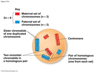
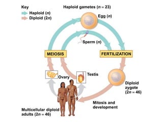
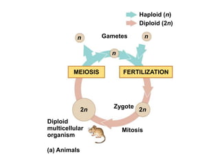
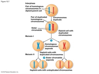
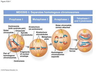
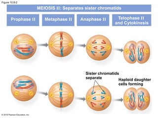
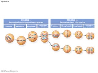
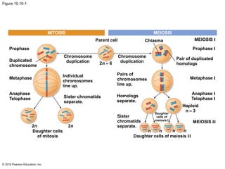
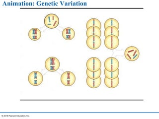
© 2016 Pearson Education, Inc.
BIOLOGY
Chapter 11 MEIOSIS AND SEXUAL REPRODUCTION
Overview
 Living organisms are distinguished by their ability to
reproduce their own kind
 Heredity is the transmission of traits from one
generation to the next
 Variation is demonstrated by the differences in
appearance that offspring show from parents and
siblings
 Genetics is the scientific study of heredity and
variation
© 2016 Pearson Education, Inc.
This OpenStax ancillary resource is © Rice University under a CC-BY 4.0 International license; it may be reproduced or modified but must be attributed to OpenStax,
Rice University and any changes must be noted. Any images credited to other sources are similarly available for reproduction, but must be attributed to their sources.
 Each of us, like these the organisms shown above, begins life as a fertilized egg
(zygote). After trillions of cell divisions, each of us develops into a complex,
multicellular organism. (credit a: modification of work by Frank Wouters; credit b:
modification of work by Ken Cole, USGS; credit c: modification of work by Martin
Pettitt)
Figure 11.1
Offspring acquire genes from parents by inheriting
chromosomes
 In a literal sense, children do not inherit particular
physical traits from their parents
 Children inherit Genes from parents
 Genes are the units of heredity and are made up of
segments of DNA
 Genes are passed to the next generation via
reproductive cells called gametes (sperm and
egg/ova)
© 2016 Pearson Education, Inc.
Inheritance of Genes
 Most DNA is packaged into
chromosomes
 Genes are located on
chromosomes.
 For example, humans have 46
chromosomes in their somatic
cells, the cells of the body
except for gametes and their
precursors
 Each gene has a specific
position, or locus, on a certain
chromosome
© 2016 Pearson Education, Inc.
Comparison of Asexual and Sexual Reproduction
 In asexual reproduction, a single individual passes
genes to its offspring without the fusion of gametes
 A clone is a group of genetically identical
individuals from the same parent
 In sexual reproduction, two parents give rise to
offspring that have unique combinations of genes
inherited from the two parents
© 2016 Pearson Education, Inc.
Figure 10.2-1
© 2016 Pearson Education, Inc.
0.5 mm
Parent
Bud
(a) Hydra
Video: Hydra Budding
© 2016 Pearson Education, Inc.
Sets of Chromosomes in Human Cells
 Human somatic cells have 23 pairs of
chromosomes
 A karyotype is an ordered display of the pairs of
chromosomes from a cell
 The two chromosomes in each pair are called
homologous chromosomes, or homologs
 Chromosomes in a homologous pair are the same
length and shape and carry genes controlling the
same inherited characters
© 2016 Pearson Education, Inc.
Figure 10.3
© 2016 Pearson Education, Inc.
Technique Results
Pair of homologous
duplicated chromosomes
Centromere 5 m
Sister
chromatids
Metaphase
chromosome
Figure 10.3-2
© 2016 Pearson Education, Inc.
Results
Pair of homologous
duplicated chromosomes
Centromere 5 m
Sister
chromatids
Metaphase
chromosome
 The sex chromosomes, which determine the sex
of the individual, are called X and Y
 Human females have a homologous pair of X
chromosomes (XX)
 Human males have one X and one Y chromosome
 The remaining 22 pairs of chromosomes are called
autosomes
© 2016 Pearson Education, Inc.
 Each pair of homologous chromosomes includes
one chromosome from each parent
 The 46 chromosomes in a human somatic cell are
two sets of 23: one from the mother and one from
the father
 A diploid cell (2n) has two sets of chromosomes
 For humans, the diploid number is 46 (2n  46)
© 2016 Pearson Education, Inc.
This OpenStax ancillary resource is © Rice University under a CC-BY 4.0 International license; it may be reproduced or modified but must be attributed to OpenStax,
Rice University and any changes must be noted. Any images credited to other sources are similarly available for reproduction, but must be attributed to their sources.
• Early in prophase I, homologous chromosomes come together to form a synapse. The
chromosomes are bound tightly together and in perfect alignment by a protein lattice called
a synaptonemal complex and by cohesin proteins at the centromere.
Figure 11.2
Figure 10.4
© 2016 Pearson Education, Inc.
Key
2n  6
Maternal set of
chromosomes (n  3)
Paternal set of
chromosomes (n  3)
Sister chromatids
of one duplicated
chromosome
Centromere
Two nonsister
chromatids in
a homologous pair
Pair of homologous
chromosomes
(one from each set)
 A gamete (sperm or egg) contains a single set of
chromosomes and is haploid (n)
 For humans, the haploid number is 23 (n = 23)
 Each set of 23 consists of 22 autosomes and a
single sex chromosome
 In an unfertilized egg (ovum), the sex chromosome
is X
 In a sperm cell, the sex chromosome may be either
X or Y
© 2016 Pearson Education, Inc.
Behavior of Chromosome Sets in the Human Life
Cycle
 Fertilization is the union of gametes (the sperm
and the egg)
 The fertilized egg is called a zygote and has one
set of chromosomes from each parent
 The zygote produces somatic cells by mitosis and
develops into an adult
© 2016 Pearson Education, Inc.
 At sexual maturity, the ovaries and testes produce
haploid gametes
 Gametes are the only types of human cells
produced by meiosis rather than mitosis
 Meiosis results in one set of chromosomes in each
gamete
 Fertilization and meiosis alternate in sexual life
cycles to maintain chromosome number
© 2016 Pearson Education, Inc.
Key
Haploid (n)
Haploid gametes (n  23)
Egg (n)
Sperm (n)
MEIOSIS FERTILIZATION
Ovary
Testis
Diploid
zygote
(2n  46)
Mitosis and
development
Multicellular diploid
adults (2n  46)
Diploid (2n)
The Sexual Life Cycle
 The alternation of meiosis and fertilization is
common to all organisms that reproduce sexually
 Gametes are the only haploid cells in animals
 They are produced by meiosis and undergo no
further cell division before fertilization
 Gametes fuse to form a diploid zygote that
divides by mitosis to develop into a multicellular
organism
© 2016 Pearson Education, Inc.
Haploid (n)
Diploid (2n)
n Gametes
n
MEIOSIS FERTILIZATION
n
2n
Diploid
multicellular
organism
(a) Animals
Zygote
2n
Mitosis
Meiosis reduces the number of chromosome sets from
diploid to haploid
 Like mitosis, meiosis is preceded by the duplication
of chromosomes
 Meiosis takes place in two sets of cell divisions,
called meiosis I and meiosis II
 The two cell divisions result in four daughter cells,
rather than the two daughter cells in mitosis
 Each daughter cell has only half as many
chromosomes as the parent cell
© 2016 Pearson Education, Inc.
The Stages of Meiosis
 For a single pair of homologous chromosomes in a
diploid cell, both members of the pair are duplicated
 The resulting sister chromatids are closely
associated all along their lengths
 Homologs may have different versions of genes,
each called an allele
 Homologs are not associated in any obvious way
except during meiosis
© 2016 Pearson Education, Inc.
Figure 10.7
© 2016 Pearson Education, Inc.
Interphase
Pair of homologous
chromosomes in
diploid parent cell
Pair of duplicated
homologous
chromosomes
Sister
chromatids
Meiosis I
Homologous
chromosomes
separate
Meiosis II
Haploid cells with
duplicated chromosomes
Sister chromatids
separate
Chromosomes
duplicate
Diploid cell with
duplicated
chromosomes
Haploid cells with unduplicated chromosomes
 Meiosis halves the total number of chromosomes very
specifically
 It reduces the number of sets from two to one, with each
daughter cell receiving one set of chromosomes
 In the first meiotic division, homologous pairs of
chromosomes pair and separate
 In the second meiotic division, sister chromatids of each
chromosome separate
 Four new haploid cells are produced as a result
© 2016 Pearson Education, Inc.
Video: Meiosis I in Sperm Formation
© 2016 Pearson Education, Inc.
Prophase I
 Chromosomes condense progressively throughout
prophase I
 Homologous chromosomes pair up, aligned gene
by gene
 In crossing over, nonsister chromatids exchange
DNA segments
 Each homologous pair has one or more X-shaped
regions called chiasmata
 Chiasmata exist at points where crossing over has
occurred.
© 2016 Pearson Education, Inc.
MEIOSIS I
This OpenStax ancillary resource is © Rice University under a CC-BY 4.0 International license; it may be reproduced or modified but must be attributed to OpenStax,
Rice University and any changes must be noted. Any images credited to other sources are similarly available for reproduction, but must be attributed to their sources.
 Crossover occurs
between non-sister
chromatids of
homologous
chromosomes. The
result is an exchange
of genetic material
between
homologous
chromosomes.
Figure 11.3
Metaphase I
 In metaphase I, homologous pairs line up at the
metaphase plate, with one chromosome facing each
pole
 Microtubules from one pole are attached to the
kinetochore of one chromosome of each tetrad
 Microtubules from the other pole are attached to the
kinetochore of the other chromosome
© 2016 Pearson Education, Inc.
Anaphase I
 In anaphase I, pairs of homologous chromosomes
separate
 One chromosome moves toward each pole, guided
by the spindle apparatus
 Sister chromatids remain attached at the
centromere and move as one unit toward the pole
© 2016 Pearson Education, Inc.
Telophase I and Cytokinesis
 In the beginning of telophase I, each half of the cell
has a haploid set of chromosomes
 Each chromosome still consists of two sister
chromatids
 Cytokinesis usually occurs simultaneously, forming
two haploid daughter cells
© 2016 Pearson Education, Inc.
 In animal cells, a cleavage furrow forms; in plant
cells, a cell plate forms
 No chromosome duplication occurs between the
end of meiosis I and the beginning of meiosis II
because the chromosomes are already replicated
© 2016 Pearson Education, Inc.
Figure 10.8-1
© 2016 Pearson Education, Inc.
MEIOSIS I: Separates homologous chromosomes
Prophase I Metaphase I Anaphase I
Sister chromatids
remain attached
Telophase I
and Cytokinesis
Centrosome
(with centriole pair)
Kinetochore
(at centromere)
Sister
chromatids
Chiasmata
Kinetochore
microtubules
Spindle
microtubules
Metaphase
plate
Pair of
homologous
chromosomes
Fragments
of nuclear
envelope
Homologous
chromosomes
separate
Cleavage
furrow
Centromere
Division in meiosis II also occurs in four phases
 Prophase II
 Metaphase II
 Anaphase II
 Telophase II and cytokinesis
 Meiosis II is very similar to mitosis
© 2016 Pearson Education, Inc.
MEIOSIS II
Prophase II
 In prophase II, a spindle apparatus forms
 In late prophase II, chromosomes (each still
composed of two chromatids) move toward the
metaphase plate
© 2016 Pearson Education, Inc.
Metaphase II
 The sister chromatids are arranged at the
metaphase plate
 Because of crossing over in meiosis I, the two sister
chromatids of each chromosome are no longer
genetically identical
 The kinetochores of sister chromatids attach to
microtubules extending from opposite poles
© 2016 Pearson Education, Inc.
Anaphase II
 In anaphase II, the sister chromatids separate
 The sister chromatids of each chromosome now
move as two newly individual chromosomes toward
opposite poles
© 2016 Pearson Education, Inc.
Telophase II and Cytokinesis
 Nuclei form, and the chromosomes begin
decondensing
 At the end of meiosis, there are four daughter cells,
each with a haploid set of unduplicated
chromosomes
 Each daughter cell is genetically distinct from the
others and from the parent cell
© 2016 Pearson Education, Inc.
Figure 10.8-2
© 2016 Pearson Education, Inc.
MEIOSIS II: Separates sister chromatids
Prophase II Metaphase II Anaphase II Telophase II
and Cytokinesis
Sister chromatids
separate Haploid daughter
cells forming
Figure 10.8
© 2016 Pearson Education, Inc.
MEIOSIS I:
Separates homologous chromosomes
Prophase I Metaphase I Anaphase I
Telophase I
and Cytokinesis
MEIOSIS II:
Separates sister chromatids
Prophase II
Telophase II
and Cytokinesis
Metaphase II Anaphase II
A Comparison of Mitosis and Meiosis
 Mitosis conserves the number of chromosome sets,
producing cells that are genetically identical to the
parent cell
 Meiosis reduces the number of chromosome sets
from two (diploid) to one (haploid), producing cells
that differ genetically from each other and from the
parent cell
 Meiosis includes two divisions after replication, each
with specific stages
© 2016 Pearson Education, Inc.
 Three events are unique to meiosis, and all three
occur in meiosis l
 Synapsis and crossing over in prophase I:
Homologous chromosomes physically connect and
exchange genetic information
 Alignment of homologous pairs at the metaphase
plate: Homologous pairs of chromosomes are
positioned there in metaphase I
 Separation of homologs during anaphase I
© 2016 Pearson Education, Inc.
Figure 10.10-1
© 2016 Pearson Education, Inc.
MITOSIS
Parent cell
Prophase
Duplicated
chromosome
Metaphase
Chromosome
duplication
Individual
chromosomes
line up.
Sister chromatids
separate.
2n  6
Chromosome
duplication
Pairs of
chromosomes
line up.
Homologs
separate.
MEIOSIS
Chiasma MEIOSIS I
Prophase I
Pair of duplicated
homologs
Metaphase I
Anaphase I
Telophase I
Daughter
cells of
meiosis I
Anaphase
Telophase
Haploid
n  3
MEIOSIS II
2n
Daughter cells
of mitosis
2n
Sister
chromatids
separate.
n nnn
Daughter cells of meiosis II
Figure 10.10-2a
© 2016 Pearson Education, Inc.
Figure 10.10-2b
© 2016 Pearson Education, Inc.
Figure 10.10-2
© 2016 Pearson Education, Inc.
Genetic variation produced in sexual life cycles
contributes to evolution
 Mutations (changes in an organism’s DNA) are the
original source of genetic diversity
 Mutations create different versions of genes called
alleles
 Reshuffling of alleles during sexual reproduction
produces genetic variation
© 2016 Pearson Education, Inc.
Origins of Genetic Variation Among Offspring
 The behavior of chromosomes during meiosis and
fertilization is responsible for most of the variation
that arises in each generation
 Three mechanisms contribute to genetic variation
 Independent assortment of chromosomes
 Crossing over
 Random fertilization
© 2016 Pearson Education, Inc.
Animation: Genetic Variation
© 2016 Pearson Education, Inc.
Animation: Meiosis
© 2016 Pearson Education, Inc.

11 lecture presentation

  • 1.
    © 2016 PearsonEducation, Inc. BIOLOGY Chapter 11 MEIOSIS AND SEXUAL REPRODUCTION
  • 2.
    Overview  Living organismsare distinguished by their ability to reproduce their own kind  Heredity is the transmission of traits from one generation to the next  Variation is demonstrated by the differences in appearance that offspring show from parents and siblings  Genetics is the scientific study of heredity and variation © 2016 Pearson Education, Inc.
  • 3.
    This OpenStax ancillaryresource is © Rice University under a CC-BY 4.0 International license; it may be reproduced or modified but must be attributed to OpenStax, Rice University and any changes must be noted. Any images credited to other sources are similarly available for reproduction, but must be attributed to their sources.  Each of us, like these the organisms shown above, begins life as a fertilized egg (zygote). After trillions of cell divisions, each of us develops into a complex, multicellular organism. (credit a: modification of work by Frank Wouters; credit b: modification of work by Ken Cole, USGS; credit c: modification of work by Martin Pettitt) Figure 11.1
  • 4.
    Offspring acquire genesfrom parents by inheriting chromosomes  In a literal sense, children do not inherit particular physical traits from their parents  Children inherit Genes from parents  Genes are the units of heredity and are made up of segments of DNA  Genes are passed to the next generation via reproductive cells called gametes (sperm and egg/ova) © 2016 Pearson Education, Inc.
  • 5.
    Inheritance of Genes Most DNA is packaged into chromosomes  Genes are located on chromosomes.  For example, humans have 46 chromosomes in their somatic cells, the cells of the body except for gametes and their precursors  Each gene has a specific position, or locus, on a certain chromosome © 2016 Pearson Education, Inc.
  • 6.
    Comparison of Asexualand Sexual Reproduction  In asexual reproduction, a single individual passes genes to its offspring without the fusion of gametes  A clone is a group of genetically identical individuals from the same parent  In sexual reproduction, two parents give rise to offspring that have unique combinations of genes inherited from the two parents © 2016 Pearson Education, Inc.
  • 7.
    Figure 10.2-1 © 2016Pearson Education, Inc. 0.5 mm Parent Bud (a) Hydra
  • 8.
    Video: Hydra Budding ©2016 Pearson Education, Inc.
  • 9.
    Sets of Chromosomesin Human Cells  Human somatic cells have 23 pairs of chromosomes  A karyotype is an ordered display of the pairs of chromosomes from a cell  The two chromosomes in each pair are called homologous chromosomes, or homologs  Chromosomes in a homologous pair are the same length and shape and carry genes controlling the same inherited characters © 2016 Pearson Education, Inc.
  • 10.
    Figure 10.3 © 2016Pearson Education, Inc. Technique Results Pair of homologous duplicated chromosomes Centromere 5 m Sister chromatids Metaphase chromosome
  • 11.
    Figure 10.3-2 © 2016Pearson Education, Inc. Results Pair of homologous duplicated chromosomes Centromere 5 m Sister chromatids Metaphase chromosome
  • 12.
     The sexchromosomes, which determine the sex of the individual, are called X and Y  Human females have a homologous pair of X chromosomes (XX)  Human males have one X and one Y chromosome  The remaining 22 pairs of chromosomes are called autosomes © 2016 Pearson Education, Inc.
  • 13.
     Each pairof homologous chromosomes includes one chromosome from each parent  The 46 chromosomes in a human somatic cell are two sets of 23: one from the mother and one from the father  A diploid cell (2n) has two sets of chromosomes  For humans, the diploid number is 46 (2n  46) © 2016 Pearson Education, Inc.
  • 14.
    This OpenStax ancillaryresource is © Rice University under a CC-BY 4.0 International license; it may be reproduced or modified but must be attributed to OpenStax, Rice University and any changes must be noted. Any images credited to other sources are similarly available for reproduction, but must be attributed to their sources. • Early in prophase I, homologous chromosomes come together to form a synapse. The chromosomes are bound tightly together and in perfect alignment by a protein lattice called a synaptonemal complex and by cohesin proteins at the centromere. Figure 11.2
  • 15.
    Figure 10.4 © 2016Pearson Education, Inc. Key 2n  6 Maternal set of chromosomes (n  3) Paternal set of chromosomes (n  3) Sister chromatids of one duplicated chromosome Centromere Two nonsister chromatids in a homologous pair Pair of homologous chromosomes (one from each set)
  • 16.
     A gamete(sperm or egg) contains a single set of chromosomes and is haploid (n)  For humans, the haploid number is 23 (n = 23)  Each set of 23 consists of 22 autosomes and a single sex chromosome  In an unfertilized egg (ovum), the sex chromosome is X  In a sperm cell, the sex chromosome may be either X or Y © 2016 Pearson Education, Inc.
  • 17.
    Behavior of ChromosomeSets in the Human Life Cycle  Fertilization is the union of gametes (the sperm and the egg)  The fertilized egg is called a zygote and has one set of chromosomes from each parent  The zygote produces somatic cells by mitosis and develops into an adult © 2016 Pearson Education, Inc.
  • 18.
     At sexualmaturity, the ovaries and testes produce haploid gametes  Gametes are the only types of human cells produced by meiosis rather than mitosis  Meiosis results in one set of chromosomes in each gamete  Fertilization and meiosis alternate in sexual life cycles to maintain chromosome number © 2016 Pearson Education, Inc.
  • 19.
    Key Haploid (n) Haploid gametes(n  23) Egg (n) Sperm (n) MEIOSIS FERTILIZATION Ovary Testis Diploid zygote (2n  46) Mitosis and development Multicellular diploid adults (2n  46) Diploid (2n)
  • 20.
    The Sexual LifeCycle  The alternation of meiosis and fertilization is common to all organisms that reproduce sexually  Gametes are the only haploid cells in animals  They are produced by meiosis and undergo no further cell division before fertilization  Gametes fuse to form a diploid zygote that divides by mitosis to develop into a multicellular organism © 2016 Pearson Education, Inc.
  • 21.
    Haploid (n) Diploid (2n) nGametes n MEIOSIS FERTILIZATION n 2n Diploid multicellular organism (a) Animals Zygote 2n Mitosis
  • 22.
    Meiosis reduces thenumber of chromosome sets from diploid to haploid  Like mitosis, meiosis is preceded by the duplication of chromosomes  Meiosis takes place in two sets of cell divisions, called meiosis I and meiosis II  The two cell divisions result in four daughter cells, rather than the two daughter cells in mitosis  Each daughter cell has only half as many chromosomes as the parent cell © 2016 Pearson Education, Inc.
  • 23.
    The Stages ofMeiosis  For a single pair of homologous chromosomes in a diploid cell, both members of the pair are duplicated  The resulting sister chromatids are closely associated all along their lengths  Homologs may have different versions of genes, each called an allele  Homologs are not associated in any obvious way except during meiosis © 2016 Pearson Education, Inc.
  • 24.
    Figure 10.7 © 2016Pearson Education, Inc. Interphase Pair of homologous chromosomes in diploid parent cell Pair of duplicated homologous chromosomes Sister chromatids Meiosis I Homologous chromosomes separate Meiosis II Haploid cells with duplicated chromosomes Sister chromatids separate Chromosomes duplicate Diploid cell with duplicated chromosomes Haploid cells with unduplicated chromosomes
  • 25.
     Meiosis halvesthe total number of chromosomes very specifically  It reduces the number of sets from two to one, with each daughter cell receiving one set of chromosomes  In the first meiotic division, homologous pairs of chromosomes pair and separate  In the second meiotic division, sister chromatids of each chromosome separate  Four new haploid cells are produced as a result © 2016 Pearson Education, Inc.
  • 26.
    Video: Meiosis Iin Sperm Formation © 2016 Pearson Education, Inc.
  • 27.
    Prophase I  Chromosomescondense progressively throughout prophase I  Homologous chromosomes pair up, aligned gene by gene  In crossing over, nonsister chromatids exchange DNA segments  Each homologous pair has one or more X-shaped regions called chiasmata  Chiasmata exist at points where crossing over has occurred. © 2016 Pearson Education, Inc. MEIOSIS I
  • 28.
    This OpenStax ancillaryresource is © Rice University under a CC-BY 4.0 International license; it may be reproduced or modified but must be attributed to OpenStax, Rice University and any changes must be noted. Any images credited to other sources are similarly available for reproduction, but must be attributed to their sources.  Crossover occurs between non-sister chromatids of homologous chromosomes. The result is an exchange of genetic material between homologous chromosomes. Figure 11.3
  • 29.
    Metaphase I  Inmetaphase I, homologous pairs line up at the metaphase plate, with one chromosome facing each pole  Microtubules from one pole are attached to the kinetochore of one chromosome of each tetrad  Microtubules from the other pole are attached to the kinetochore of the other chromosome © 2016 Pearson Education, Inc.
  • 30.
    Anaphase I  Inanaphase I, pairs of homologous chromosomes separate  One chromosome moves toward each pole, guided by the spindle apparatus  Sister chromatids remain attached at the centromere and move as one unit toward the pole © 2016 Pearson Education, Inc.
  • 31.
    Telophase I andCytokinesis  In the beginning of telophase I, each half of the cell has a haploid set of chromosomes  Each chromosome still consists of two sister chromatids  Cytokinesis usually occurs simultaneously, forming two haploid daughter cells © 2016 Pearson Education, Inc.
  • 32.
     In animalcells, a cleavage furrow forms; in plant cells, a cell plate forms  No chromosome duplication occurs between the end of meiosis I and the beginning of meiosis II because the chromosomes are already replicated © 2016 Pearson Education, Inc.
  • 33.
    Figure 10.8-1 © 2016Pearson Education, Inc. MEIOSIS I: Separates homologous chromosomes Prophase I Metaphase I Anaphase I Sister chromatids remain attached Telophase I and Cytokinesis Centrosome (with centriole pair) Kinetochore (at centromere) Sister chromatids Chiasmata Kinetochore microtubules Spindle microtubules Metaphase plate Pair of homologous chromosomes Fragments of nuclear envelope Homologous chromosomes separate Cleavage furrow Centromere
  • 34.
    Division in meiosisII also occurs in four phases  Prophase II  Metaphase II  Anaphase II  Telophase II and cytokinesis  Meiosis II is very similar to mitosis © 2016 Pearson Education, Inc. MEIOSIS II
  • 35.
    Prophase II  Inprophase II, a spindle apparatus forms  In late prophase II, chromosomes (each still composed of two chromatids) move toward the metaphase plate © 2016 Pearson Education, Inc.
  • 36.
    Metaphase II  Thesister chromatids are arranged at the metaphase plate  Because of crossing over in meiosis I, the two sister chromatids of each chromosome are no longer genetically identical  The kinetochores of sister chromatids attach to microtubules extending from opposite poles © 2016 Pearson Education, Inc.
  • 37.
    Anaphase II  Inanaphase II, the sister chromatids separate  The sister chromatids of each chromosome now move as two newly individual chromosomes toward opposite poles © 2016 Pearson Education, Inc.
  • 38.
    Telophase II andCytokinesis  Nuclei form, and the chromosomes begin decondensing  At the end of meiosis, there are four daughter cells, each with a haploid set of unduplicated chromosomes  Each daughter cell is genetically distinct from the others and from the parent cell © 2016 Pearson Education, Inc.
  • 39.
    Figure 10.8-2 © 2016Pearson Education, Inc. MEIOSIS II: Separates sister chromatids Prophase II Metaphase II Anaphase II Telophase II and Cytokinesis Sister chromatids separate Haploid daughter cells forming
  • 40.
    Figure 10.8 © 2016Pearson Education, Inc. MEIOSIS I: Separates homologous chromosomes Prophase I Metaphase I Anaphase I Telophase I and Cytokinesis MEIOSIS II: Separates sister chromatids Prophase II Telophase II and Cytokinesis Metaphase II Anaphase II
  • 41.
    A Comparison ofMitosis and Meiosis  Mitosis conserves the number of chromosome sets, producing cells that are genetically identical to the parent cell  Meiosis reduces the number of chromosome sets from two (diploid) to one (haploid), producing cells that differ genetically from each other and from the parent cell  Meiosis includes two divisions after replication, each with specific stages © 2016 Pearson Education, Inc.
  • 42.
     Three eventsare unique to meiosis, and all three occur in meiosis l  Synapsis and crossing over in prophase I: Homologous chromosomes physically connect and exchange genetic information  Alignment of homologous pairs at the metaphase plate: Homologous pairs of chromosomes are positioned there in metaphase I  Separation of homologs during anaphase I © 2016 Pearson Education, Inc.
  • 43.
    Figure 10.10-1 © 2016Pearson Education, Inc. MITOSIS Parent cell Prophase Duplicated chromosome Metaphase Chromosome duplication Individual chromosomes line up. Sister chromatids separate. 2n  6 Chromosome duplication Pairs of chromosomes line up. Homologs separate. MEIOSIS Chiasma MEIOSIS I Prophase I Pair of duplicated homologs Metaphase I Anaphase I Telophase I Daughter cells of meiosis I Anaphase Telophase Haploid n  3 MEIOSIS II 2n Daughter cells of mitosis 2n Sister chromatids separate. n nnn Daughter cells of meiosis II
  • 44.
    Figure 10.10-2a © 2016Pearson Education, Inc.
  • 45.
    Figure 10.10-2b © 2016Pearson Education, Inc.
  • 46.
    Figure 10.10-2 © 2016Pearson Education, Inc.
  • 47.
    Genetic variation producedin sexual life cycles contributes to evolution  Mutations (changes in an organism’s DNA) are the original source of genetic diversity  Mutations create different versions of genes called alleles  Reshuffling of alleles during sexual reproduction produces genetic variation © 2016 Pearson Education, Inc.
  • 48.
    Origins of GeneticVariation Among Offspring  The behavior of chromosomes during meiosis and fertilization is responsible for most of the variation that arises in each generation  Three mechanisms contribute to genetic variation  Independent assortment of chromosomes  Crossing over  Random fertilization © 2016 Pearson Education, Inc.
  • 49.
    Animation: Genetic Variation ©2016 Pearson Education, Inc.
  • 50.
    Animation: Meiosis © 2016Pearson Education, Inc.