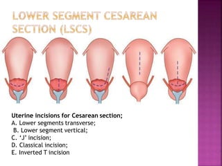
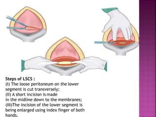
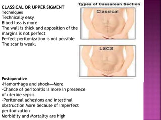
IBRAHIM MOHAMED
HIRSI AWALE
JU
DEFINITION : It is an operative procedure
whereby the fetuses after the end of 28th
week are delivered through an incision on
the abdominal and uterine walls.
The first operation performed on a patientis
referred to as a primary cesarean section.
When the operation is performed in
subsequent pregnancies, it is called repeat
cesarean section.
INCIDENCE: The incidence of cesarean
section is steadily rising. During the last
decade there has been two to three fold rise
in the incidence from the initial rate of
about 10%. due to improved anesthesia,
availability of blood transfusion and
antibiotics,
Cesarean delivery is done when labor is
contraindicated (central placenta previa)
and/or vaginal delivery is found unsafe for
the fetus and/or mother.
The indications are broadly divided into
two categories :• Absolute • Relative (CMN)
Vaginal delivery is not possible. Cesarean is needed
even with a dead fetus
Indications:
1. Central placenta previa
2.Contracted pelvis or cephalopelvic
disproportion (absolute)
3.Pelvic mass causing obstruction (cervical or
broad ligament fibroid)
4. Advanced carcinoma cervix
5. Vaginal obstruction (atresia, stenosis)
Primigravidae :
(1) Failed induction
(2) Fetal distress (no reassuring FHR)
(3) Cephalo pelvic disproportion (CPD)
(4)Dystocia (dysfunctional labour) no progress of
labour
(5)Malposition and malpresentation
(occipitoposterior, breech).
Mutigravidae :
(1) Previous Caesarean delivery
(2)Antepartum haemorrhage (placenta praevia,
placental abruption)
(3) Malpresentation (breech, transverse lie).
Vaginal delivery may be possible but risks to the mother
and/ or to the baby are high
1. Cephalopelvic disproportion (relative)
2. Previous cesarean delivery
(a)when primary CS was due to recurrent indication
(contracted pelvis).
(b) Previous two CS (c) Features of scar dehiscence.
(d) Previous classical CS
3. Non-reassuring FHR (fetal distress)
4. Dystocia may be due to (three Ps) relatively large
fetus (passenger), small pelvis (passage) or inefficient
uterine contractions (Power)
5. Antepartum hemorrhage
6. Malpresentation—Breech, shoulder (transverse
lie), brow
7. Failure to progress in labor after induction
8. Hypertensive disorders—(a) Severe pre-
eclampsia, (b) Eclampsia—uncontrolled fits even
with antiseizure therapy (
9. Medical-gynecological disorders—(a) Diabetes
(uncontrolled), heart disease (coarctation of aorta,
Marfan’s syndrome. (b) Mechanical obstruction (due
to benign or malignant pelvic tumors (carcinoma
cervix), or following repair of vesicovaginal fistula
• Elective
• Emergency (An arbitrary time limit of 30
minutes)
•A) Lower segment
•B) Classical or upper segment
A) Lower Segment Cesarean Section (LSCS):
In this operation, the extraction of the baby is
done through an incision made in the lower
segment through a transperitoneal approach. It
is the only method practiced in present day
obstetrics and unless specified, cesarean section
means lower segment operation. The operation
done through an extra- peritoneal approach to
the lower segment in infected cases is obsolete.
According to the opening of peritoneal
cavity:-
A) transperitoneal: The ordinary
operation where the peritoneal cavity is
opened before incising the uterus
B) Extraperitoneal: the peritoneal
cavity is not opened and the lower uterine
segment is reached either laterally or
inferiorly by reflecting the peritoneum of
the vesico-uterine pouch (Space of
RETZIUS). Its indicated in case of infected
uterine contents as chorioamnionitis
B) Classical: In this operation, the baby is extracted through
an incision made in the upper segment of the uterus.
INDICATIONS:
Lower segment approach is difficult—
(1)Dense adhesions due to previous abdominal
operation
(2)Severe contracted pelvis (osteomalacic or rachitic)
with pendulous abdomen.
• Lower segment approach is risky—
(1)Big fibroid on the lower segment—Blood loss is more
and contemplating myomectomy may end in
hysterectomy
(2)Carcinoma cervix—To prevent dissemination of the
growth and postoperative sepsis
(3) Repair of high VVF
(4)Complete anterior placenta previa—risk of
hemorrhage.
Perimortem cesarean section— is done to
have a live baby (rare). Perimortem section
is an extreme emergency procedure.
Classical section is done in a woman who has
suffered a cardiac arrest. The infant may
survive if delivery is done within 10 minutes
of maternal death.
PREOPERATIVE PREPARATION
Informed Consent, anesthesia and blood transfusion is
obtained.
1) Abdomen is scrubbed with soap and nonorganic iodidelotion.
Hair may be clipped.
2) Premedicative sedative must not be given.
3) Ranitidine (H2 blocker) 150 mg is given orally night before
(elective procedure) and it is repeated (50 mg IM or IV) one hour
before the surgery to raise the gastric pH.
4) Metoclopramide (10 mg IV) is given to increase the tone of the
lower esophageal sphincter as well as to reduce the stomach
contents. It is administered after about 3 minutes of pre-
oxygenation in the theater.
5) The stomach should be emptied,
6) Bladder should be emptied by a Foley catheter
7) FHS should be checked
8) Neonatologist should be made available.
9) Cross match blood when above average blood loss (placenta
previa, prior multiple cesarean delivery) is anticipated.
Uterine incisions for Cesarean section;
A. Lower segments transverse;
B. Lower segment vertical;
C. ‘J’ incision;
D. Classical incision;
E. Inverted T incision
Steps of LSCS :
(I) The loose peritoneum on the lower
segment is cut transversely;
(II) A short incision is made
in the midline down to the membranes;
(III)The incision of the lower segment is
being enlarged using index finger of both
hands.
(IV)Sagittal section showing
insinuation of the fingers between the
lower uterine flap and the fetal head
until the posterior surface is reached;
(V) Methods of delivery of the head;
(VI) Placenta is being delivered
VII VIII IX
(VII)Inserting the continuous catgut
suture taking deeper muscles
excluding the decidua
(VIII)Similar method of continuous
suture taking superficial muscles and
fascia down to the first layer of
suture
(IX)Continuous peritoneal catgut
suture.
1)Observation for the first 6–8 hours is
important. Periodic check up of pulse, BP
,
amount of vaginal bleeding and behavior of the
uterus (in low transverse incision) is done and
recorded.
2)Fluid—Sodium chloride (0.9%) or Ringer’s
lactate drip is continued until at least 2 – 2.5
liters of the solution are infused. Blood
transfusion is helpful in anemic mothers for a
speedy post-operative recovery. Blood
transfusion is required if the blood loss is more
than average during the operation (average
blood loss in cesarean section is approximately
0.5 to 1 liter).
3)Oxytocics: Injection oxytocin 5 units IM or IV (slow) is
given and may be repeated.
4)Prophylactic antibiotic (cephalosporins,
metronidazole) for all cesarean delivery is given for 2–3
days But Therapeutic antibiotic is given when
indicated.
5)Analgesics in the form of pethidine hydrochloride 75–
100 mg is administered and may have to be repeated.
6)Ambulation—The patient can sit on the bed or even
get out of bed to evacuate the bladder, provided
the general condition permits. She is encouraged to
move her legs and ankles and to breathe deeply to
minimize leg vein thrombosis and pulmonary embolism.
7)Baby- is put to the breast for feeding after 3–4 hours
when mother is stable and relieved of pain.
CLASSICAL OR UPPER SIGMENT
Techniques
Technically easy
Blood loss is more
The wall is thick and apposition of the
margins is not perfect
Perfect peritonization is not possible
The scar is weak.
Postoperative
-Hemorrhage and shock—More
-Chance of peritonitis is more in presence
of uterine sepsis
-Peritoneal adhesions and intestinal
obstruction More because of imperfect
peritonization
Morbidity and Mortality are high
INTRAOPERATIVE COMPLICATIONS
1 Extension of uterine incision: to one or both the
sides. This may involve the uterine vessels to cause
severe hemorrhage, may lead to broad ligament
hematoma formation.
2 Bladder injury may occur in a repeat procedure
3 Morbid adherent placenta (placenta accreta) is
commonly seen in cases with placenta previa who had
prior cesarean delivery. Total hysterectomy is often
needed for such a case to control hemorrhage.
4 Hemorrhage may be due to uterine atony or uterine
lacerations.
POSTOPERATIVE COMPLICATIONS
MATERNAL : • Immediate • Remote
A) IMMEDIATE
1 Postpartum hemorrhage
2 Shock
3 Anesthetic hazards; particularly (Mendelson’s
syndrome)
4 Infections
5 Intestinal obstruction
6 DVT
B) REMOTE:
Gynecological: Menstrual excess or irregularities,
chronic pelvic pain or backache.
General surgical: Incisional hernia, Intestinal
obstruction due to adhesions and bands.
Future pregnancy—There is risk of scar rupture
FETAL :
1-Intracranial hemorrhage
2- RDS
3- Prematurity; This is seen when fetal maturity is
uncertain.
Cesarean hysterectomy refers to an
operation where cesarean section is followed
by removal of the uterus. The common
conditions are :
(1) Morbid adherent placenta
(2)Atonic uterus and uncontrolled
postpartum hemorrhage
(3) Big fibroid (parous)
(4)Extensive lacerations due to extension of
tears with broad ligament hematoma
(5) Grossly infected uterus
(6) Rupture uterus.
A) Breech presentation
-Assisted vaginal Delivery of breech
B) Dystocia
- Partographic monitoring in labor management
- Active management of labor
C) Fetal distress, Vaginal birth after cesarean
section
- Confirm fetal acidosis by fetal blood sampling
D) Amnioinfusion
- Management of cases with variable or early FHR
Deceleration due to oligohydramnios, Meconium
stained liquor
DC Dutta's Textbook of
Obstetrics_7E
c-section-180515193200.pptx

c-section-180515193200.pptx

  • 1.
  • 2.
    DEFINITION : Itis an operative procedure whereby the fetuses after the end of 28th week are delivered through an incision on the abdominal and uterine walls.
  • 3.
    The first operationperformed on a patientis referred to as a primary cesarean section. When the operation is performed in subsequent pregnancies, it is called repeat cesarean section.
  • 4.
    INCIDENCE: The incidenceof cesarean section is steadily rising. During the last decade there has been two to three fold rise in the incidence from the initial rate of about 10%. due to improved anesthesia, availability of blood transfusion and antibiotics,
  • 5.
    Cesarean delivery isdone when labor is contraindicated (central placenta previa) and/or vaginal delivery is found unsafe for the fetus and/or mother. The indications are broadly divided into two categories :• Absolute • Relative (CMN)
  • 6.
    Vaginal delivery isnot possible. Cesarean is needed even with a dead fetus Indications: 1. Central placenta previa 2.Contracted pelvis or cephalopelvic disproportion (absolute) 3.Pelvic mass causing obstruction (cervical or broad ligament fibroid) 4. Advanced carcinoma cervix 5. Vaginal obstruction (atresia, stenosis)
  • 7.
    Primigravidae : (1) Failedinduction (2) Fetal distress (no reassuring FHR) (3) Cephalo pelvic disproportion (CPD) (4)Dystocia (dysfunctional labour) no progress of labour (5)Malposition and malpresentation (occipitoposterior, breech). Mutigravidae : (1) Previous Caesarean delivery (2)Antepartum haemorrhage (placenta praevia, placental abruption) (3) Malpresentation (breech, transverse lie).
  • 8.
    Vaginal delivery maybe possible but risks to the mother and/ or to the baby are high 1. Cephalopelvic disproportion (relative) 2. Previous cesarean delivery (a)when primary CS was due to recurrent indication (contracted pelvis). (b) Previous two CS (c) Features of scar dehiscence. (d) Previous classical CS 3. Non-reassuring FHR (fetal distress) 4. Dystocia may be due to (three Ps) relatively large fetus (passenger), small pelvis (passage) or inefficient uterine contractions (Power)
  • 9.
    5. Antepartum hemorrhage 6.Malpresentation—Breech, shoulder (transverse lie), brow 7. Failure to progress in labor after induction 8. Hypertensive disorders—(a) Severe pre- eclampsia, (b) Eclampsia—uncontrolled fits even with antiseizure therapy ( 9. Medical-gynecological disorders—(a) Diabetes (uncontrolled), heart disease (coarctation of aorta, Marfan’s syndrome. (b) Mechanical obstruction (due to benign or malignant pelvic tumors (carcinoma cervix), or following repair of vesicovaginal fistula
  • 10.
    • Elective • Emergency(An arbitrary time limit of 30 minutes)
  • 11.
    •A) Lower segment •B)Classical or upper segment A) Lower Segment Cesarean Section (LSCS): In this operation, the extraction of the baby is done through an incision made in the lower segment through a transperitoneal approach. It is the only method practiced in present day obstetrics and unless specified, cesarean section means lower segment operation. The operation done through an extra- peritoneal approach to the lower segment in infected cases is obsolete.
  • 12.
    According to theopening of peritoneal cavity:- A) transperitoneal: The ordinary operation where the peritoneal cavity is opened before incising the uterus B) Extraperitoneal: the peritoneal cavity is not opened and the lower uterine segment is reached either laterally or inferiorly by reflecting the peritoneum of the vesico-uterine pouch (Space of RETZIUS). Its indicated in case of infected uterine contents as chorioamnionitis
  • 13.
    B) Classical: Inthis operation, the baby is extracted through an incision made in the upper segment of the uterus. INDICATIONS: Lower segment approach is difficult— (1)Dense adhesions due to previous abdominal operation (2)Severe contracted pelvis (osteomalacic or rachitic) with pendulous abdomen. • Lower segment approach is risky— (1)Big fibroid on the lower segment—Blood loss is more and contemplating myomectomy may end in hysterectomy (2)Carcinoma cervix—To prevent dissemination of the growth and postoperative sepsis (3) Repair of high VVF (4)Complete anterior placenta previa—risk of hemorrhage.
  • 14.
    Perimortem cesarean section—is done to have a live baby (rare). Perimortem section is an extreme emergency procedure. Classical section is done in a woman who has suffered a cardiac arrest. The infant may survive if delivery is done within 10 minutes of maternal death.
  • 15.
    PREOPERATIVE PREPARATION Informed Consent,anesthesia and blood transfusion is obtained. 1) Abdomen is scrubbed with soap and nonorganic iodidelotion. Hair may be clipped. 2) Premedicative sedative must not be given. 3) Ranitidine (H2 blocker) 150 mg is given orally night before (elective procedure) and it is repeated (50 mg IM or IV) one hour before the surgery to raise the gastric pH. 4) Metoclopramide (10 mg IV) is given to increase the tone of the lower esophageal sphincter as well as to reduce the stomach contents. It is administered after about 3 minutes of pre- oxygenation in the theater. 5) The stomach should be emptied, 6) Bladder should be emptied by a Foley catheter 7) FHS should be checked 8) Neonatologist should be made available. 9) Cross match blood when above average blood loss (placenta previa, prior multiple cesarean delivery) is anticipated.
  • 16.
    Uterine incisions forCesarean section; A. Lower segments transverse; B. Lower segment vertical; C. ‘J’ incision; D. Classical incision; E. Inverted T incision
  • 17.
    Steps of LSCS: (I) The loose peritoneum on the lower segment is cut transversely; (II) A short incision is made in the midline down to the membranes; (III)The incision of the lower segment is being enlarged using index finger of both hands.
  • 18.
    (IV)Sagittal section showing insinuationof the fingers between the lower uterine flap and the fetal head until the posterior surface is reached; (V) Methods of delivery of the head; (VI) Placenta is being delivered
  • 19.
    VII VIII IX (VII)Insertingthe continuous catgut suture taking deeper muscles excluding the decidua (VIII)Similar method of continuous suture taking superficial muscles and fascia down to the first layer of suture (IX)Continuous peritoneal catgut suture.
  • 20.
    1)Observation for thefirst 6–8 hours is important. Periodic check up of pulse, BP , amount of vaginal bleeding and behavior of the uterus (in low transverse incision) is done and recorded. 2)Fluid—Sodium chloride (0.9%) or Ringer’s lactate drip is continued until at least 2 – 2.5 liters of the solution are infused. Blood transfusion is helpful in anemic mothers for a speedy post-operative recovery. Blood transfusion is required if the blood loss is more than average during the operation (average blood loss in cesarean section is approximately 0.5 to 1 liter).
  • 21.
    3)Oxytocics: Injection oxytocin5 units IM or IV (slow) is given and may be repeated. 4)Prophylactic antibiotic (cephalosporins, metronidazole) for all cesarean delivery is given for 2–3 days But Therapeutic antibiotic is given when indicated. 5)Analgesics in the form of pethidine hydrochloride 75– 100 mg is administered and may have to be repeated. 6)Ambulation—The patient can sit on the bed or even get out of bed to evacuate the bladder, provided the general condition permits. She is encouraged to move her legs and ankles and to breathe deeply to minimize leg vein thrombosis and pulmonary embolism. 7)Baby- is put to the breast for feeding after 3–4 hours when mother is stable and relieved of pain.
  • 22.
    CLASSICAL OR UPPERSIGMENT Techniques Technically easy Blood loss is more The wall is thick and apposition of the margins is not perfect Perfect peritonization is not possible The scar is weak. Postoperative -Hemorrhage and shock—More -Chance of peritonitis is more in presence of uterine sepsis -Peritoneal adhesions and intestinal obstruction More because of imperfect peritonization Morbidity and Mortality are high
  • 23.
    INTRAOPERATIVE COMPLICATIONS 1 Extensionof uterine incision: to one or both the sides. This may involve the uterine vessels to cause severe hemorrhage, may lead to broad ligament hematoma formation. 2 Bladder injury may occur in a repeat procedure 3 Morbid adherent placenta (placenta accreta) is commonly seen in cases with placenta previa who had prior cesarean delivery. Total hysterectomy is often needed for such a case to control hemorrhage. 4 Hemorrhage may be due to uterine atony or uterine lacerations.
  • 24.
    POSTOPERATIVE COMPLICATIONS MATERNAL :• Immediate • Remote A) IMMEDIATE 1 Postpartum hemorrhage 2 Shock 3 Anesthetic hazards; particularly (Mendelson’s syndrome) 4 Infections 5 Intestinal obstruction 6 DVT
  • 25.
    B) REMOTE: Gynecological: Menstrualexcess or irregularities, chronic pelvic pain or backache. General surgical: Incisional hernia, Intestinal obstruction due to adhesions and bands. Future pregnancy—There is risk of scar rupture FETAL : 1-Intracranial hemorrhage 2- RDS 3- Prematurity; This is seen when fetal maturity is uncertain.
  • 26.
    Cesarean hysterectomy refersto an operation where cesarean section is followed by removal of the uterus. The common conditions are : (1) Morbid adherent placenta (2)Atonic uterus and uncontrolled postpartum hemorrhage (3) Big fibroid (parous) (4)Extensive lacerations due to extension of tears with broad ligament hematoma (5) Grossly infected uterus (6) Rupture uterus.
  • 27.
    A) Breech presentation -Assistedvaginal Delivery of breech B) Dystocia - Partographic monitoring in labor management - Active management of labor C) Fetal distress, Vaginal birth after cesarean section - Confirm fetal acidosis by fetal blood sampling D) Amnioinfusion - Management of cases with variable or early FHR Deceleration due to oligohydramnios, Meconium stained liquor
  • 28.
    DC Dutta's Textbookof Obstetrics_7E