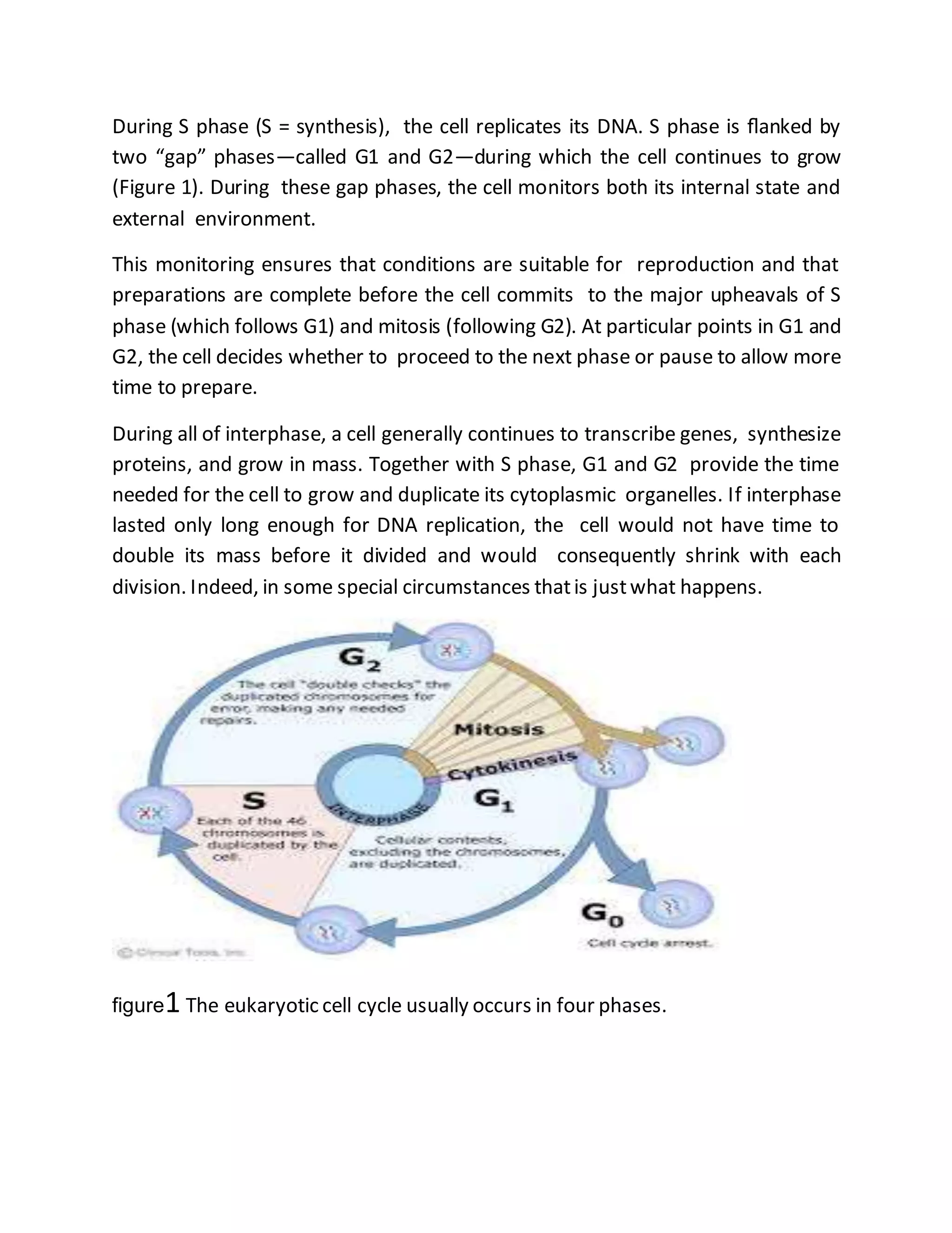
CELL CYCLE
“Where a cell arises, there must be a previous cell, just as animals can only arise
from animals and plants from plants.” A cell reproduces by carrying out an orderly
sequence of events in which it duplicates its contents and then divides in two.
This cycle of duplication and division, known as the cell cycle, is the essential
mechanism by which all living things reproduce. The details of the cell cycle vary
fromorganismto organismand at different times in an individual organism’s life.
Certain features of the cell cycle, however, are universal, as they allow every cell
to perform the fundamental task of copying and passing on its genetic
information to the next generation of cells.
The most basic function of the cell cycle is to duplicate accurately the vast
amount of DNA in the chromosomes and then to segregate the DNA into
genetically identical daughter cells such that each cell receives a complete copy
of the entire genome . In most cases, a cell also duplicates its other
macromolecules and organelles and doubles in size before it divides; otherwise,
each time a cell split it would get smaller and smaller.
Thus, to maintain their size, dividing cells coordinate their growth with their
division. The duration of the cell cycle varies greatly from one cell type to another.
The Eukaryotic Cell Cycle Usually Includes Four Phases
Seen in a microscope, the two most dramatic events in the cell cycle are when
the nucleus divides, a process called mitosis, and when the cell later splits in two,
a process called cytokinesis. These two processes together constitute the M
phase of the cycle. In a typical mammalian cell, the whole of M phase takes about
an hour, which is only a small fraction of the total cell-cycle time .
The period between one M phase and the next is called interphase. Viewed with
a microscope, it appears, deceptively, as an uneventful interlude during which
the cell simply increases in size. Interphase, however, is a very busy time for a
proliferating cell, and it encompasses the remaining three phases of the cell cycle.
During S phase (S = synthesis), the cell replicates its DNA. S phase is flanked by
two “gap” phases—called G1 and G2—during which the cell continues to grow
(Figure 1). During these gap phases, the cell monitors both its internal state and
external environment.
This monitoring ensures that conditions are suitable for reproduction and that
preparations are complete before the cell commits to the major upheavals of S
phase (which follows G1) and mitosis (following G2). At particular points in G1 and
G2, the cell decides whether to proceed to the next phase or pause to allow more
time to prepare.
During all of interphase, a cell generally continues to transcribe genes, synthesize
proteins, and grow in mass. Together with S phase, G1 and G2 provide the time
needed for the cell to grow and duplicate its cytoplasmic organelles. If interphase
lasted only long enough for DNA replication, the cell would not have time to
double its mass before it divided and would consequently shrink with each
division. Indeed, in some special circumstances thatis justwhat happens.
figure1 The eukaryotic cell cycle usually occurs in four phases.
A Cell-Cycle Control System Triggers the Major Processes
of the Cell Cycle
To ensure that they replicate all their DNA and organelles, and divide in an
orderly manner, eukaryotic cells possess a complex network of regulatory
proteins known as the cell-cycle control system. This system guarantees that the
events of the cell cycle—DNA replication, mitosis, and so on—occur in a set
sequence and that each process has been completed beforethe next one begins.
If DNA synthesis is slowed down or stalled, mitosis and cell division must also be
delayed. Similarly, if DNA is damaged, the cycle must arrest in G1, S, or G2 so that
the cell can repair the damage, either before DNA replication is started or
completed or before the cell enters M phase. The cell-cycle control system
achieves all of this by employing molecular brakes, sometimes called checkpoints,
to pause the cycle at certain transition points. In this way, the control system
does not trigger the next step in the cycle unless the cell is properly prepared.
The cell-cycle control system regulates progression through the cell cycle at three
main transition points (Figure 1). At the transition from G1 to S phase, the control
system confirms that the environment is favorable for proliferation before
committing to DNA replication. Cell proliferation in animals requires both
sufficient nutrients and specific signal molecules in the extracellular environment;
if these extracellular conditions are unfavorable, cells can delay progress through
G1 and may even enter a specialized resting state known as G0 (G zero).
At the transition from G2 to M phase, the control system confirms that the DNA
is undamaged and fully replicated, ensuring that the cell does not enter mitosis
unless its DNA is intact. Finally, during mitosis, the cell-cycle control machinery
ensures that the duplicated chromosomes are properly attached to a cytoskeletal
machine, called the mitotic spindle, before the spindle pulls the chromosomes
apart and segregates them into the two daughter cells.
In animals, the transition from G1 to S phase is especially important as a point in
the cell cycle where the control system is regulated. Signals from other cells
stimulate cell proliferation when more cells are needed—and block it when they
are not. The cell-cycle control system therefore plays a central part in the
regulation of cell numbers in the tissues of the body; if the control system
malfunctions such that cell division is excessive, cancer can result.
G1 PHASE
In addition to being a bustling period of metabolic activity, cell growth, and repair,
G1 is an important point of decision-making for the cell. Based on intracellular
signals that provide information about the size of the cell and extracellular signals
reflecting the environment, the cell-cycle control machinery can either hold the
cell transiently in G1 (or in a more prolonged nonproliferative state, G0), or allow
it to prepare for entry into the S phase of another cell cycle. Once past this critical
G1-to-S transition, a cell usually continues all the way through the rest of the cell
cycle quickly—typically within 12–24 hours in mammals.
S PHASE
Before a cell divides, it must replicate its DNA. this replication must occur with
extreme accuracy to minimize the risk of mutations in the next cell generation. Of
equal importance, every nucleotide in the genome must be copied once—and
only once—to prevent the damaging effects of gene amplification.Once a cell has
successfully replicated its DNA in S phase, and progressed through G2, it is ready
to enter M phase. During this relatively brief period, the cell will divide its nucleus
(mitosis) and then its cytoplasm(cytokinesis; seeFigure 1).
M PHASE
Although M phase (mitosis plus cytokinesis) occurs over a relatively short amount
of time—about one hour in a mammalian cell that divides once a day, or even
once a year—it is by far the most dramatic phase of the cell cycle. During this
brief period, the cell reorganizes virtually all of its components and distributes
them equally into the two daughter cells. The earlier phases of the cell cycle, in
effect, serveto set the stage for the drama of M phase.
The central problem for a cell in M phase is to accurately segregate the
chromosomes that were duplicated in the preceding S phase, so that each new
daughter cell receives an identical copy of the genome. With minor variations, all
eukaryotes solve this problem in a similar way: they assemble two specialized
cytoskeletal machines, one that pulls the duplicated chromosomes apart (during
mitosis) and another that divides the cytoplasminto two halves (cytokinesis).
MITOSIS
Before nuclear division, or mitosis, begins, each chromosome has been
duplicated and consists of two identical sister chromatids, held together along
their length by cohesin proteins . During mitosis, the cohesin proteins are cleaved,
the sister chromatids split apart, and the resulting daughter chromosomes are
pulled to opposite poles of the cell by the mitotic spindle.
The Mitotic Spindle Starts to Assemble in Prophase
The mitotic spindle begins to form in prophase. This assembly of the highly
dynamic spindle depends on the remarkable properties of microtubules.
microtubules continuously polymerize and depolymerize by the addition and loss
of their tubulin subunits, and individual filaments alternate between growing and
shrinking—a process called dynamic instability . At the start of mitosis, the
stability of microtubules decreases.As a result, during prophase, rapidly growing
and shrinking microtubules extend in all directions from the two centrosomes,
exploring the interior of the cell.
Some of the microtubules growing from one centrosome interact with the
microtubules from the other centrosome . This interaction stabilizes the
microtubules, preventing them from depolymerizing, and it joins the two sets of
microtubules together to form the basic framework of the mitotic spindle, with its
characteristic bipolar shape . The two centrosomes that give rise to these
microtubules are now called spindle poles, and the interacting microtubules are
called interpolar microtubules .
The assembly of the spindle is driven, in part, by motor proteins associated with
the interpolar microtubules that help to cross-link the two sets of microtubules.
In the next stage of mitosis, the duplicated chromosomes attach to the spindle
microtubules in such a way that, when the sister chromatids separate, they will be
drawn to opposite poles of the cell.figure 2
figure2 THE PRINCIPAL STAGES OF MPHASEINAN ANIMAL CELL
Chromosomes Attach to the Mitotic Spindle at
Prometaphase
Prometaphase starts abruptly with the disassembly of the nuclear envelope,
which breaks up into small membrane vesicles. This process is triggered by the
phosphorylation and consequent disassembly of nuclear pore proteins and the
intermediate filament proteins of the nuclear lamina, the network of fibrous
proteins that underlies and stabilizes the nuclear envelope. The spindle
microtubules, which have been lying in wait outside the nucleus, now gain access
to the duplicated chromosomes and capturethem (figure 2).
The spindle microtubules grab hold of the chromosomes at kinetochores, protein
complexes that assemble on the centromere of each condensed chromosome
during late prophase (Figure 3). Each duplicated chromosome has two
kinetochores—one on each sister chromatid—which face in opposite directions.
Kinetochores recognize the special DNA sequence present at the centromere: if
this sequence is altered, kinetochores fail to assemble and, consequently, the
chromosomes failto segregateproperly during mitosis.
Chromosomes are more than passive passengers in the process of spindle
assembly: they can themselves stabilize and organize microtubules into
functional mitotic spindles. In cells without centrosomes—including all plant cells
and some animal cell types—the chromosomes nucleate microtubule assembly,
and motor proteins then move and arrange the microtubules and chromosomes
into a bipolar spindle. Even in animal cells that normally have centrosomes, a
bipolar spindle can still be formed in this way if the centrosomes are removed . In
cells with chromosomes, motor proteins, and centrosomes work together to form
the mitotic spindle.
figure3Kinetochores attach chromosomes to the mitotic spindle.
Chromosomes Line Up at the Spindle Equator at
Metaphase
During prometaphase, the duplicated chromosomes, now attached to the mitotic
spindle ,Eventually, they align at the equator of the spindle, halfway between the
two spindle poles, thereby forming the metaphase plate. This event defines the
beginning of metaphase( see figure 2).
Although the forces that act to bring the chromosomes to the equator are not
completely understood, both the continual growth and shrinkage of the
microtubules and the action of microtubule motor proteins are required. A
continuous balanced addition and loss of tubulin subunits is also required to
maintain the metaphase spindle: when tubulin addition to the ends of
microtubules is blocked by the drug colchicine, tubulin loss continues until the
metaphase spindle disappears.
The chromosomes gathered at the equator of the metaphase spindle oscillate
back and forth, continually adjusting their positions, indicating that the tug-of-
war between the microtubules attached to opposite poles of the spindle
continues to operate after the chromosomes areall aligned.
If one of the pair of kinetochore attachments is artificially severed with a laser
beam during metaphase, the entire duplicated chromosome immediately moves
toward the pole to which it remains attached. Similarly, if the attachment
between sister chromatids is cut, the two chromosomes separate and move
toward opposite poles. These experiments show that the duplicated
chromosomes are not simply deposited at the metaphase plate. They are
suspended there under tension. In anaphase, that tension will pull the sister
chromatids apart.
Proteolysis Triggers Sister-Chromatid Separation at
Anaphase
Anaphase begins abruptly with the breakage of the cohesin linkages that hold
sister chromatids together . This release allows each chromatid—now considered
a chromosome—to be pulled to the spindle pole to which it is attached . This
movement segregates the two identical sets of chromosomes to opposite ends of
the spindle (seefigure 2).
Once the sister chromatids separate, the resulting chromosomes are pulled to
the spindle pole to which they are attached. They all move at the same speed,
which is typically about 1 μm per minute. The movement is the consequence of
two independent processes thatdepend on different parts of the mitotic spindle.
The two processes are called anaphase A and anaphase B, and they occur more
or less simultaneously. In anaphase A, the kinetochore microtubules shorten and
the attached chromosomes move poleward. In anaphase B, the spindle poles
themselves move apart, further segregating the two sets of chromosomes (Figure
4).
figure4Two processes segregate daughter chromosomes atanaphase.
The Nuclear Envelope Re-forms at Telophase
By the end of anaphase, the daughter chromosomes have separated into two
equal groups, one at each pole of the spindle. During telophase, the final stage of
mitosis, the mitotic spindle disassembles, and a nuclear envelope reassembles
around each group of chromosomes to form the two daughter nuclei . Vesicles of
nuclear membrane first cluster around individual chromosomes and then fuse to
re-formthe nuclear envelope (seefigure 2).
During this process, the nuclear pore proteins and nuclear lamins that were
phosphorylated during prometaphase are now dephosphorylated, which allows
them to reassemble and the nuclear envelope and lamina to re-form(Figure5).
Once the nuclear envelope has re-formed, the pores pump in nuclear proteins,
the nucleus expands, and the condensed chromosomes decondense into their
interphase state. As a consequence of this decondensation, gene transcription is
able to resume. A new nucleus has been created, and mitosis is complete. All that
remains is for the cell to complete its division into two separatedaughter cells.
figure 5The nuclear envelope breaks down and re-forms during mitosis.
CYTOKINESIS
Cytokinesis, the process by which the cytoplasm is cleaved in two, completes M
phase (see figure 2 ) .It usually begins in anaphase but is not completed until the
two daughter nuclei have formed in telophase. Whereas mitosis depends on a
transient microtubule-based structure, the mitotic spindle, cytokinesis in animal
cells depends on a transient structure based on actin and myosin filaments, the
contractile ring . Both the plane of cleavage and the timing of cytokinesis,
however, aredetermined by the mitotic spindle.
The Mitotic Spindle Determines the Plane of Cytoplasmic
Cleavage
The first visible sign of cytokinesis in animal cells is a puckering and furrowing of
the plasma membrane that occurs during anaphase.The furrowing invariably
occurs in a plane that runs perpendicular to the long axis of the mitotic spindle.
This positioning ensures that the cleavage furrow cuts between the two groups of
segregated chromosomes, so that each daughter cell receives an identical and
complete set of chromosomes. If the mitotic spindle is deliberately displaced
(using a fine glass needle) as soon as the furrow appears, the furrow disappears
and a new one develops at a site corresponding to the new spindle location and
orientation. (figure6)
Once the furrowing process is well under way, however, cleavage proceeds even
if the mitotic spindle is artificially sucked out of the cell or depolymerized using
the drug colchicine. How does the mitotic spindle dictate the position of the
cleavage furrow? The mechanism is still uncertain, but it appears that, during
anaphase, the overlapping interpolar microtubules that form the central spindle
recruit and activate proteins that signal to the cell cortex to initiate the assembly
of the contractile ring at a position midway between the spindle poles. Because
these signals originate in the anaphase spindle, this mechanism also contributes
to the timing of cytokinesis in late mitosis.
When the mitotic spindle is located centrally in the cell—the usual situation in
most dividing cells—the two daughter cells produced will be of equal size. During
embryonic development, however, there are some instances in which the
dividing cell moves its mitotic spindle to an asymmetrical position, and,
consequently, the furrow creates two daughter cells that differ in size. In most of
these asymmetric divisions, the daughters also differ in the molecules they inherit,
and they usually develop into differentcell types.
figure 6 The contractile ring divides the cell in two

Cell cycle and mitosis

  • 1.
    CELL CYCLE “Where acell arises, there must be a previous cell, just as animals can only arise from animals and plants from plants.” A cell reproduces by carrying out an orderly sequence of events in which it duplicates its contents and then divides in two. This cycle of duplication and division, known as the cell cycle, is the essential mechanism by which all living things reproduce. The details of the cell cycle vary fromorganismto organismand at different times in an individual organism’s life. Certain features of the cell cycle, however, are universal, as they allow every cell to perform the fundamental task of copying and passing on its genetic information to the next generation of cells. The most basic function of the cell cycle is to duplicate accurately the vast amount of DNA in the chromosomes and then to segregate the DNA into genetically identical daughter cells such that each cell receives a complete copy of the entire genome . In most cases, a cell also duplicates its other macromolecules and organelles and doubles in size before it divides; otherwise, each time a cell split it would get smaller and smaller. Thus, to maintain their size, dividing cells coordinate their growth with their division. The duration of the cell cycle varies greatly from one cell type to another. The Eukaryotic Cell Cycle Usually Includes Four Phases Seen in a microscope, the two most dramatic events in the cell cycle are when the nucleus divides, a process called mitosis, and when the cell later splits in two, a process called cytokinesis. These two processes together constitute the M phase of the cycle. In a typical mammalian cell, the whole of M phase takes about an hour, which is only a small fraction of the total cell-cycle time . The period between one M phase and the next is called interphase. Viewed with a microscope, it appears, deceptively, as an uneventful interlude during which the cell simply increases in size. Interphase, however, is a very busy time for a proliferating cell, and it encompasses the remaining three phases of the cell cycle.
  • 2.
    During S phase(S = synthesis), the cell replicates its DNA. S phase is flanked by two “gap” phases—called G1 and G2—during which the cell continues to grow (Figure 1). During these gap phases, the cell monitors both its internal state and external environment. This monitoring ensures that conditions are suitable for reproduction and that preparations are complete before the cell commits to the major upheavals of S phase (which follows G1) and mitosis (following G2). At particular points in G1 and G2, the cell decides whether to proceed to the next phase or pause to allow more time to prepare. During all of interphase, a cell generally continues to transcribe genes, synthesize proteins, and grow in mass. Together with S phase, G1 and G2 provide the time needed for the cell to grow and duplicate its cytoplasmic organelles. If interphase lasted only long enough for DNA replication, the cell would not have time to double its mass before it divided and would consequently shrink with each division. Indeed, in some special circumstances thatis justwhat happens. figure1 The eukaryotic cell cycle usually occurs in four phases.
  • 3.
    A Cell-Cycle ControlSystem Triggers the Major Processes of the Cell Cycle To ensure that they replicate all their DNA and organelles, and divide in an orderly manner, eukaryotic cells possess a complex network of regulatory proteins known as the cell-cycle control system. This system guarantees that the events of the cell cycle—DNA replication, mitosis, and so on—occur in a set sequence and that each process has been completed beforethe next one begins. If DNA synthesis is slowed down or stalled, mitosis and cell division must also be delayed. Similarly, if DNA is damaged, the cycle must arrest in G1, S, or G2 so that the cell can repair the damage, either before DNA replication is started or completed or before the cell enters M phase. The cell-cycle control system achieves all of this by employing molecular brakes, sometimes called checkpoints, to pause the cycle at certain transition points. In this way, the control system does not trigger the next step in the cycle unless the cell is properly prepared. The cell-cycle control system regulates progression through the cell cycle at three main transition points (Figure 1). At the transition from G1 to S phase, the control system confirms that the environment is favorable for proliferation before committing to DNA replication. Cell proliferation in animals requires both sufficient nutrients and specific signal molecules in the extracellular environment; if these extracellular conditions are unfavorable, cells can delay progress through G1 and may even enter a specialized resting state known as G0 (G zero). At the transition from G2 to M phase, the control system confirms that the DNA is undamaged and fully replicated, ensuring that the cell does not enter mitosis unless its DNA is intact. Finally, during mitosis, the cell-cycle control machinery ensures that the duplicated chromosomes are properly attached to a cytoskeletal machine, called the mitotic spindle, before the spindle pulls the chromosomes apart and segregates them into the two daughter cells. In animals, the transition from G1 to S phase is especially important as a point in the cell cycle where the control system is regulated. Signals from other cells
  • 4.
    stimulate cell proliferationwhen more cells are needed—and block it when they are not. The cell-cycle control system therefore plays a central part in the regulation of cell numbers in the tissues of the body; if the control system malfunctions such that cell division is excessive, cancer can result. G1 PHASE In addition to being a bustling period of metabolic activity, cell growth, and repair, G1 is an important point of decision-making for the cell. Based on intracellular signals that provide information about the size of the cell and extracellular signals reflecting the environment, the cell-cycle control machinery can either hold the cell transiently in G1 (or in a more prolonged nonproliferative state, G0), or allow it to prepare for entry into the S phase of another cell cycle. Once past this critical G1-to-S transition, a cell usually continues all the way through the rest of the cell cycle quickly—typically within 12–24 hours in mammals. S PHASE Before a cell divides, it must replicate its DNA. this replication must occur with extreme accuracy to minimize the risk of mutations in the next cell generation. Of equal importance, every nucleotide in the genome must be copied once—and only once—to prevent the damaging effects of gene amplification.Once a cell has successfully replicated its DNA in S phase, and progressed through G2, it is ready to enter M phase. During this relatively brief period, the cell will divide its nucleus (mitosis) and then its cytoplasm(cytokinesis; seeFigure 1). M PHASE Although M phase (mitosis plus cytokinesis) occurs over a relatively short amount of time—about one hour in a mammalian cell that divides once a day, or even once a year—it is by far the most dramatic phase of the cell cycle. During this brief period, the cell reorganizes virtually all of its components and distributes them equally into the two daughter cells. The earlier phases of the cell cycle, in effect, serveto set the stage for the drama of M phase.
  • 5.
    The central problemfor a cell in M phase is to accurately segregate the chromosomes that were duplicated in the preceding S phase, so that each new daughter cell receives an identical copy of the genome. With minor variations, all eukaryotes solve this problem in a similar way: they assemble two specialized cytoskeletal machines, one that pulls the duplicated chromosomes apart (during mitosis) and another that divides the cytoplasminto two halves (cytokinesis). MITOSIS Before nuclear division, or mitosis, begins, each chromosome has been duplicated and consists of two identical sister chromatids, held together along their length by cohesin proteins . During mitosis, the cohesin proteins are cleaved, the sister chromatids split apart, and the resulting daughter chromosomes are pulled to opposite poles of the cell by the mitotic spindle. The Mitotic Spindle Starts to Assemble in Prophase The mitotic spindle begins to form in prophase. This assembly of the highly dynamic spindle depends on the remarkable properties of microtubules. microtubules continuously polymerize and depolymerize by the addition and loss of their tubulin subunits, and individual filaments alternate between growing and shrinking—a process called dynamic instability . At the start of mitosis, the stability of microtubules decreases.As a result, during prophase, rapidly growing and shrinking microtubules extend in all directions from the two centrosomes, exploring the interior of the cell. Some of the microtubules growing from one centrosome interact with the microtubules from the other centrosome . This interaction stabilizes the microtubules, preventing them from depolymerizing, and it joins the two sets of microtubules together to form the basic framework of the mitotic spindle, with its characteristic bipolar shape . The two centrosomes that give rise to these microtubules are now called spindle poles, and the interacting microtubules are called interpolar microtubules .
  • 6.
    The assembly ofthe spindle is driven, in part, by motor proteins associated with the interpolar microtubules that help to cross-link the two sets of microtubules. In the next stage of mitosis, the duplicated chromosomes attach to the spindle microtubules in such a way that, when the sister chromatids separate, they will be drawn to opposite poles of the cell.figure 2 figure2 THE PRINCIPAL STAGES OF MPHASEINAN ANIMAL CELL
  • 7.
    Chromosomes Attach tothe Mitotic Spindle at Prometaphase Prometaphase starts abruptly with the disassembly of the nuclear envelope, which breaks up into small membrane vesicles. This process is triggered by the phosphorylation and consequent disassembly of nuclear pore proteins and the intermediate filament proteins of the nuclear lamina, the network of fibrous proteins that underlies and stabilizes the nuclear envelope. The spindle microtubules, which have been lying in wait outside the nucleus, now gain access to the duplicated chromosomes and capturethem (figure 2). The spindle microtubules grab hold of the chromosomes at kinetochores, protein complexes that assemble on the centromere of each condensed chromosome during late prophase (Figure 3). Each duplicated chromosome has two kinetochores—one on each sister chromatid—which face in opposite directions. Kinetochores recognize the special DNA sequence present at the centromere: if this sequence is altered, kinetochores fail to assemble and, consequently, the chromosomes failto segregateproperly during mitosis. Chromosomes are more than passive passengers in the process of spindle assembly: they can themselves stabilize and organize microtubules into functional mitotic spindles. In cells without centrosomes—including all plant cells and some animal cell types—the chromosomes nucleate microtubule assembly, and motor proteins then move and arrange the microtubules and chromosomes into a bipolar spindle. Even in animal cells that normally have centrosomes, a bipolar spindle can still be formed in this way if the centrosomes are removed . In cells with chromosomes, motor proteins, and centrosomes work together to form the mitotic spindle.
  • 8.
    figure3Kinetochores attach chromosomesto the mitotic spindle. Chromosomes Line Up at the Spindle Equator at Metaphase During prometaphase, the duplicated chromosomes, now attached to the mitotic spindle ,Eventually, they align at the equator of the spindle, halfway between the two spindle poles, thereby forming the metaphase plate. This event defines the beginning of metaphase( see figure 2). Although the forces that act to bring the chromosomes to the equator are not completely understood, both the continual growth and shrinkage of the microtubules and the action of microtubule motor proteins are required. A continuous balanced addition and loss of tubulin subunits is also required to maintain the metaphase spindle: when tubulin addition to the ends of microtubules is blocked by the drug colchicine, tubulin loss continues until the metaphase spindle disappears. The chromosomes gathered at the equator of the metaphase spindle oscillate back and forth, continually adjusting their positions, indicating that the tug-of- war between the microtubules attached to opposite poles of the spindle continues to operate after the chromosomes areall aligned.
  • 9.
    If one ofthe pair of kinetochore attachments is artificially severed with a laser beam during metaphase, the entire duplicated chromosome immediately moves toward the pole to which it remains attached. Similarly, if the attachment between sister chromatids is cut, the two chromosomes separate and move toward opposite poles. These experiments show that the duplicated chromosomes are not simply deposited at the metaphase plate. They are suspended there under tension. In anaphase, that tension will pull the sister chromatids apart. Proteolysis Triggers Sister-Chromatid Separation at Anaphase Anaphase begins abruptly with the breakage of the cohesin linkages that hold sister chromatids together . This release allows each chromatid—now considered a chromosome—to be pulled to the spindle pole to which it is attached . This movement segregates the two identical sets of chromosomes to opposite ends of the spindle (seefigure 2). Once the sister chromatids separate, the resulting chromosomes are pulled to the spindle pole to which they are attached. They all move at the same speed, which is typically about 1 μm per minute. The movement is the consequence of two independent processes thatdepend on different parts of the mitotic spindle. The two processes are called anaphase A and anaphase B, and they occur more or less simultaneously. In anaphase A, the kinetochore microtubules shorten and the attached chromosomes move poleward. In anaphase B, the spindle poles themselves move apart, further segregating the two sets of chromosomes (Figure 4).
  • 10.
    figure4Two processes segregatedaughter chromosomes atanaphase. The Nuclear Envelope Re-forms at Telophase By the end of anaphase, the daughter chromosomes have separated into two equal groups, one at each pole of the spindle. During telophase, the final stage of mitosis, the mitotic spindle disassembles, and a nuclear envelope reassembles around each group of chromosomes to form the two daughter nuclei . Vesicles of nuclear membrane first cluster around individual chromosomes and then fuse to re-formthe nuclear envelope (seefigure 2). During this process, the nuclear pore proteins and nuclear lamins that were phosphorylated during prometaphase are now dephosphorylated, which allows them to reassemble and the nuclear envelope and lamina to re-form(Figure5).
  • 11.
    Once the nuclearenvelope has re-formed, the pores pump in nuclear proteins, the nucleus expands, and the condensed chromosomes decondense into their interphase state. As a consequence of this decondensation, gene transcription is able to resume. A new nucleus has been created, and mitosis is complete. All that remains is for the cell to complete its division into two separatedaughter cells. figure 5The nuclear envelope breaks down and re-forms during mitosis. CYTOKINESIS
  • 12.
    Cytokinesis, the processby which the cytoplasm is cleaved in two, completes M phase (see figure 2 ) .It usually begins in anaphase but is not completed until the two daughter nuclei have formed in telophase. Whereas mitosis depends on a transient microtubule-based structure, the mitotic spindle, cytokinesis in animal cells depends on a transient structure based on actin and myosin filaments, the contractile ring . Both the plane of cleavage and the timing of cytokinesis, however, aredetermined by the mitotic spindle. The Mitotic Spindle Determines the Plane of Cytoplasmic Cleavage The first visible sign of cytokinesis in animal cells is a puckering and furrowing of the plasma membrane that occurs during anaphase.The furrowing invariably occurs in a plane that runs perpendicular to the long axis of the mitotic spindle. This positioning ensures that the cleavage furrow cuts between the two groups of segregated chromosomes, so that each daughter cell receives an identical and complete set of chromosomes. If the mitotic spindle is deliberately displaced (using a fine glass needle) as soon as the furrow appears, the furrow disappears and a new one develops at a site corresponding to the new spindle location and orientation. (figure6) Once the furrowing process is well under way, however, cleavage proceeds even if the mitotic spindle is artificially sucked out of the cell or depolymerized using the drug colchicine. How does the mitotic spindle dictate the position of the cleavage furrow? The mechanism is still uncertain, but it appears that, during anaphase, the overlapping interpolar microtubules that form the central spindle recruit and activate proteins that signal to the cell cortex to initiate the assembly of the contractile ring at a position midway between the spindle poles. Because these signals originate in the anaphase spindle, this mechanism also contributes to the timing of cytokinesis in late mitosis. When the mitotic spindle is located centrally in the cell—the usual situation in most dividing cells—the two daughter cells produced will be of equal size. During embryonic development, however, there are some instances in which the
  • 13.
    dividing cell movesits mitotic spindle to an asymmetrical position, and, consequently, the furrow creates two daughter cells that differ in size. In most of these asymmetric divisions, the daughters also differ in the molecules they inherit, and they usually develop into differentcell types. figure 6 The contractile ring divides the cell in two